. 2017 Jan 30:8:14310.
doi: 10.1038/ncomms14310.

Portal protein functions akin to a DNA-sensor that couples genome-packaging to icosahedral capsid maturation

Affiliations

Portal protein functions akin to a DNA-sensor that couples genome-packaging to icosahedral capsid maturation

Ravi K Lokareddy et al. Nat Commun. .

Abstract

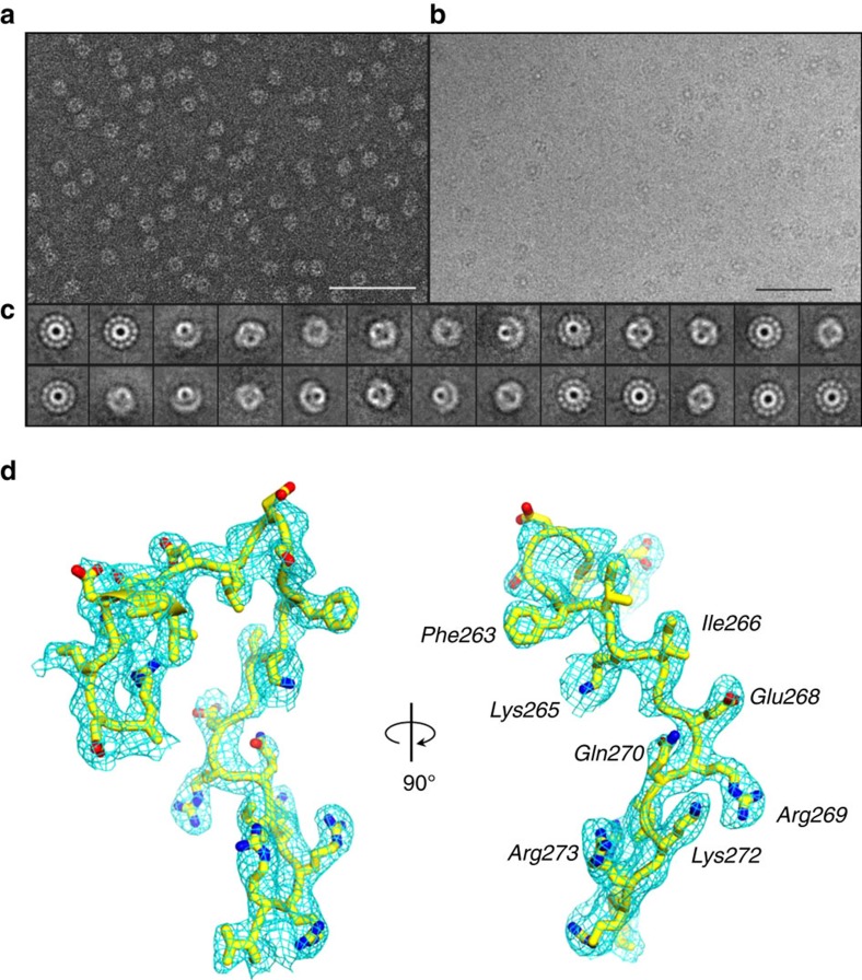
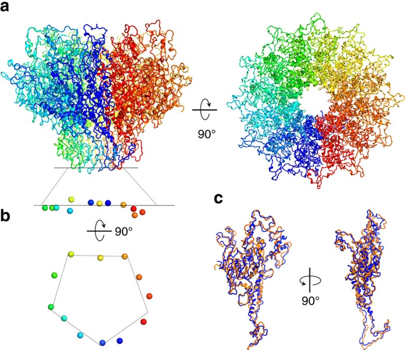
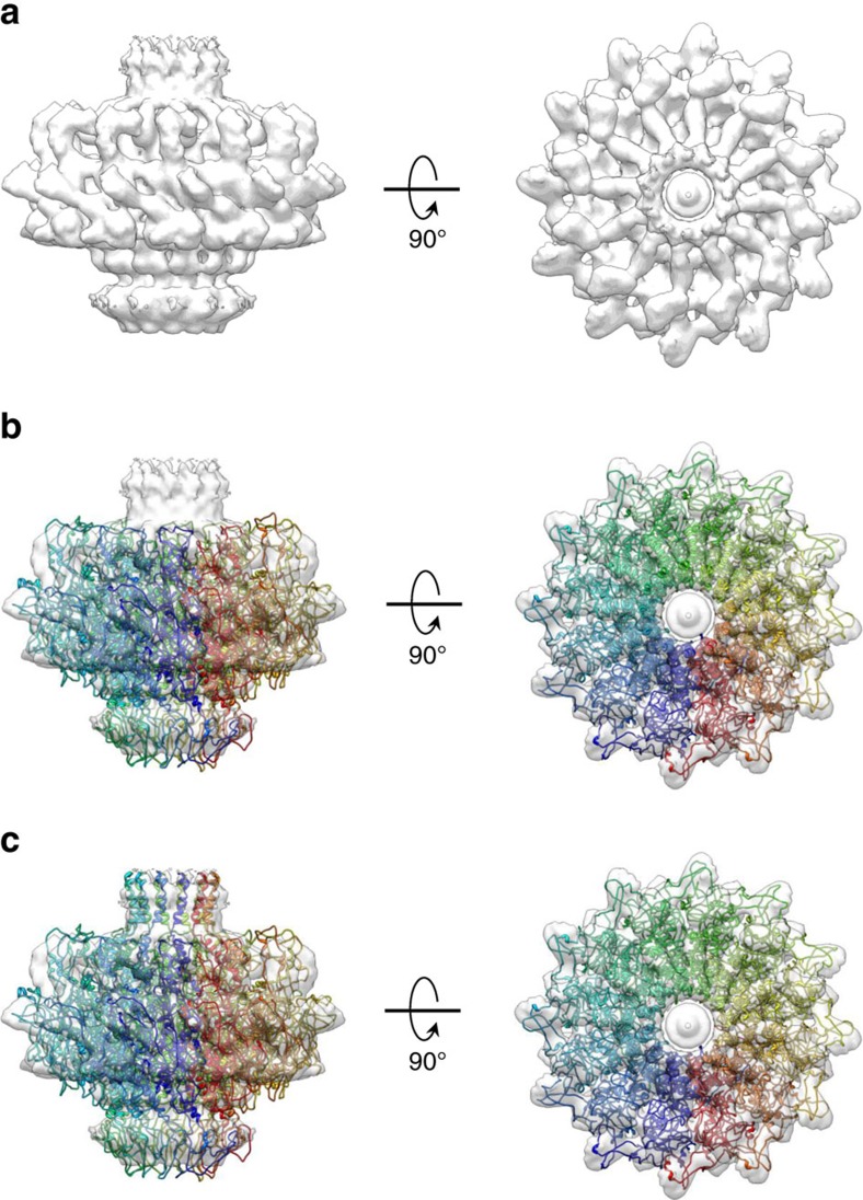
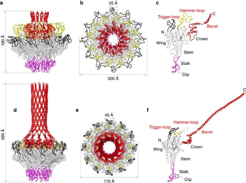
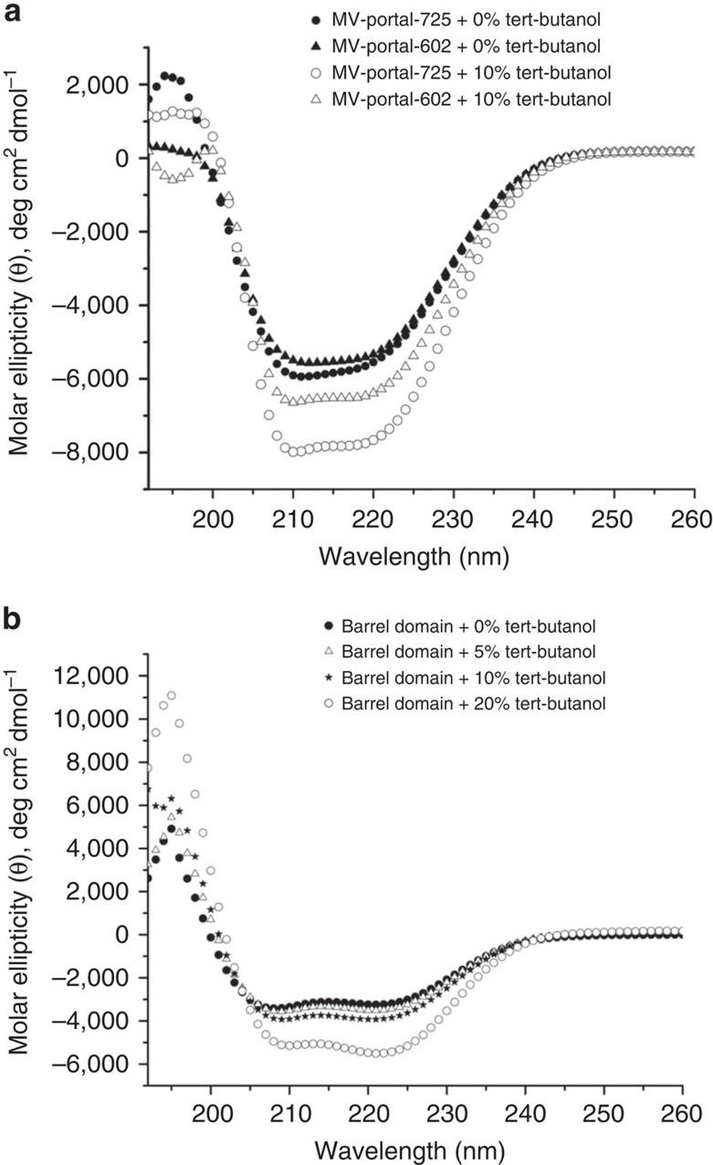
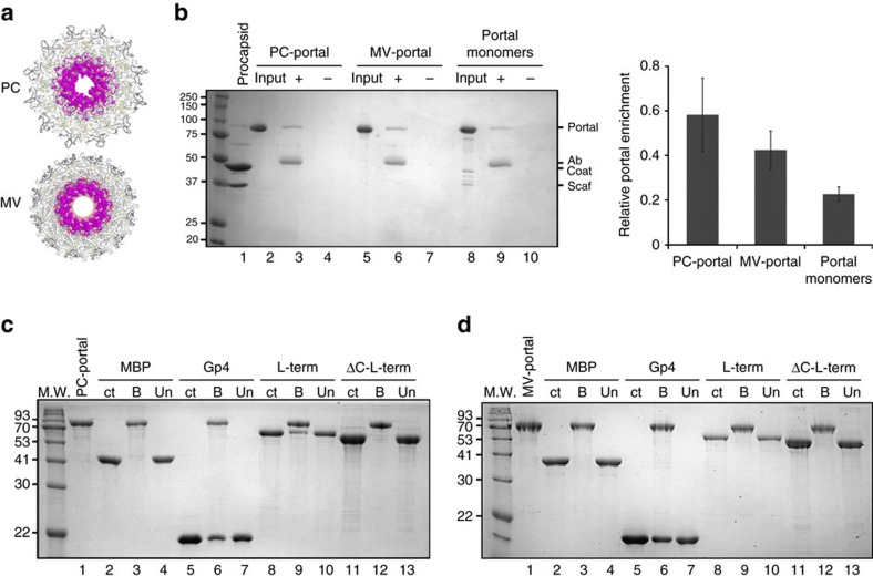
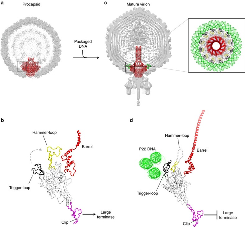
Tailed bacteriophages and herpesviruses assemble infectious particles via an empty precursor capsid (or 'procapsid') built by multiple copies of coat and scaffolding protein and by one dodecameric portal protein. Genome packaging triggers rearrangement of the coat protein and release of scaffolding protein, resulting in dramatic procapsid lattice expansion. Here, we provide structural evidence that the portal protein of the bacteriophage P22 exists in two distinct dodecameric conformations: an asymmetric assembly in the procapsid (PC-portal) that is competent for high affinity binding to the large terminase packaging protein, and a symmetric ring in the mature virion (MV-portal) that has negligible affinity for the packaging motor. Modelling studies indicate the structure of PC-portal is incompatible with DNA coaxially spooled around the portal vertex, suggesting that newly packaged DNA triggers the switch from PC- to MV-conformation. Thus, we propose the signal for termination of 'Headful Packaging' is a DNA-dependent symmetrization of portal protein.


Conflict of interest statement

The authors declare no competing financial interests.

Figures

Figure 1
Figure 1. Evidence for a conformation of P22 portal protein by cryo-EM.
(a) Representative micrograph of PC-portal negatively stained with 1% uranyl formate (Scale bar, 1 μm). (b) Representative micrograph of frozen-hydrated PC-portals (Scale bar, 1 μm). (c) Selected, reference-free 2D class averages of frozen-hydrated particles showing top and side projection views of PC-portal. (d) A σA-weighted 2Fo–Fc difference electron density map computed at 3.30 Å resolution is displayed around a portion of the PC-portal protein model (Arg249Val276), which is shown as sticks. The density is displayed as cyan mesh contoured at 1.65σ above background.
Figure 2
Figure 2. Crystal structure of PC-portal core at 3.30 Å resolution.
(a) Ribbon diagram of the crystal structure of PC-portal protein core shown in side (left panel) and top (right panel) view. (b) Asymmetry in portal protein subunits. Side and bottom view of the side chain oxygen atom of Asn380 (shown as spheres) from each of the 12 subunits. (c) Superimposition of two most dissimilar subunits of PC-portal core, namely chain A (in blue) and chain J (in orange) (RMSD=3.4 Å).
Figure 3
Figure 3. Architecture of the full length PC-portal protein.
(a) 12-fold averaged cryo-EM map of portal protein (coloured in semi-transparent grey) extracted from the 8.7 Å asymmetric reconstruction of P22 procapsid (EMD-1828). (b) Crystal structure of PC-portal core overlaid to the cryo-EM map shown in (a). (c) A complete model of PC-portal protein that including the crystal structure of PC-portal protein core and modelled C-terminus spanning residues 602–631.
Figure 4
Figure 4. Structural comparison of PC- versus MV-portal protein.
Ribbon diagram of P22 portal protein ring and protomer in procapsid (ac) and mature virion (df) conformation. The portal oligomer is coloured in grey with stalk, trigger-loop, hammer-loop and crown-barrel coloured in magenta, black, yellow and red, respectively. The hammer-loop, invisible in MV-portal, is shown as dashes.
Figure 5
Figure 5. The barrel domain is unfolded in solution.
(a) The far-UV CD spectra of portal-725 (black circle) and portal-602 (black triangle) dissolved at 1 μM final concentration, in 10 mM HEPES, pH 7.4 and 70 mM NaCl. Significant effect of 10% tert-butanol was observed for portal-725 (open black circle) as compared with portal-602 (open black triangle). (b) The far-UV CD spectra of the isolated barrel domain dissolved at 5 μM final concentration in 10 mM HEPES, pH 7.4 and 70 mM NaCl in presence and absence of tert-butanol. For each sample, CD spectra were measured at 10 °C in the presence of 0% (black circle), 5% (open black triangle), 10% (black star) and 20% (open black circle) tert-butanol.
Figure 6
Figure 6. Large terminase binds exclusively to PC-portal.
(a) Bottom views of PC-portal (top) and MV-portal (bottom) with residues 375–385 in the stalk loop shown as spheres. (b) Portal protein immunoprecipitation assay. Left panel: Coomassie blue stained SDS–PAGE of a representative portal protein immunoprecipitation. ‘Input': 2.5 μg of portal proteins; ‘+': proteins immunoprecipitated by the anti-stalk antibody; ‘−': proteins incubated with Protein A agarose beads without the antibody. The migration of portal protein, antibody IgG band (Ab), coat protein and scaffolding protein (Scaf) is indicated. Right panel: quantification of portal protein band relative to the antibody band for three experiments, as quantified by densitometry (the error bar represents standard deviation). (c,d) SDS–PAGE analysis of L-terminase binding to PC- (c) and MV-portal protein (d) immobilized on CNBr beads. Portal-beads (lane 1) were used to selectively pull-down MBP (lane 3), gp4 (lane 6), L-terminase (lane 9), ΔC-L-terminase (lane 12). ‘ct': control, ‘B' and ‘Un' are fractions bound and unbound to portal beads.
Figure 7
Figure 7. Modelling portal protein maturation.
(a) Cut-open representation of P22 procapsid (EMD-1827) with the ribbon structure of portal protein (in red) overlaid to the cryo-EM density. (b) Magnified side view of the PC-portal protomer found in procapsid. (c) Cut-open representation of P22 mature virion (EMD-1220) with the ribbon structure of portal protein (in red) overlaid to the cryo-EM density. Magnified on the right is a top view of MV-portal protein surrounded by three rings of DNA visible in the cryo-EM reconstruction. (d) Magnified side view of the MV-portal protomer surrounded by the three rings of DNA.

References

    1. Suhanovsky M. M. & Teschke C. M. Nature's favorite building block: deciphering folding and capsid assembly of proteins with the HK97-fold. Virology 479–480, 487–497 (2015). - PMC - PubMed
    1. Cardone G., Heymann J. B., Cheng N., Trus B. L. & Steven A. C. Procapsid assembly, maturation, nuclear exit: dynamic steps in the production of infectious herpesvirions. Adv. Exp. Med. Biol. 726, 423–439 (2012). - PMC - PubMed
    1. Guo P. et al. Common mechanisms of DNA translocation motors in bacteria and viruses using one-way revolution mechanism without rotation. Biotechnol. Adv. 32, 853–872 (2014). - PMC - PubMed
    1. Bhardwaj A., Olia A. S. & Cingolani G. Architecture of viral genome-delivery molecular machines. Curr. Opin. Struct. Biol. 25, 1–8 (2014). - PMC - PubMed
    1. Chen D. H. et al. Structural basis for scaffolding-mediated assembly and maturation of a dsDNA virus. Proc. Natl Acad. Sci. USA 108, 1355–1360 (2011). - PMC - PubMed

Publication types

MeSH terms