. 2017 Jan 30:8:14011.
doi: 10.1038/ncomms14011.

Specialized interfaces of Smc5/6 control hinge stability and DNA association

Affiliations

Specialized interfaces of Smc5/6 control hinge stability and DNA association

Aaron Alt et al. Nat Commun. .

Abstract

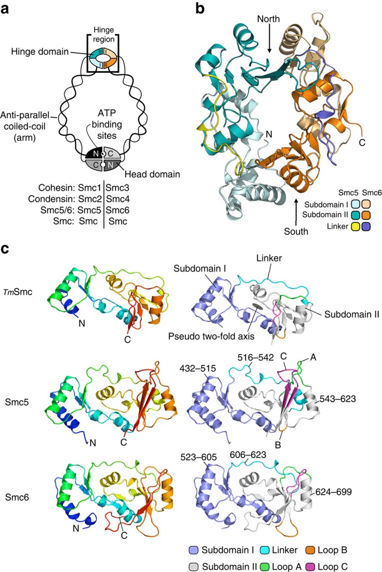
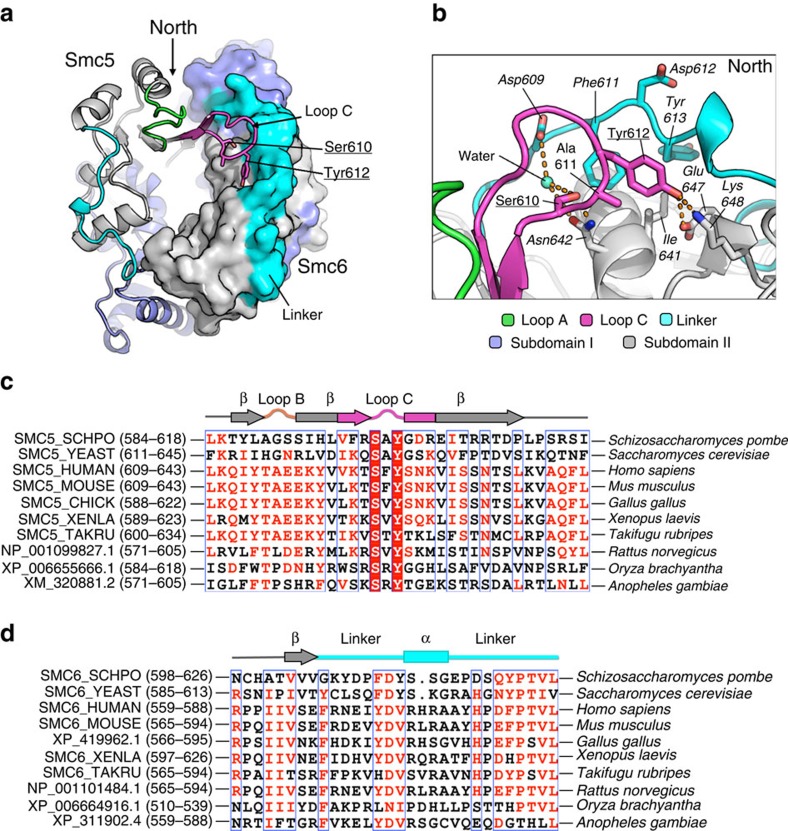
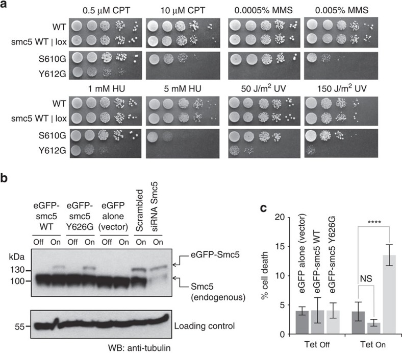
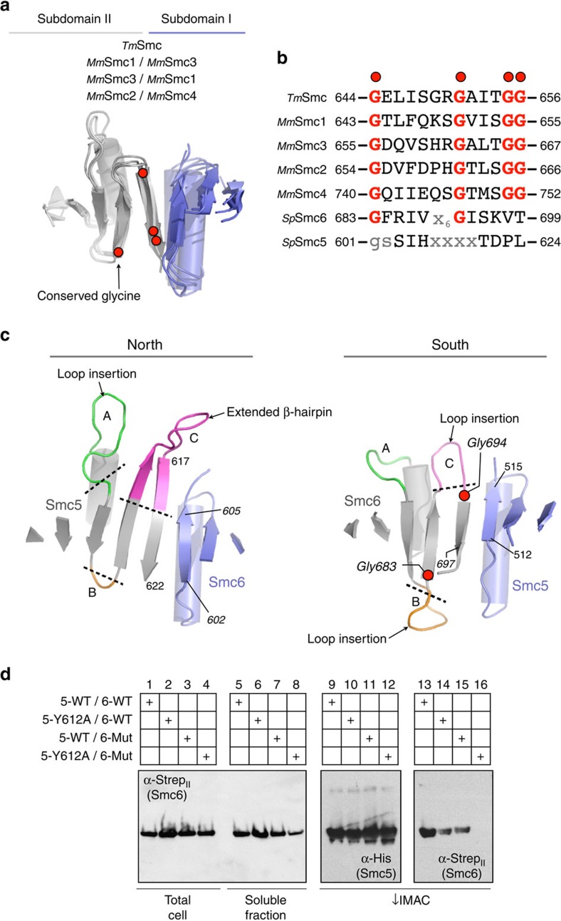
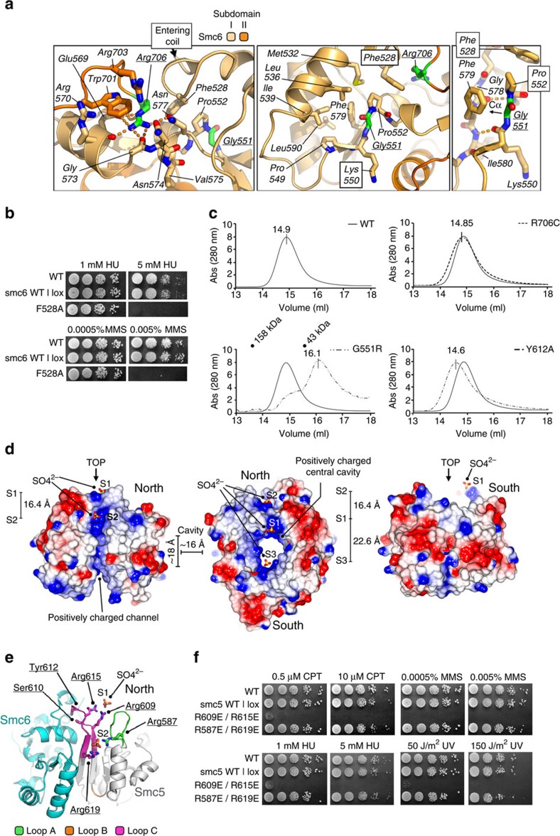
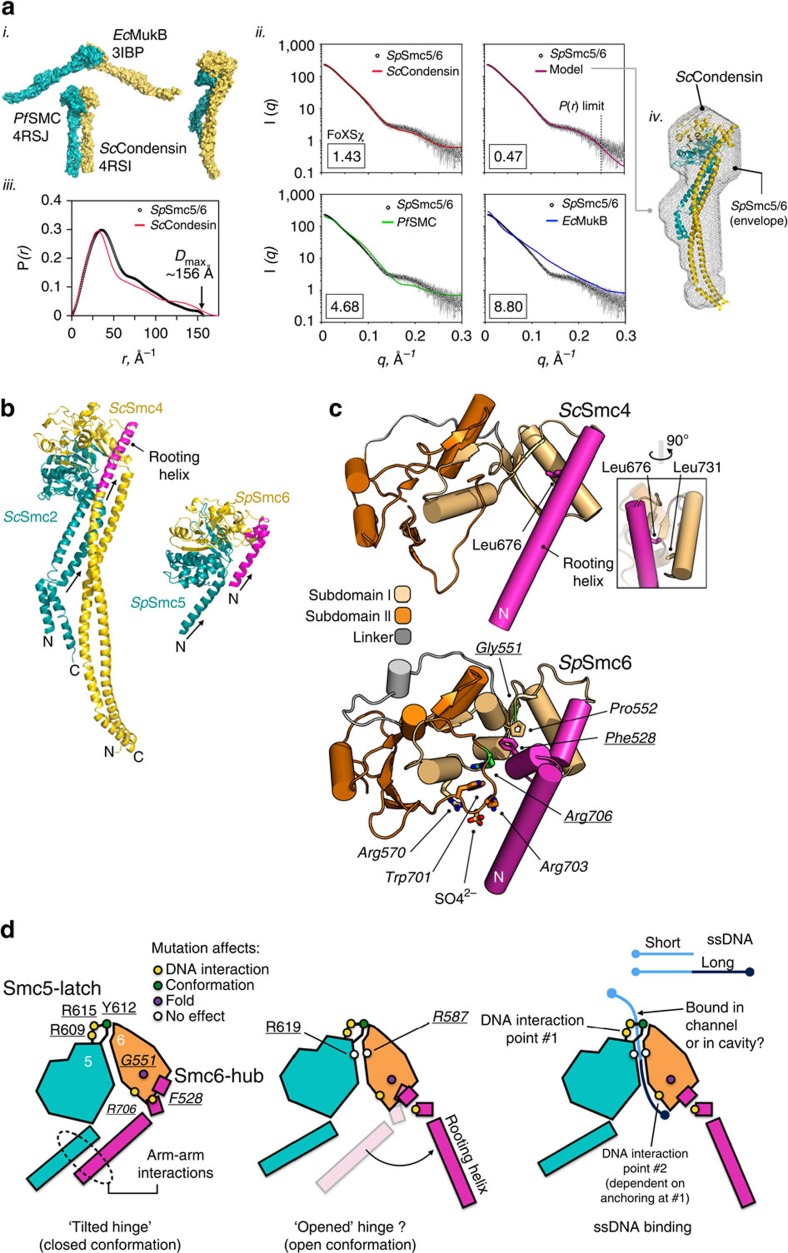
The Structural Maintenance of Chromosomes (SMC) complexes: cohesin, condensin and Smc5/6 are involved in the organization of higher-order chromosome structure-which is essential for accurate chromosome duplication and segregation. Each complex is scaffolded by a specific SMC protein dimer (heterodimer in eukaryotes) held together via their hinge domains. Here we show that the Smc5/6-hinge, like those of cohesin and condensin, also forms a toroidal structure but with distinctive subunit interfaces absent from the other SMC complexes; an unusual 'molecular latch' and a functional 'hub'. Defined mutations in these interfaces cause severe phenotypic effects with sensitivity to DNA-damaging agents in fission yeast and reduced viability in human cells. We show that the Smc5/6-hinge complex binds preferentially to ssDNA and that this interaction is affected by both 'latch' and 'hub' mutations, suggesting a key role for these unique features in controlling DNA association by the Smc5/6 complex.


Figures

Figure 1
Figure 1. The heterodimeric hinge of S. pombe Smc5/6.
(a) Schematic diagram highlighting the conserved architecture and domain composition of the SMC family of proteins. (b) Molecular-cartoon depiction of the S. pombe Smc5/6 heterodimeric hinge, indicating component subdomains, and North and South interfaces; see associated key for details. (c) Comparison of the hinge-domains of Smc5 and Smc6 with a prototypical SMC protein from T. maritima (PDB: 1GXL). (Left) Molecular-cartoon depictions coloured blue→red, from N→C-terminus. (Right) Cartoons coloured according to subdomain, connecting loops and linker regions; see associated key for details. Amino acid boundaries for Subdomain I, Subdomain II and inter-connecting linker region are indicated.
Figure 2
Figure 2. Smc5-Loop C is a conserved feature.
(a) Combined molecular cartoon (Smc5) and molecular surface (Smc6) highlighting the position of Smc5-Loop C and key amino acids. (b) Representative view of the molecular interactions made by Smc5-Loop C with the linker and Subdomain II regions of Smc6; see associated key for details. Selected amino acid residues are labelled. In this, and all subsequent figures residues from Smc5 are shown in plain type, and those from Smc6 in italic type. Amino acids mutated in this study are additionally underlined. (c) Multiple amino acid sequence alignment generated with MultAlin (http://multalin.toulouse.inra.fr/multalin/) for the Loop C region of Smc5. Highly conserved residues are indicated by a red background and white text. (d) MultAlin alignment for the linker region of Smc6. Amino acid residues conserved in physiological properties are coloured in red. Regions of conservation are indicated by the blue outline.
Figure 3
Figure 3. Mutation of Smc5-Loop C in yeast and human cells.
(a) DNA damage sensitivity of S. pombe strains containing Smc5-Loop C mutations S610G and Y612G. Dose and type of treatment is as indicated. WT 501 and Smc5 WT | lox strains are included as controls. (b) Anti-Smc5 western blot confirming induction of eGFP-fused proteins upon addition of doxycycline to the cell culture medium. Identities of species detected by the antibody were confirmed by treatment of cells with siRNA targeting Smc5, reducing the total amount of endogenous WT protein, but not affecting levels of the siRNA-resistant eGFP-fused Smc5. (c) Cell viability assay for U2OS cells stably transfected with doxycycline-inducible constructs expressing eGFP-fused wild-type or Loop C mutant (Y626G) forms of human Smc5. Results are the mean of three independent experiments, each in triplicate, with error bars representing 1 s.d. ****P<0.0001, two-way ANOVA.
Figure 4
Figure 4. The heterodimer interfaces of Smc5/6 are highly divergent.
(a) Schematic secondary structure molecular cartoon, highlighting both the 8-stranded β-sheet and the position of conserved glycine residues, found at the core of each hinge interface in murine cohesin (Smc1/3), murine condensin (Smc2/4) and TmSmc. (b) Amino acid sequence alignment highlighting the conserved set of glycine residues found in the last two β-strands of Subdomain II of SMC-family hinge-domains. Smc6 contains a partial match to the consensus sequence, but Smc5 does not. (c) Molecular cartoon, as in a but for the North and South interfaces of Smc5/6. The loops connecting the last three β-strands of Subdomain II are additionally highlighted, and labelled consecutively from A to C. Amino acids at the start and end of the β-strands that pair to form each interface are numbered. (d) Assessing hinge stability by co-expression/co-purification assay. His-tagged Smc5-hinge was co-expressed with StrepII-tagged Smc6-hinge in E. coli. After lysis, and clarification, the soluble fraction was passed through an IMAC column, capturing the Smc5-hinge. After successive washes, to remove any unbound material, the amount of co-purified Smc6-hinge was assessed by western blot. WT=wild-type, 5-Y612A=Smc5-hinge containing the Loop C mutation, 6-Mut=Smc6-hinge containing S692E, G694K and S696E mutations.
Figure 5
Figure 5. Alleles of smc6 and sulphate-ion coordinating arginine-pairs
(a, left) Molecular secondary structure cartoon, highlighting the relative positions of the Arg706 and Gly551 residues (coloured green) mutated in the S. pombe smc6-X and smc6-T2 alleles, respectively. (a, middle) Alternative view of the Gly551 residue, showing its proximity to Arg706. (a, right) Mutation of Gly551 to any residue would cause disruption of the protein fold, due to the Cα position being conformationally constrained to point into the core of the protein, rather than out to solvent. Potential hydrogen bonds are indicated by the orange dashed-lines. See associated key for additional details. (b) DNA damage sensitivity of S. pombe strains containing the single-point mutant F528A. Dose and type of treatment is as indicated. (b) Spot tests showing HU and MMS sensitivity of Smc6-F528A strain. (c) Analytical size exclusion chromatograms for wild-type or the indicated mutant forms of Smc5/6-hinge. ‘Truncated-hinge' constructs were used in these experiments (see Supplementary Table 1) as we found that elution volume differences were more pronounced than in equivalent experiments with extended hinges; this is consistent with a dominant effect of the long flexible ‘arms' on hydrodynamic radius, masking subtler changes in hinge conformation. The elution peak positions of a molecular mass calibration are also shown for the G551R (smc6-T2) chromatogram. (d) Molecular surface representations, coloured by electrostatic potential. (Left) view of the North interface, (Middle) top down view, (Right) view of the South interface. Bound sulphate ions are shown in ball-and-stick representation, and consecutively labelled S1 – S3. (e) Molecular secondary structure cartoon highlighting the position of the Smc5 Arg587/Arg619 and Arg609/Arg615 pairs which each coordinate a sulphate ion. See associated key for details. (f) DNA damage sensitivity of S. pombe strains containing the charge-reversal mutants R587E/R619E and R609E/R615E. Dose and type of treatment is as indicated.
Figure 6
Figure 6. The Smc5/6-hinge preferentially binds to ssDNA.
(a) Fluorescence polarization assay. Wild-type (WT) Smc5/6-hinge binds with higher affinity to a 45 nt ssDNA (open circle) substrate than to a 45 bp dsDNA substrate (closed circle). (b) Representative EMSA gel, imaged by laser-scanning, for the interaction of wild-type Smc5/6-hinge with a 45 nt ssDNA substrate. The position of a fully resolved protein:DNA species is indicated by the ‘Complex' label. At higher protein concentrations a second species, which does not enter the gel, is also observed (labelled with an asterisk). (c) Biotinylated-oligonucleotide pull-down assay. WT Smc5/6-hinge was incubated with magnetic beads coated with the indicated length of oligonucleotide. M=molecular mass marker. (d) Fluorescence polarization assays for binding of the indicated Smc5/6-hinge mutant to a 15mer oligonucleotide. (e) Sensorgrams for SwitchSENSE ‘Fdown' experiments. Representative association (left) and dissociation curves (right), at a protein concentration of 5 μM, are shown for binding of WT and mutant forms of the Smc5/6-hinge (top) to immobilized ssDNA. The solid black lines indicate the fit of the indicated binding model to the experimental data. Fluorescence polarization data are the mean of either 4 (a) or 3 replicates (d,e) with error bars indicating 1 standard deviation. Dissociation constants (Kd) were calculated by least-squares fitting of a one-site binding model. ND, not determined.
Figure 7
Figure 7. Conformation of the Smc5/6-hinge.
(a) (i) Molecular surface representations for ‘extended-hinge' structures of E. coli MukB (EcMukB), P. furiosis SMC (PfSMC) and S. cerevisiae Smc2/Smc4 (ScCondensin). One protomer of the hinge-dimer is coloured in cyan, the other in yellow. (ii) P(r)-distribution for the extended-hinge of Smc5/6 (open circles) and calculated P(r)-distribution for ScCondensin (red line) (iii) Fits of calculated SAXS profiles for ScCondensin, PfSMC and EcMukB, as well as the ab initio DAMMIF dummy atom model, to the experimental data collected for Smc5/6 (open circles). Goodness-of-fit values (chi, χ) determined with FoXS are shown in each case. (iv) Overlay of the extended-hinge structure of ScCondesin with the molecular envelope (grey mesh) defined by the DAMMIF dummy atom model. (b) Molecular cartoon representations of the heterodimeric hinges from ScCondensin and Smc5/6-hinge (this manuscript), highlighting the positions of the incoming N-terminal coils (marked with arrows), and the ‘rooting helix' (coloured magenta). (c) Schematic molecular cartoon of the hinge-domain of ScSmc4 (top) and SpSmc6 (bottom). The rooting helix of ScSmc4 is anchored by the side chain of Leu676, which makes interactions with a hydrophobic cluster that includes Leu731 (inset). Mutation of either residue, to glutamic acid, is lethal in budding yeast. The rooting helix equivalent in SpSmc6 is interrupted by a short helical element, which contains amino acid Phe528 that interacts with amino acids of the Smc6 ‘hub'. Positions of Arg706 and Gly551, mutated in the fission yeast alleles smc6-X and smc6-T2, respectively, are additionally highlighted (stick representation, with carbon atoms coloured green). See associated key for additional details. (d) A schematic and speculative model for DNA-mediated conformational changes at the Smc5/6-hinge, based upon the experimental work presented here, and that of Soh et al.. The positions of key amino acids, forming the latch and hub features of Smc5/6 are highlighted, and colour-coded according to their observed effect on Smc5/6-hinge function or fold (see associated key).

References

    1. Lehmann A. R. et al. The rad18 gene of Schizosaccharomyces pombe defines a new subgroup of the SMC superfamily involved in DNA repair. Mol. Cell Biol. 15, 7067–7080 (1995). - PMC - PubMed
    1. Ju L. et al. SMC6 is an essential gene in mice, but a hypomorphic mutant in the ATPase domain has a mild phenotype with a range of subtle abnormalities. DNA Repair (Amst) 12, 356–366 (2013). - PubMed
    1. Ampatzidou E., Irmisch A., O'Connell M. J. & Murray J. M. Smc5/6 is required for repair at collapsed replication forks. Mol. Cell Biol. 26, 9387–9401 (2006). - PMC - PubMed
    1. Branzei D. et al. Ubc9- and mms21-mediated sumoylation counteracts recombinogenic events at damaged replication forks. Cell 127, 509–522 (2006). - PubMed
    1. De Piccoli G. et al. Smc5-Smc6 mediate DNA double-strand-break repair by promoting sister-chromatid recombination. Nat. Cell Biol. 8, 1032–1034 (2006). - PMC - PubMed

Publication types

MeSH terms