. 2017 Jan 30:7:41533.
doi: 10.1038/srep41533.

Contributions of age-related alterations of the retinal pigment epithelium and of glia to the AMD-like pathology in OXYS rats

Affiliations

Contributions of age-related alterations of the retinal pigment epithelium and of glia to the AMD-like pathology in OXYS rats

Darya V Telegina et al. Sci Rep. .

Abstract

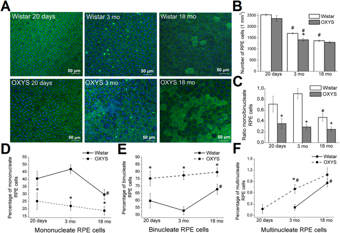
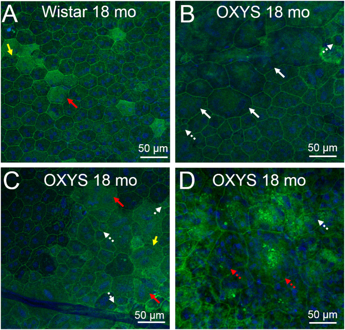
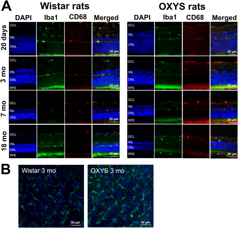
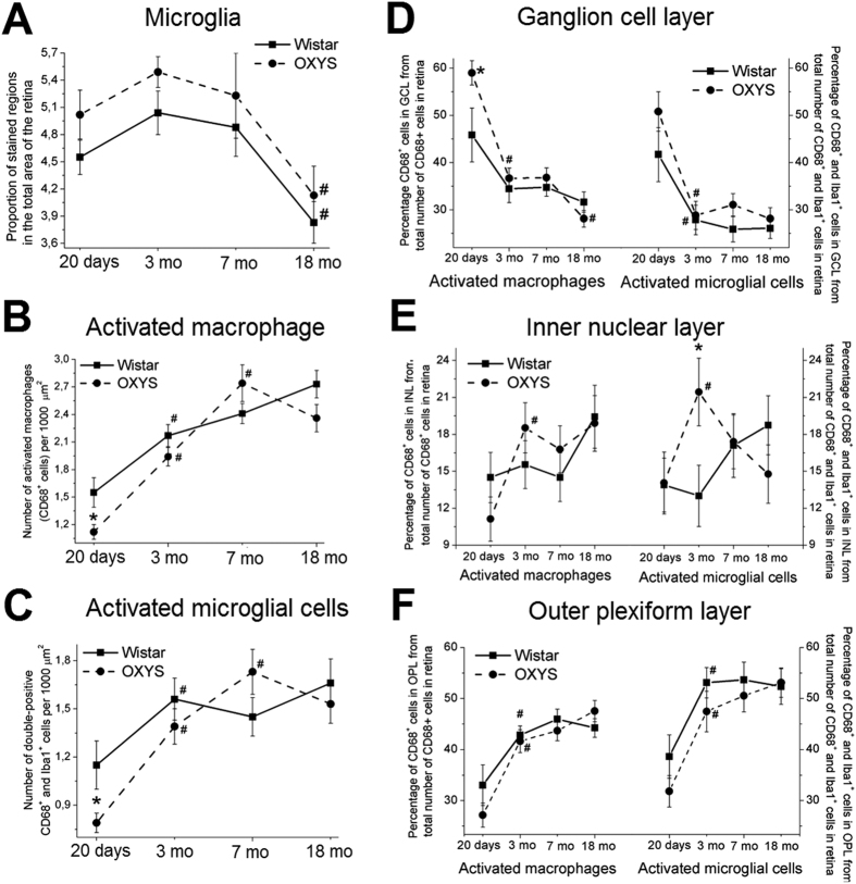
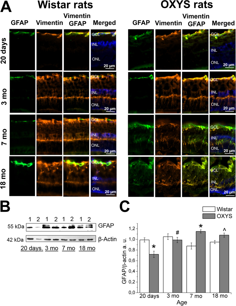
Age-related macular degeneration (AMD) is a major cause of blindness in developed countries, and the molecular pathogenesis of early events of AMD is poorly understood. It is known that age-related alterations of retinal pigment epithelium (RPE) cells and of glial reactivity are early hallmarks of AMD. Here we evaluated contributions of the age-related alterations of the RPE and of glia to the development of AMD-like retinopathy in OXYS rats. We showed that destructive alterations in RPE cells are a primary change during the development of retinopathy in OXYS rats. Furthermore, a defect of retinal maturation and decreased immune function at the preclinical stage of retinopathy were observed in OXYS rats in addition to the impairment of RPE cell proliferation and of their capacity for division. At the active stage of the disease, the atrophic alterations increased, and reactive gliosis was observed when disease progressed, but immune function stayed weakened. Unexpectedly, we did not observe migration of microglia and macrophages into the photoreceptor layer. These results and the wide spectrum of age-related retinal alterations in humans as well as individual differences in the risk of AMD may be attributed to genetic factors and to differences in the underlying molecular events.


Conflict of interest statement

The authors declare no competing financial interests.

Figures

Figure 1
Figure 1. Analysis of alterations of RPE cells as a function of age in the retina of Wistar and OXYS rats.
(A) Confocal images of a flat mount of Wistar (upper panel) and OXYS rat RPE (lower panel), showing the changes in RPE cells during normal aging (Wistar rats) and during the development of retinopathy (OXYS rats). Phalloidin was used for the RPE staining. Quantitative analysis of cell density of RPE (B), the ratio of the numbers of mono-/binucleate RPE cells (C), the percentages of mononucleate (D), binucleate (E), and multinucleate cells (F) per 1 mm2 in the central zone of the retina of OXYS and Wistar rats. The data are shown as mean ± SEM; *p < 0.05 for differences between the strains; #p < 0.05 for age-related differences within a strain.
Figure 2
Figure 2. The destructive and atrophic alterations of RPE cells in Wistar and OXYS rats at the age of 18 months.
Phalloidin was used for RPE staining. (A) Confocal images of a flat-mount of a Wistar rat RPE. The yellow arrows indicate polyploid cells. (B) The hypertrophy (white arrows) and hyperplasia (white dotted arrows) in OXYS rats during active progression of the disease. (С) Accumulation of lipofuscin granules in the disintegrating RPE cells in OXYS rats (red arrows). (D) Changes of the cell shape (red dotted arrow) disrupt an orderly mosaic in OXYS rats at age 18 months.
Figure 3
Figure 3
(A) Representative immunofluorescent images of co-localisation of microglia (Iba1, green), activated macrophages (CD68, red) and cell nuclei (DAPI, blue) in the retina of OXYS and Wistar rats at various ages. (B) Confocal images of a flat mount of the retinas (from 3-month-old OXYS and Wistar rats) showing the ramified Iba1+ microglial morphology.
Figure 4
Figure 4. Analysis of alterations of Iba1+ microglia and CD68+ macrophage density as a function of age in the retina of OXYS and Wistar rats.
(A) The data on immunofluorescence levels of the microglia marker Iba1 in retinal cryosections of 20-day-old and 3-, 7- and 18-month-old Wistar and OXYS rats. We calculated the proportion of stained regions in the total area of the retina. (B) The change in the number of activated macrophages (CD68+ cells) per 1000 μm2 in OXYS and Wistar retinas at different ages. (C) The change in the number of activated microglial cells (CD68+ and Iba1+ cells) per 1000 μm2 in OXYS and Wistar rats at various ages. The distribution of activated macrophages and microglial cells in various layers of the retina: ganglion layer (D), inner nuclear layer (E) and outer plexiform layer (F). The data are shown as mean ± SEM; *p < 0.05 for differences between the strains; #p < 0.05 for age-related differences within a strain. Abbreviations: GCL, ganglion layer; INL, inner nuclear layer; ONL, outer plexiform layer; RPE, retinal pigment epithelium.
Figure 5
Figure 5. Age-related alterations of vimentin and GFAP expression in the rat retina.
(А) Representative immunofluorescent images of co-localisation of GFAP (green), vimentin (yellow) and cell nuclei (DAPI, blue) in the retina of OXYS and Wistar rats at ages 20 days and 3, 7 and 18 months. Abbreviations: GCL, ganglion layer; INL, inner nuclear layer; ONL, outer nuclear layer. (B) Levels of GFAP protein in the retina of Wistar rats (lane 1) and OXYS rats (lane 2; 20-day-old and 3-, 7- and 18-month-old rats) according to immunoblot analysis. The relative amount of the GFAP protein was calculated as intensity of a GFAP band divided by intensity of a β-actin band in the retina (C). The data are shown as mean ± SEM; *p < 0.05 for differences between the strains; #p < 0.05 for age-related differences within a strain; ^an insignificant difference between the strains.

References

    1. Kinnunen K., Petrovski G., Moe M. C., Berta A. & Kaarniranta K. Molecular mechanisms of retinal pigment epithelium damage and development of age-related macular degeneration. Acta Ophthalmol. 90, 299–309 (2012). - PubMed
    1. Rickman C., Farsiu S., Toth C. & Klingeborn M. Dry Age-Related Macular Degeneration: Mechanisms, Therapeutic Targets, and Imaging. Invest Ophthalmol Vis Sci. 5, ORSF68–ORSF80 (2013). - PMC - PubMed
    1. Curcio C. A., Presley J. B., Millican C. L. & Medeiros N. E. Basal deposits and drusen in eyes with age-related maculopathy: evidence for solid lipid particles. Exp Eye Res. 80, 761–775 (2005). - PubMed
    1. Coorey N. J., Shen W., Chung S. H., Zhu L. & Gillies M. C. The role of glia in retinal vascular disease. Clin Exp Optom. 95, 266–281 (2012). - PubMed
    1. Madeira M. H., Boia R., Santos P. F., Ambrósio A. F. & Santiago A. R. Contribution of microglia-mediated neuroinflammation to retinal degenerative diseases. Mediators Inflamm. 2015, 673090, 10.1155/2015/673090 (2015). - DOI - PMC - PubMed

Publication types

MeSH terms