. 2017 Jan 30:6:e21392.
doi: 10.7554/eLife.21392.

The paranodal cytoskeleton clusters Na+ channels at nodes of Ranvier

Affiliations

The paranodal cytoskeleton clusters Na+ channels at nodes of Ranvier

Veronique Amor et al. Elife. .

Abstract

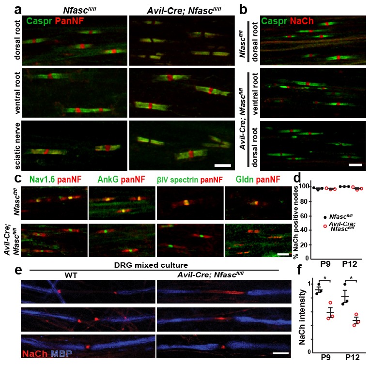
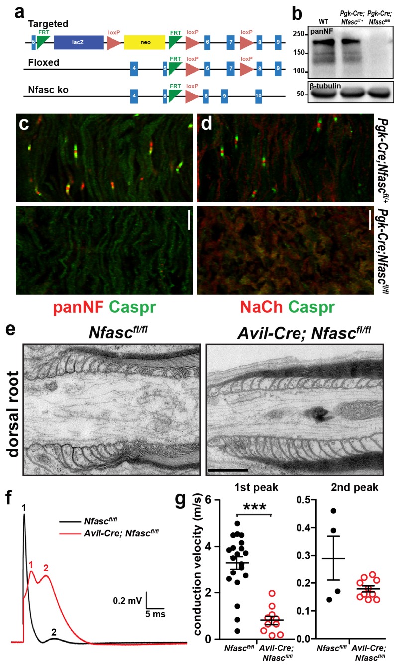
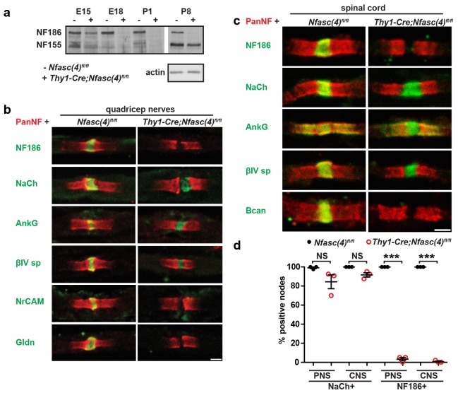
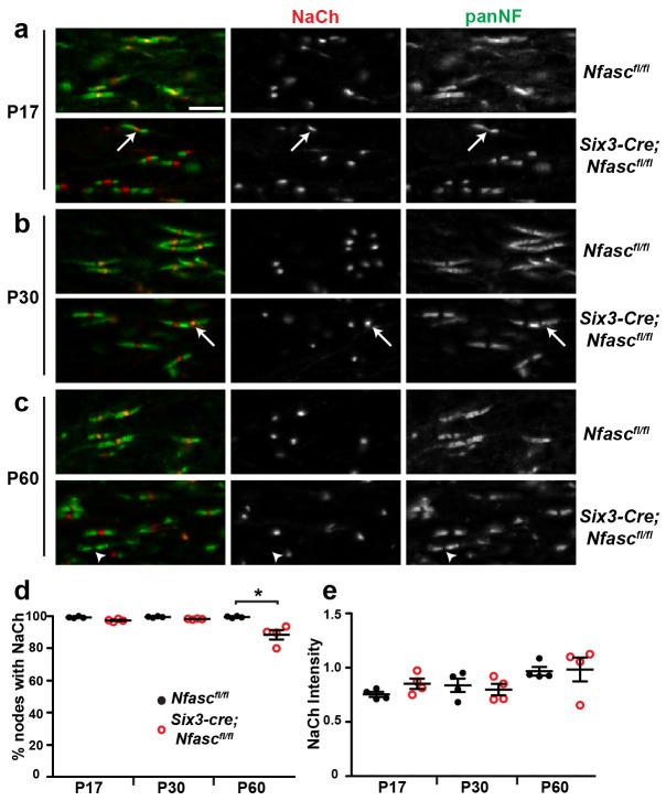
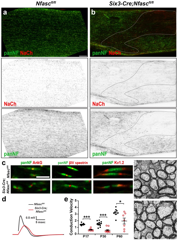
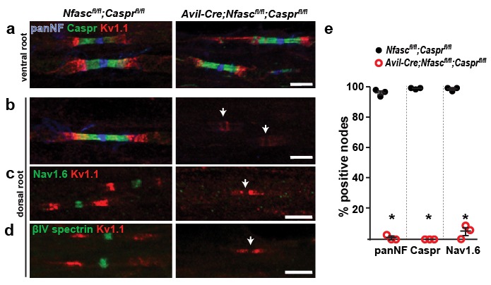
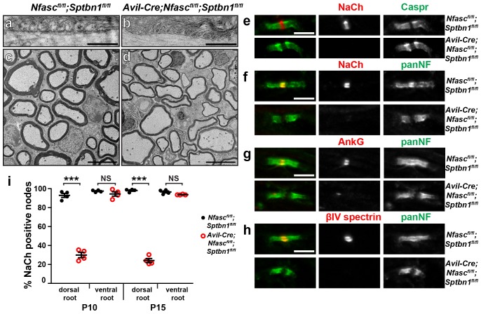
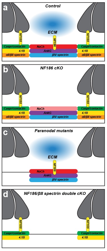
A high density of Na+ channels at nodes of Ranvier is necessary for rapid and efficient action potential propagation in myelinated axons. Na+ channel clustering is thought to depend on two axonal cell adhesion molecules that mediate interactions between the axon and myelinating glia at the nodal gap (i.e., NF186) and the paranodal junction (i.e., Caspr). Here we show that while Na+ channels cluster at nodes in the absence of NF186, they fail to do so in double conditional knockout mice lacking both NF186 and the paranodal cell adhesion molecule Caspr, demonstrating that a paranodal junction-dependent mechanism can cluster Na+ channels at nodes. Furthermore, we show that paranode-dependent clustering of nodal Na+ channels requires axonal βII spectrin which is concentrated at paranodes. Our results reveal that the paranodal junction-dependent mechanism of Na+channel clustering is mediated by the spectrin-based paranodal axonal cytoskeleton.

Keywords: axon; ion channel; mouse; myelin; neuroscience.


Conflict of interest statement

The authors declare that no competing interests exist.

Figures

Figure 1.
Figure 1.. NF155-dependent paranodal junctions can cluster Na+ channels in the PNS.
(a) Immunostaining of P12 dorsal roots, ventral roots, and sciatic nerve from Nfascfl/fl and Avil-Cre;Nfascfl/fl mice using a pan Nfasc (Pan-NF, red) and Caspr (green) antibodies. Note the yellow overlap of the two labels at the paranodes in both genotypes. Scale bar, 5 µm. (b) Immunostaining of P12 dorsal and ventral roots from Nfascfl/fl and Avil-Cre;Nfascfl/fl mice using pan Na+ channel (NaCh, red) and Caspr (green) antibodies. Scale bar, 5 µm. (c) Immunostaining of P12 dorsal roots from Nfascfl/fl and Avil-Cre;Nfascfl/fl mice using panNF (red) to label nodes and paranodes, and antibodies against Nav1.6, AnkG, βIV spectrin, or Gldn to label nodes (green). Note that the nodal panNF staining observed in the Nfascfl/fl mice is much stronger than the paranodal immunoreactivity. Thus, the paranodal panNF staining is more pronounced in the Avil-Cre;Nfascfl/fl mice. Scale bar, 5 µm. (d) Quantification of the percentage of dorsal root nodes of Ranvier with nodal Na+ channels at P9 and P12. N = 3 mice at each time point for each genotype. (e) Immunostaining of dorsal root ganglion/Schwann cell mixed cultures from wild-type and Avil-Cre;Nfascfl/fl mice using antibodies against NaCh (red) and myelin basic protein (MBP, blue). Scale bar = 10 µm. (f) Quantification of the relative Na+ channel fluorescence intensity at P9 and P12. N = 3 mice at each time point for each genotype. *p=0.016 at P9; p=0.026 at P12. The number of nodes measured at P9 were 263 and 250, at P12 were 337 and 269 in the Nfascfl/fl and Avil-Cre;Nfascfl/fl dorsal roots, respectively. DOI: http://dx.doi.org/10.7554/eLife.21392.002
Figure 1—figure supplement 1.
Figure 1—figure supplement 1.. Characterization of Pgk-Cre;Nfascfl/fl and Avil-Cre;Nfascfl/fl mice.
(a) Targeting strategy to generate Nfasc conditional knockout mice. (b) Immunoblot of P6 control and Pgk-Cre;Nfascfl/fl mouse brain lysates using PanNF antibodies and β-tubulin antibodies as a loading control. (c, d) Immunostaining of P4 Pgk-Cre;Nfascfl/+ and Pgk-Cre;Nfascfl/fl mouse sciatic nerves using PanNF (c, red), Nach (d, red), and Caspr (green) antibodies. Scale bar = 10 µm. (e) Transmission electron microscopy of dorsal root paranodes from P12 Nfascfl/fl and Avil-Cre;Nfascfl/fl mice. Scale bar = 0.5 µm. (f) Example of a dorsal root compound action potential from P10 Nfascfl/fl and Avil-Cre;Nfascfl/fl mice. (g) Conduction velocities calculated from the first and second peaks of the compound action potential recorded from P10 dorsal roots. In many recordings, a clear second peak was not always detectable and this was usually the case in the Nfascfl/fl roots. We have only included the second peak where it was clearly resolved in the recordings. ***p=2.62E−08. DOI: http://dx.doi.org/10.7554/eLife.21392.003
Figure 1—figure supplement 2.
Figure 1—figure supplement 2.. NF155-dependent paranodal junctions can cluster Na+ channels in the PNS and CNS.
(a) Immunoblot of embryonic day 15 (E15), E18, P1, and P8 spinal cords from Nfasc(4)fl/fl and Thy1-Cre;Nfasc(4)fl/fl mice using PanNF antibodies and actin as a loading control. (bc) Immunostaining of P5 quadriceps nerves (b) and P5 spinal cord (c) from Nfasc(4)fl/fl and Thy1-Cre;Nfasc(4)fl/fl mice using PanNF (red) and other node-specific antibodies (green) as indicated in the figure. Scale bars = 2 µm. (d) The percentage of P5 PNS and CNS nodes from Nfasc(4)fl/fl and Thy1-Cre;Nfasc(4)fl/fl mice that were labeled using antibodies against Na+ channels or NF186. N = 3 mice/genotype. ***p=4.18E−08 (PNS NF186+); p=4.18E−08 (CNS NF186+). DOI: http://dx.doi.org/10.7554/eLife.21392.004
Figure 2.
Figure 2.. NF155-dependent paranodal junctions can cluster Na+ channels in the CNS.
(ac) Immunostaining of P17, P30, and P60 optic nerves from Nfascfl/fl and Six3-Cre;Nfascfl/fl mice using chicken panNF (green) and mouse monoclonal pan Na+ channel (NaCh, red) antibodies. Scale bar, 10 µm. (d) The percentage of panNF nodes that have Na+ channels. N = 4 animals per time point per genotype. *p=0.03. (e) Nodal Na+ channel immunofluorescence intensity at P17, P30, and P60 in optic nerves from Nfascfl/fl and Six3-Cre;Nfascfl/fl mice. N = 4 animals per time point per genotype. DOI: http://dx.doi.org/10.7554/eLife.21392.005
Figure 2—figure supplement 1.
Figure 2—figure supplement 1.. Characterization of Six3-Cre;Nfascfl/fl mice.
(a, b) Immunostaining of Nfascfl/fl (a) and Six3-Cre;Nfascfl/fl (b) optic nerves using antibodies against panNF (green) and Na+ channels (red). The dotted line in (b) indicates a region where recombination occurred in oligodendrocytes. Scale bar = 50 µm. (c) Immunostaining of Nfascfl/fl and Six3-Cre;Nfascfl/fl nodes of Ranvier using antibodies against panNF (green) and AnkG, βIV spectrin, or Kv1.2 (red). Scale bar = 5 µm. (d) Optic nerve compound action potential recordings from P17 Nfascfl/fl and Six3-Cre;Nfascfl/fl mice. (e) Conduction velocities calculated from the peak of the compound action potential recorded from P17, P30, and P60 optic nerves. P17: ***p=2.75E−07. P30: ***p=2.47E−06. P60: *p=0.03. (f, g) Transmission electron microscopy of Nfascfl/fl(F) and Six3-Cre;Nfascfl/fl (G) optic nerves. Scale bar = 1 µm. DOI: http://dx.doi.org/10.7554/eLife.21392.006
Figure 3.
Figure 3.. Paranodes can cluster Na+ channels at nodes of Ranvier.
(a, b) Immunostaining of P14 Nfascfl/fl;Casprfl/fl and Avil-Cre;Nfascfl/fl;Casprfl/fl ventral (a) and dorsal (b) roots using antibodies against panNF (blue), Caspr (green), and Kv1.1 (red). Arrows in (b) indicate the location of nodes. Scale bars, 10 µm. (c, d) Immunostaining of P14 Nfascfl/fl;Casprfl/fl and Avil-Cre;Nfascfl/fl;Casprfl/fl dorsal roots using antibodies against Nav1.6 Na+ channels (c, green), βIV spectrin (d, green), and Kv1.1 (red). Arrows indicate the location of nodes. Scale bars, 10 µm. (e) The percentage of nodes or paranodes labeled with the indicated antibodies in P14 Nfascfl/fl;Casprfl/fl and Avil-Cre;Nfascfl/fl;Casprfl/fl dorsal roots. N = 3 animals per genotype. Number of nodes analyzed in Nfascfl/fl;Casprfl/fl dorsal root: PanNF, 288; Caspr, 298; Nav1.6, 301. Number of nodes analyzed in Avil-Cre;Nfascfl/fl;Casprfl/fl dorsal root: PanNF, 365; Caspr, 365; Nav1.6, 336. *PanNF, p=0.0002; Caspr, p=8.07E-06; Nav1.6, p=0.0012. DOI: http://dx.doi.org/10.7554/eLife.21392.007
Figure 4.
Figure 4.. The paranodal spectrin-based cytoskeleton can assemble nodes of Ranvier.
(a, b) TEM of longitudinal sections from dorsal roots of Nfascfl/fl;Sptbn1fl/fl (a) and Avil-Cre;Nfascfl/fl;Sptbn1fl/fl mice (b). Scale bars, 0.5 µm. (c, d) TEM of cross sections through dorsal roots of Nfascfl/fl;Sptbn1fl/fl (c) and Avil-Cre;Nfascfl/fl;Sptbn1fl/fl mice (d). Scale bars, 4 µm. (e) Immunostaining of P10 Nfascfl/fl;Sptbn1fl/fl and Avil-Cre;Nfascfl/fl;Sptbn1fl/fl dorsal roots using antibodies against Na+ channels and Caspr shows intact paranodal junctions. Scale bar, 5 µm. (fh) Immunostaining of P15 Nfascfl/fl;Sptbn1fl/fl and Avil-Cre;Nfascfl/fl;Sptbn1fl/fl dorsal roots using antibodies against Na+ channels (e, red), AnkG (f, red), βIV spectrin (g, red) and panNF (green). Scale bar, 5 µm. (i) The percentage of nodes labeled for Na+ channels in P10 and P15 Nfascfl/fl;Sptbn1fl/fl and Avil-Cre;Nfascfl/fl;Sptbn1fl/fl dorsal and ventral roots. N = 4 animals per age and genotype. ***p=2.0E−06 at P10; p=6.26E−08 at P15. DOI: http://dx.doi.org/10.7554/eLife.21392.008
Figure 5.
Figure 5.. Two glia-dependent mechanisms can cluster Na+ channels at nodes of Ranvier.
(a) Cartoon of control node of Ranvier. (b) Na+ channels are clustered in NF186-deficient mice by a paranodal spectrin cytoskeleton. However, the density of Na+ channels is reduced in the PNS. (c) Na+ channels are clustered in NF155-deficient and other paranodal mutant mice by NF186-ECM interactions. (d) Na+ channels fail to cluster at nodes of Ranvier when both NF186 and the paranodal cytoskeleton are lost. DOI: http://dx.doi.org/10.7554/eLife.21392.010
Author response image 1.
Author response image 1.. Nodal NaCh intensity in the Six3Cre;Nfascfl/flCNS optic nerve.
DOI: http://dx.doi.org/10.7554/eLife.21392.011

Similar articles

Cited by

References

    1. Amor V, Feinberg K, Eshed-Eisenbach Y, Vainshtein A, Frechter S, Grumet M, Rosenbluth J, Peles E. Long-term maintenance of Na+ channels at nodes of Ranvier depends on glial contact mediated by gliomedin and NrCAM. Journal of Neuroscience. 2014;34:5089–5098. doi: 10.1523/JNEUROSCI.4752-13.2014. - DOI - PMC - PubMed
    1. Bekku Y, Vargová L, Goto Y, Vorísek I, Dmytrenko L, Narasaki M, Ohtsuka A, Fässler R, Ninomiya Y, Syková E, Oohashi T. Bral1: its role in diffusion barrier formation and conduction velocity in the CNS. Journal of Neuroscience. 2010;30:3113–3123. doi: 10.1523/JNEUROSCI.5598-09.2010. - DOI - PMC - PubMed
    1. Bhat MA, Rios JC, Lu Y, Garcia-Fresco GP, Ching W, St Martin M, Li J, Einheber S, Chesler M, Rosenbluth J, Salzer JL, Bellen HJ. Axon-glia interactions and the domain organization of myelinated axons requires neurexin IV/Caspr/Paranodin. Neuron. 2001;30:369–383. doi: 10.1016/S0896-6273(01)00294-X. - DOI - PubMed
    1. Boyle ME, Berglund EO, Murai KK, Weber L, Peles E, Ranscht B. Contactin orchestrates assembly of the septate-like junctions at the paranode in myelinated peripheral nerve. Neuron. 2001;30:385–397. doi: 10.1016/S0896-6273(01)00296-3. - DOI - PubMed
    1. Chang KJ, Susuki K, Dours-Zimmermann MT, Zimmermann DR, Rasband MN. Oligodendrocyte myelin glycoprotein does not influence node of ranvier structure or assembly. Journal of Neuroscience. 2010;30:14476–14481. doi: 10.1523/JNEUROSCI.1698-10.2010. - DOI - PMC - PubMed

Publication types

MeSH terms

Substances

LinkOut - more resources