. 2017 Mar 1;127(3):857-873.
doi: 10.1172/JCI90025. Epub 2017 Jan 30.

Autophagy facilitates macrophage depots of sustained-release nanoformulated antiretroviral drugs

Autophagy facilitates macrophage depots of sustained-release nanoformulated antiretroviral drugs

Divya Prakash Gnanadhas et al. J Clin Invest. .

Abstract

Long-acting anti-HIV products can substantively change the standard of care for patients with HIV/AIDS. To this end, hydrophobic antiretroviral drugs (ARVs) were recently developed for parenteral administration at monthly or longer intervals. While shorter-acting hydrophilic drugs can be made into nanocarrier-encased prodrugs, the nanocarrier encasement must be boosted to establish long-acting ARV depots. The mixed-lineage kinase 3 (MLK-3) inhibitor URMC-099 provides this function by affecting autophagy. Here, we have shown that URMC-099 facilitates ARV sequestration and its antiretroviral responses by promoting the nuclear translocation of the transcription factor EB (TFEB). In monocyte-derived macrophages, URMC-099 induction of autophagy led to retention of nanoparticles containing the antiretroviral protease inhibitor atazanavir. These nanoparticles were localized within macrophage autophagosomes, leading to a 4-fold enhancement of mitochondrial and cell vitality. In rodents, URMC-099 activation of autophagy led to 50-fold increases in the plasma drug concentration of the viral integrase inhibitor dolutegravir. These data paralleled URMC-099-mediated induction of autophagy and the previously reported antiretroviral responses in HIV-1-infected humanized mice. We conclude that pharmacologic induction of autophagy provides a means to extend the action of a long-acting, slow, effective release of antiretroviral therapy.


Conflict of interest statement

Conflict of interest: H.A. Gelbard and H.E. Gendelman are members of the scientific advisory board for WavoDyne Therapeutics Inc., which is developing URMC-099 as a first-in-class MLK-3 inhibitor for clinical trials. URMC-099 is owned by URMC (patent nos. US 8,846,909 B2; 8,877,772; and 9,181,247 and associated international patents).

Figures

Figure 1
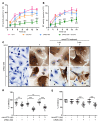
Figure 1. URMC-099 potentiates antiretroviral activity of ARV nanoformulations.
HIV-1ADA–infected human MDMs were treated with 1 μM nanoATV on (A) day 1 or (B) day 3 after infection in the presence or absence of 400 ng/ml URMC-099. Supernatants were collected on different days after infection to measure HIV-1 RT activity. Values represent the mean ± SD (n = 5). The same HIV control plot is presented in A and B. For a better comparison, see the HIV control plot in Figure 5, A and B, and in Supplemental Figure 5, A, B, E, and F. The mean was compared by 2-way ANOVA, which showed a time-dependent effect (P < 0.0001). Pairwise comparisons were performed with Bonferroni’s post-hoc test for P < 0.05 compared with acontrol, bNanoATV, and cURMC-099. (C) On day 7 after infection, the cells were fixed and stained for HIV-1 p24 antigen and counterstained with hematoxylin. Multi- and mononucleated cells are marked with white and black arrowheads, respectively. Scale bar: 20 μm. On (D) day 7 and (E) day 14 after infection, the percentage of MNGCs was quantified from the total number of cells in 20 different fields. Values represent the mean ± SD. **P ≤ 0.01 and ***P ≤ 0.001, by Student’s t test. Multiple comparisons were corrected for the FDR by the Benjamini-Hochberg method, and data are representative of 3 independent experiments. dpi, days post infection.
Figure 2
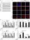
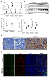
Figure 2. URMC-099 regulates TFEB nuclear localization.
Human MDMs were treated for 14 days with 100 (+*) or 400 ng/ml (+#) URMC-099 in the presence or absence of 100 μM nanoATV, with or without HIV-1ADA infection. (A) MDMs were fractionated and analyzed by Western blotting. (B) TFEB and (C) mTOR quantification of Western blots. Protein bands were quantified and normalized to actin β (ACTB) or histone H3 using ImageJ2 software. (D) Human MDMs were stained for TFEB (red) and DAPI (blue) and analyzed using a confocal microscope to visualize nuclear localization of TFEB. Scale bar: 20 μm. (E) Quantification of colocalization coefficient for TFEB and DAPI. (F) Total RNA was isolated on day 14, and real-time qPCR was performed to determine TFEB expression. Values represent the mean ± SD (n = 3). *P ≤ 0.05, **P ≤ 0.01, and ***P ≤ 0.001, by Student’s t test. (B, C, E, and F) Data were corrected for multiple comparisons using the Benjamini-Hochberg method. Data are representative of 3 independent experiments. All URMC-099 treatments not specified used 400 ng/ml.
Figure 3
Figure 3. URMC-099 induces autophagy in macrophages.
Human MDMs were treated for 14 days with 100 (+*) or 400 ng/ml (+#) URMC 099, in the presence or absence of 100 μM nanoATV, with or without HIV-1ADA infection. (A) Total cell lysates were analyzed for different autophagy markers by Western blotting. (B and C) Quantification of Western blots. (B) LC3BII/LC3B1 ratio and (C) quantification of protein expression of LC3BI (white bars), LC3BII (light gray bars), BECN1 (dark gray bars), and SQSTM1 (black bars). Protein bands were quantified and normalized to ACTB using ImageJ2 software (n = 3). (D) Total RNA was isolated on day 14, and real-time qPCR was performed to determine MAP1LC3B, BECN1, and SQSTM1 expression (n = 3). (E) On day 14, an MTT assay was performed to assess mitochondrial activity (n = 5). Values represent the mean ± SD. *P ≤ 0.05, **P ≤ 0.01, ***P ≤ 0.001, and P ≤ 0.05, acompared with uninfected control MDMs and bcompared with HIV-1–infected MDMs; Student’s t test. (CE) Data were corrected for multiple comparisons using the Benjamini-Hochberg method. Data are representative of 3 independent experiments. All URMC-099 treatments not specified used 400 ng/ml.
Figure 4
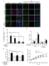
Figure 4. URMC-099 increases nanoATV retention in macrophage autophagosomes.
(A) Human MDMs were treated for 14 days with 400 ng/ml URMC-099 in the presence of fluorescently tagged nanoATV (10 μM) (purple), with or without HIV-1ADA infection. MDMs were transfected with an LC3B-tagged GFP construct (green), stained with DAPI (blue), and analyzed by confocal microscopy. Scale bar: 20 μm. (B) Quantification of LC3B puncta that colocalized with nanoATV. (C) Human MDMs were treated with nanoATV (10 μM or 100 μM) in the presence or absence of 400 ng/ml URMC-099. After 14 days, the ATV concentration was determined in total cells, and LC3B autophagosomal compartments were isolated using magnetic bead separation (n = 3). (D) Human MDMs were treated with 100 μM nanoART and 400 ng/ml URMC-099. Cells were transfected with siRNA for TFEB or ATG13, and on day 14, the ATV concentration was determined in total cells (n = 3). (E) URMC-099 regulated the release of nanoATV. Human MDMs were treated with 100 μM nanoATV for 16 hours, washed with PBS, and incubated with or without 400 ng/ml URMC-099. On different days, supernatant was collected, ATV concentration was quantified using HPLC, and the cumulative ATV release was plotted according to treatment duration (n = 3). Comparison of the means by 2-way factorial ANOVA showed a time-dependent treatment effect (P = 0.0002), with pairwise comparisons made using Bonferroni’s post-hoc test (#P < 0.05). Data are representative of 3 independent experiments. (BD) Values represent the mean ± SD. *P ≤ 0.05 and ***P ≤ 0.001, by Student’s t test. Multiple comparisons were corrected for the FDR using the Benjamini-Hochberg method.
Figure 5
Figure 5. URMC-099–induced autophagy affects HIV-1 clearance.
HIV-1ADA–infected human MDMs were treated with 1 μM nanoATV on day 1 or day 3 after infection and incubated with or without 400 ng/ml URMC-099 and in the presence of the autophagy inhibitors (A) 3-MA (100 μM) or (B) chloroquine (10 μM). Supernatants were collected on different days after infection and analyzed for HIV-1 RT activity (n = 5). The same HIV-1 infection control plot is presented in A and B. (A and B) The mean values of RT activity were assessed by 2-way factorial ANOVA, which showed a significant time-dependent treatment effect (P < 0.02). Pairwise comparisons using Bonferroni’s post-hoc test were assessed for URMC-099–treated cultures, with P < 0.05 compared with HIV-infected controls in the absence (acontrol) or presence (bHIV) of an autophagy inhibitor. (CE) MDMs were treated in the presence or absence of 400 ng/ml URMC-099 for 14 days. Twenty-four hours or twelve hours before harvesting, cells were treated with 10 μM cycloheximide (CHX) to inhibit translation. Total cell lysates were analyzed by Western blotting. (D) Values represent the mean ± SEM of LC3BII/LC3BI ratios and were compared by Student’s t test and adjusted for multiple comparisons using the Benjamini-Hochberg method. *P ≤ 0.05 and **P ≤ 0.01 (n = 3). (E) Differences in mean fold changes were assessed by 2-way ANOVA and pairwise comparison with the respective proteins was done using Bonferroni’s post-hoc test. P ≤ 0.05 for ano URMC-099/no CHX control and bURMC-099/no CHX control (n = 3). (F) Human MDMs treated with 400 ng/ml URMC-099 were transfected with either TFEB siRNA or ATG13 siRNA on days 3 and 7. On day 14, cell lysates were analyzed for by Western blotting. (G) URMC-099–treated (400 ng/ml) and untreated (control) MDMs were transfected with LC3B-GFP on day 12, and 48 hours later were stained and imaged with a confocal microscope. Scale bars: 20 μm. Data are representative of 3 independent experiments.
Figure 6
Figure 6. URMC-099 and autophagy in tissues.
Mice were given twice-daily i.p. injections of buffer (control) or URMC-099 (10 mg/kg), and after 21 days, mice were sacrificed and tissues collected. (A) Total RNA was isolated from liver, and real-time qPCR was performed. (BD) Liver homogenate was analyzed for different autophagy markers using Western blotting (each lane is from 1 mouse) and is presented as (C) the LC3BII/LC3BI ratio and (D) the quantification normalized to ACTB using ImageJ2 software. Paraffin-embedded, 5-μm-thick splenic sections were stained for (E) LC3B, developed with DAB and counterstained with hematoxylin, and for (F) LC3B (green) and mouse CD68 in macrophages (red). DAPI was used to stain nuclei. Values represent the mean ± SEM. *P ≤ 0.05 and **P ≤ 0.01, and ***P ≤ 0.001, by Mann-Whitney U test. Multiple comparisons were assessed for the FDR using the Benjamini-Hochberg method. Five and four mice were included in the control and URMC-099 groups, respectively. Scale bars: 20 μm.
Figure 7
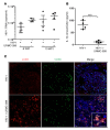
Figure 7. URMC-099 treatment in humanized mice.
(A and B) Humanized NSG mice were infected with HIV-1ADA and treated with URMC-099 (n = 4 per group). Ten weeks after infection, (A) plasma viral load and (B) plasma IL-1β concentration were determined (n = 4 mice per group). Values represent the mean ± SEM. ***P ≤ 0.001, by Mann–Whitney U test. (C) Paraffin-embedded spleen tissue sections from URMC-099–treated and control HIV-1–infected humanized mice were stained for LC3B (red) in autophagosomes and CD68 (green) in macrophages. DAPI was used to stain nuclei. The merged panel shows colocalization of LC3B and CD68 in spleens from URMC-099–treated mice. Scale bars: 20 μm.
Figure 8
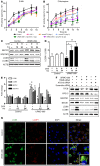
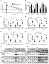
Figure 8. URMC-099 and nanoART parenteral coadministration sustains plasma DTG levels.
Mice were injected i.m. with a single dose (45 mg/kg) of nanoDTG and treated with or without daily i.p. injections of URMC-099 (10 mg/kg). (A) On different days, blood was collected, and the plasma DTG concentration was determined by UPLC-MS/MS. (B) On day 14 (D14) and day 28 (D28), mice were sacrificed and DTG levels quantified in different tissues by UPLC-MS/MS. (CF) On days 14 and 28, total RNA was isolated from splenic and liver tissues, and real-time qPCR was performed for different genes. Values represent the mean ± SEM. *P ≤ 0.05 and **P ≤ 0.01, by Mann-Whitney U test (n = 6 mice per group). (G and H) On days 14 and 28, splenic and liver tissues were collected, and total tissue lysate was analyzed by Western blotting for different autophagy markers. Each lane is representative of 5 animals from each of the groups. Multiple comparisons were corrected for the FDR using the Benjamini-Hochberg method. Six mice were assessed per group.
Figure 9
Figure 9. URMC-099 facilitates depots of nanoART in macrophage autophagosomes.
Nanoformulated ARVs enter the MDM via clathrin-coated pits and are transported to the early endosomes. Parts of the nanoARV are recycled by getting into recycling endosomes by Rab14-mediated fast recycling or trafficking regulated by endosomal sorting complexes required for transport machinery (ESCRT) and Rab7. The particles reach late endosomes via ESCRT and Rab11 and eventually fuse with lysosomes. HIV-1 fuses with the cell and releases its contents to the cytoplasm. Partial core shell uncoating and reverse transcription lead to the formation of a preintegration complex that enters the nucleus, and provirus is formed following integration. After proviral transcription and translation, viral assembly and maturation take place. HIV-1 Nef inhibits autophagy by sequestering TFEB in the cytoplasm. A subtherapeutic dose of nanoART limits antiretroviral responses. URMC-099–assisted nuclear translocation of TFEB overcomes HIV-1 Nef–mediated inhibition of autophagy. This affects autophagosome formation and retention of nanoformulated drugs specifically in the autophagosomes. In the presence of URMC-099, nanoformulated ARV retention is increased, and release is decreased even with a subtherapeutic drug dose, ultimately attenuating HIV-1 infection.

Similar articles

Cited by

References

    1. Das MK, Sarma A, Chakraborty T. Nano-ART and NeuroAIDS. Drug Deliv Transl Res. 2016;6(5):452–472. doi: 10.1007/s13346-016-0293-z. - DOI - PubMed
    1. Kimata JT, Rice AP, Wang J. Challenges and strategies for the eradication of the HIV reservoir. Curr Opin Immunol. 2016;42:65–70. doi: 10.1016/j.coi.2016.05.015. - DOI - PMC - PubMed
    1. Nowacek A, Gendelman HE. NanoART, neuroAIDS and CNS drug delivery. Nanomedicine (Lond) 2009;4(5):557–574. doi: 10.2217/nnm.09.38. - DOI - PMC - PubMed
    1. Araínga M, Guo D, Wiederin J, Ciborowski P, McMillan J, Gendelman HE. Opposing regulation of endolysosomal pathways by long-acting nanoformulated antiretroviral therapy and HIV-1 in human macrophages. Retrovirology. 2015;12:5. - PMC - PubMed
    1. Dash PK, et al. Long-acting nanoformulated antiretroviral therapy elicits potent antiretroviral and neuroprotective responses in HIV-1-infected humanized mice. AIDS. 2012;26(17):2135–2144. doi: 10.1097/QAD.0b013e328357f5ad. - DOI - PMC - PubMed

MeSH terms