. 2017 May;158(5):931-944.
doi: 10.1097/j.pain.0000000000000860.

A central role for R7bp in the regulation of itch sensation

Affiliations

A central role for R7bp in the regulation of itch sensation

Mritunjay Pandey et al. Pain. 2017 May.

Abstract

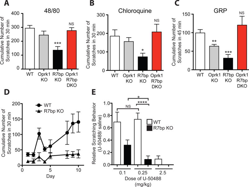
Itch is a protective sensation producing a desire to scratch. Pathologic itch can be a chronic symptom of illnesses such as uremia, cholestatic liver disease, neuropathies and dermatitis, however current therapeutic options are limited. Many types of cell surface receptors, including those present on cells in the skin, on sensory neurons and on neurons in the spinal cord, have been implicated in itch signaling. The role of G protein signaling in the regulation of pruriception is poorly understood. We identify here 2 G protein signaling components whose mutation impairs itch sensation. R7bp (a.k.a. Rgs7bp) is a palmitoylated membrane anchoring protein expressed in neurons that facilitates Gαi/o -directed GTPase activating protein activity mediated by the Gβ5/R7-RGS complex. Knockout of R7bp diminishes scratching responses to multiple cutaneously applied and intrathecally-administered pruritogens in mice. Knock-in to mice of a GTPase activating protein-insensitive mutant of Gαo (Gnao1 G184S/+) produces a similar pruriceptive phenotype. The pruriceptive defect in R7bp knockout mice was rescued in double knockout mice also lacking Oprk1, encoding the G protein-coupled kappa-opioid receptor whose activation is known to inhibit itch sensation. In a model of atopic dermatitis (eczema), R7bp knockout mice showed diminished scratching behavior and enhanced sensitivity to kappa opioid agonists. Taken together, our results indicate that R7bp is a key regulator of itch sensation and suggest the potential targeting of R7bp-dependent GTPase activating protein activity as a novel therapeutic strategy for pathological itch.


Conflict of interest statement

The authors declare no competing financial interests.

Figures

Fig. 1
Fig. 1. Nociceptive somatosensation is largely preserved in mice lacking R7bp
(A) Schematic illustration of R7bp, in complex with R7-RGS and Gβ5 proteins, and heterotrimeric G protein Gαi/o subunit, the target of Gβ5/R7-RGS mediated GTPase activating protein (GAP) activity, positioned near the inner face of the plasma membrane. R7bp facilitates the GAP activity of the Gβ5/R7-RGS complex through its membrane-anchoring function. Following Gi/o activation by agonist-activated Gi/o-coupled G protein coupled receptor (GPCR), the GAP activity of the Gβ5/R7-RGS complex (arrow) helps to terminate signaling through Gαi/o and Gβγ. R7bp is shown anchored to the plasma membrane by dual palmitoylation (two squiggles), Gαi/o by myristoylation (single squiggle), and Gβγ by γ-isoprenylation (single squiggle). The R7-RGS subfamily of RGS proteins (RGS6, RGS7, RGS9 and RGS11) shares a similar domain architecture: an N-terminal DEP/DHEX domain (the putative binding site of R7bp), followed by a G-gamma-like domain (GGL) that imparts the ability to bind Gβ5, and a C-terminal RGS core domain that possesses GAP activity directed against Gαi/o (arrow). (B) Sections through lumbar dorsal root ganglia (DRG) harvested from wild type (WT) (upper panel) and R7bp knockout (KO) (lower panel) mice and hybridized in situ with antisense probes against Gβ5, R7bp, and the R7-RGS family members Rgs7 and Rgs9 showed no obvious differences in transcript expression. (C) Sections through DRG from WT and R7bp KO mice hybridized in situ with an antisense probe against the capsaicin-binding transient receptor potential cation channel subfamily V member 1 (Trpv1) showed no obvious differences in transcript expression. (D) No significant differences between WT and R7bp KO littermates were observed in the eye wipe assay using the Trpv1 agonist capsaicin (CAP). Results using phosphate-buffered saline (PBS) are shown for comparison. (E) No significant differences between WT and R7bp KO littermates were noted in the tail immersion test of thermal nociception. (F) R7bp KO mice exhibited an increased latency in 53° C hot plate testing compared to WT littermates. (G) No obvious differences in mas-related receptor Mrgprd transcript expression in DRG from WT versus R7bp KO mice were observed by in situ hybridization. (H) No significant differences between WT and R7bp KO littermates were seen with Von Frey filament behavioral testing of mechanical nociception. (I) Results of the tail withdrawal assay using the Randall-Selitto test showed no significant difference between WT and R7bp KO littermates. (J) No significant differences between WT and R7bp KO littermates were observed in the eye wipe test of chemical nociception using the Trpa1 agonist mustard oil (MO). Results using phosphate-buffered saline (PBS) are shown for comparison. Statistics and representative figure reporting: For panels C and G, the hybridization experiment was repeated twice with similar results, using previously published probes[32]. For B, the hybridization experiment was repeated three times for each probe with similar results. In D–F, and H–J, the evaluator was blind to the genotype of the mouse being tested. In D–F, and H–J, the two-tailed unpaired Student’s t-test was employed, with bars indicating mean ± S.E.M. For panels D, H, and J, n= number of mice from each genotype tested (for panel D, PBS WT n= 6; CAP WT n= 9; PBS KO n= 5; CAP KO n= 8; for panel H, n=8; for panel J, PBS WT n= 6; MO WT n= 7; PBS KO n= 6; MO KO n= 7). For panels E, F, and I, n= number of mice tested (for panel E, WT n= 7, KO n= 8, with mice tested 2 or 3 times, with results from individual mice averaged; for panel F, WT n= 14, KO n= 10, with mice tested 3 times, with results from individual mice averaged; for panel I, WT n= 8, KO n= 7, with mice tested 3 times, with results from individual mice averaged.) P values: D, for PBS, P= 0.95, for CAP, P= 0.91; E, P =0.1; F, *** P=0.0002; H, P =0.63; I, P= 0.27; J, for PBS, P= 0.84, for MO, P= 0.80.
Fig. 2
Fig. 2. R7bp-knockout mice exhibit impaired pruriception but retain peripheral sensory neuronal responsiveness to pruritogens
(A,B) Cumulative scratching behavior induced by intradermal histamine, a prototypical pruritogen that signals through the H1 and H4 subclasses of histamine GPCR (A) and compound 48/80, which acts indirectly via histamine release from mast cells (B) over 30 minutes was significantly decreased in R7bp knockout (KO) mice (filled bars) compared to their wild-type (WT) littermates (open bars). (C, D, and E) Cumulative scratching behavior to the pruritogens serotonin (5-HT) (C), chloroquine, a 4-aminoquinoline antimalarial drug that signals itch through MrpgrA3 and Trpa1 [25; 43] (D), and SLIGRL-NH2, a synthetic peptide that signals itch through MrpgrC11 and Trpa1 [26; 43] (E) over 30 minutes was significantly decreased in R7bp KO mice compared to their WT littermates. (F) In wild-type mice, intradermal application of the TLR3 receptor agonist and double stranded RNA viral mimetic polyinosinic: polycytidylic acid (poly (I:C)) induced more cumulative scratching behavior (open bars) relative to intradermal injection of the TLR3-inactive double stranded DNA analog deoxypolyinosinic: deoxypolycytidylic acid (poly (dI:dC)) (filled bars) when observed for 30 minutes. (G) R7bp KO mice had significantly reduced cumulative scratching behavior in response to intradermal poly (I:C) (filled bars) compared to their WT littermates (open bars) when observed for 30 minutes. (H) The cumulative scratching response over 30 minutes following intradermal injection of the pruritogenic cytokine thymic stromal lymphopoietin (TSLP) was significantly lower in R7bp KO mice (filled bar) compared to their WT littermates (open bar). (I) R7bp KO mice (filled bar) showed significantly reduced cumulative scratching behavior in response to the intradermal injection of endothelin-1, a paracrine peptide released from endothelial cells, compared to their WT littermates (open bar) when observed for 30 minutes. (J, K) The cumulative scratching response over 30 minutes following intradermal injection of the pruritogenic bile salts deoxycholic acid (DCA) (J) or taurolithocholic acid (TLCA) (K) was significantly lower in R7bp KO mice (filled bar) compared with their WT littermates (open bar). (L) Dorsal root ganglion cells from WT (upper panel) and R7bp KO (lower panel) mice hybridized in situ with antisense probes against Nppb (left) and the mas-related G protein-coupled receptor MrgprA3 (right), specific markers of primary pruriceptive neurons, showed no obvious differences in transcript expression. (M, N) Quantification of Nppb-positive (M) and MrgprA3-positive (N) neurons in sections from DRGs harvested from WT and R7bp KO mice, following in situ hybridization, showed no significant differences. (O,P) Time course of intracellular calcium mobilization in isolated DRG cells harvested from WT (blue symbols) or R7bp KO (red symbols) mice in response to the in vitro application of histamine (O) or chloroquine (P). Statistics and representative figure reporting: For A-K the evaluator was blind to the genotype of the mouse being tested. For the cell counting experiments shown in M and N, the evaluator was blind to the genotype of the mouse from which the DRG section originated. In A–K, and M, N the two-tailed unpaired Student’s t-test was employed, with bars indicating mean ± S.E.M. The hybridization experiment in panel L was repeated twice with similar results, using previously published probes[32]. For O and P, two-way ANOVA was employed with data points indicating mean ± S.E.M. For panels A–K, n= number of mice from each genotype tested (for panel A, WT n= 10, KO n= 9; for panel B, WT n= 8, KO n= 9; for panel C, WT n= 10, KO n= 11; for panel D, WT n= 8, KO n= 10; for panel E, n= 8; for panel F, n= 8 for each compound; for panel G, n= 8; for panel H, n=7; for panel I, n= 9; for panel J, WT n= 8, KO n =6; for panel K, n= 8). For M and N, n = the number of sections per genotype counted (n = 6). For O and P, n = 4 calcium mobilization experiments per genotype per drug. P values: A, *** P= 0.0001; B, *** P =0.0004; C, **** P < 0.0001; D, *** P = 0.0004; E, ** P = 0.004; F, *** P = 0.0005; G, *** P= 0.0002; H, * P =0.04; I, ** P=0.002; J, * P = 0.04; K, * P =0.014; M, P = 0.23; N, P = 0.06; O, P = 0.73; P, P = 0.24.
Fig. 3
Fig. 3. Mice heterozygous for an RGS protein-insensitive mutant of Gαo (Gnao1 G184S/+) show impaired responsiveness to cutaneous pruritogens
(A) In neurons expressing the Gβ5/R7-RGS/R7bp complex and wild-type G protein signaling components, a normal signal results from the balance between agonist-stimulated, G protein-coupled receptor (GPCR*)-mediated Gi/o activation, and R7-RGS protein RGS domain-mediated Gαi/o-directed GTPase-activated protein (GAP) activity that de-activates Gi/o (cf. Fig. 1A). For simplicity, only the Gαi/o component, and not the Gβγ complex, of the Gi/o heterotrimer is shown. (B) An enhanced signaling phenotype is predicted from knockout of R7bp, resulting in loss of R7bp-facilitated, Gβ5/R7-RGS complex-mediated GAP activity directed against Gαi/o. (C) A similar enhanced signaling phenotype is predicted from introduction of a mutation in Gαi/o that blocks RGS protein binding, renders the subunit RGS protein-insensitive, and prevents GAP activity. (D, E) Intradermal administration of the histaminergic pruritogen compound 48/80 (D) and the non-histaminergic pruritogen chloroquine (E) evoked significantly fewer scratching responses in Gnao1 G184S/+ mice (Het) (filled bars), expressing an RGS protein-insensitive mutant of Gαo, compared to their wild type (WT) littermates (open bars). Statistics reporting: In D and E, the evaluator was blind to the genotype of the mouse being tested. In D and E, the two-tailed unpaired Student’s t-test was employed, with bars indicating mean ± S.E.M. For panels D and E, n= number of mice from each genotype tested (n = 8). P values: D, *** P=0.0001; E, **** P < 0.0001.
Fig. 4
Fig. 4. Mice lacking R7bp, or heterozygous for the RGS protein-insensitive Gnao1 G184S mutation, show impaired scratching responses to intrathecal pruritogens
(A) Illustration of the relationship between primary peripheral pruriceptors, and the secondary (Npra+) and tertiary (Grpr+) pruriceptors located in the grey matter of the spinal cord dorsal horn (boundary of the spinal cord indicated by dashed line) [32]. The itch-specific peptides natriuretic peptide type B (Nppb) (released from the primary pruriceptors) and gastrin-releasing peptide (GRP) (released from the secondary pruriceptors) are indicated. Drugs and peptides injected into the cerebrospinal fluid within the thecal sac can act directly on neurons in the dorsal horn grey matter, including secondary and tertiary pruriceptors. (B) Intrathecal injection of Nppb induced significantly more scratching behavior (open bar) than did the PBS vehicle control (grey bar) in wild type (WT) mice. Intrathecal Nppb induced significantly less scratching behavior in R7bp knockout (KO) mice (filled bar) as compared to their WT littermates (open bar). (C) Intrathecal injection of GRP induced significantly more scratching behavior (open bar) than did the saline vehicle control (grey bar) in WT mice. Intrathecal GRP induced significantly less scratching behavior in R7bp KO mice (filled bar) as compared to their WT littermates (open bar). (D) Intrathecal GRP produced significant fewer scratching responses in mice heterozygous for the Gnao1 G184S mutation (Het) (filled bar) compared to their WT littermates (open bar). Statistics reporting: In B–D, the evaluator was blind to the genotype of the mouse being tested. In B–D, the two-tailed unpaired Student’s t-test was employed, with bars indicating mean ± S.E.M. For panels B–D, n= number of mice from each genotype tested (for panel B, Nppb WT n= 8, Nppb KO n= 8; PBS WT n = 6; for panel C, GRP WT n= 8, GRP KO n= 8; Saline WT n = 8; for panel D, n= 8). P values: B, Nppb WT vs. Nppb KO *** P < 0.0001, for Nppb WT vs. PBS WT *** P <0.0001; C, GRP WT vs. GRP KO ** P =0.0046 GRP WT vs. Saline WT * P =0.02; D, **** P < 0.0001.
Fig. 5
Fig. 5. Mice lacking R7bp show impaired activation of spinal cord dorsal horn neurons in response to cutaneous pruritogens
(A) Sections through lumbar spinal cord of R7bp knockout (KO) and wild-type (WT) mice showed no obvious differences in the expression of Gβ5, Rgs7 and Rgs9 by in situ hybridization (ISH) with the corresponding antisense probes. R7bp expression was lacking in R7bp KO mice. (Shown are lateral, dorsal quadrants of coronal lumbar spinal cord sections following ISH with the indicated probes.) (B) Intradermal nuchal injection of compound 48/80 induced a lower expression of c-Fos, a marker of neuronal activation, in the cervical spinal lateral dorsal horn of in R7bp KO mice when compared to WT littermates. Staining for the neuronal marker NeuN was used to identify neurons in the dorsal horn of cervical spinal cord and is indicated by green fluorescence. Red fluorescence indicates c-Fos expression in 48/80-responsive neurons. The dashed yellow ellipses in the rightmost panels demarcate the region of the lateral dorsal horn within which c-Fos positive cells were counted for quantification. (C, D) Quantification of c-Fos expression when analyzed after intradermal application of compound 48/80 (C) or chloroquine (D) revealed a significant decrease in c-Fos expression in R7bp KO mice (filled bars) compared to WT controls (open bars). Intradermal injection of the PBS vehicle control in WT mice was also analyzed for comparison purposes (stippled bar in C). Statistics and representative figure reporting: For panel A, the hybridization experiment was repeated three times with similar results, and no limitation on reproducibility. For panel B, the experiment was repeated in each of 5 mice per genotype (5 to 9 sections per mouse) with no limitation on reproducibility. In panels C and D, the evaluator was blind to the genotype of the mouse from which the spinal sections being analyzed were derived. In panels C and D, the two-tailed unpaired Student’s t-test was employed, with bars indicating mean ± S.E.M. For panels C and D, n= number of cell counts (for panel C, WT 48/80 n= 73 cell counts from sections from 5 mice (5 to 9 sections per mouse), WT PBS n= 25 cell counts from sections from 4 mice (4 to 10 sections per mouse), KO 48/80 n= 78 cell counts from sections from 5 mice (5 to 9 sections per mouse); for panel D, WT CQ n= 79 cell counts from sections from 4 mice (8 to 11 sections per mouse), KO CQ n= 70 cell counts from sections from 4 mice (8 to 11 sections per mouse)). P values: C, WT 48/80 vs. KO 48/80 *** P < 0.0001, WT 48/80 vs. WT PBS *** P < 0.0001; D, *** P < 0.0001.
Fig. 6
Fig. 6. Knockout of Oprk1 reverses the loss-of-pruriception phenotype in R7bp knockout mice and mice lacking R7bp show diminished scratching behavior and enhanced sensitivity to kappa opioid agonists in a model of atopic dermatitis
(A, B) Intradermal application of pruritogens 48/80 (A) and chloroquine (B) in R7bp knockout (KO) mice (black bars) led to a significant loss of scratching behavior compared to wild type (WT) littermates (open bars). However, Oprk1 single knockout mice (grey bars) and double knockout (Oprk1 R7bp DKO) mice (red bars), missing both R7bp and the kappa-opioid receptor, responded like WT mice to the cutaneous pruritogens. (C) Mice lacking R7bp alone (R7bp KO) (black bar) and the kappa-opioid receptor alone (Oprk1 KO) (grey bar) showed significantly less scratching behavior compared to wild WT littermates (open bar) following intrathecal gastrin-releasing peptide (GRP) administration. However, double knockout (Oprk1 R7bp DKO) mice missing both R7bp and the kappa-opioid receptor responded normally to intrathecal GRP (red bar). (D) Mice lacking R7bp (R7bp KO)(filled triangles) or their wild-type littermates (WT)(filled circles) were evaluated for scratching behavior during a ten-day period of daily topical application of diphenylcyclopropenone (DCP) in a model of atopic dermatitis, over a period of 30 minutes on days 1–5 and 8–10, as described in Methods. (E) The relative scratching behavior of wild-type (open bars) and R7bp KO mouse littermates (filled bars) was calculated as the ratio of scratching behavior following injection of the specific kappa opioid receptor agonist U50,488 injection to that following a preceding saline injection in the same mouse on day 5, day 8, and day 10 of topical DCP treatment in a model of atopic dermatitis (see panel D) as described in Methods. Mice were given an intraperitoneal injection of U50,488 at a dose of 0.1, 0.25 and 2.5 mg/kg on day 5, day 8, and day 10 respectively. Statistics reporting: For panels A–E, the evaluator was blind to the genotype of the mouse being tested. In A–C and E, the two-tailed unpaired Student’s t-test was employed, with bars indicating mean ± S.E.M. In D, two-way ANOVA was employed with bars indicating mean ± S.E.M. For panels A–E, n= number of mice from each genotype tested (A–C, WT n= 6, R7BP KO n= 7, DKO n= 6; D and E, WT n= 10, R7BP KO n= 8). P values: panel A, WT vs. OPRK1 KO P = 0.2, WT vs. R7bp KO *** P= 0.0007, WT vs. DKO P = 0.57; panel B, WT vs. OPRK1 KO P = 0.46, WT vs. R7bp KO * P= 0.01, WT vs. DKO P = 0.69; panel C, WT vs. OPRK1 KO ** P= 0.004, WT vs. R7bp KO *** P= 0.0004, WT vs. DKO P = 0.42; for A–C, NS = not significant vs. the wild-type; panel D, WT vs. R7BP KO, P < 0.0001; panel E, WT 0.1 mg/kg vs. 0.25 mg/kg P = 0.82 (NS = not significant between doses), R7BP KO 0.1 mg/kg vs. 0.25 mg/kg * P = 0.04, WT vs. R7BP KO for 0.25 mg/kg dose **** P < 0.0001.
Fig. 7
Fig. 7. R7bp and Oprk1 are coexpressed in a subset of spinal dorsal horn neurons and proposed model of R7bp regulation of the pruriceptive signaling pathway
(A) Microscopic sections through lumbar spinal cord were collected from Oprk1/R7bp double knockout control (DKO) (A–D) and wild type (WT) (E–H) mice and hybridized in situ with antisense probes against Oprk1 (red signal) (A, E), R7bp (green signal) (B, F) (cf. B,F to Fig. 5A), or both (C, D, G, H) as described in Methods (ISH = in situ hybridization). Shown is the portion of the section containing the spinal dorsal horn. White arrows in H indicate a subset of cells in WT sections positive for both R7bp and Oprk1 probes (yellow signal). (B) Schematic diagram showing a proposed model in which R7bp regulates pruriception. Primary pruriceptors activated by cutaneous pruritogens activate Npra+ secondary pruriceptors (blue) that in turn activate Grpr+ tertiary pruriceptors (yellow). The itch signal then traverses a yet-to-be-defined spinal circuit before it ascends via the contralateral spino-thalamic tract to higher brain centers resulting in conscious pruriception. Our model hypothesizes the presence of Oprk1+ neurons containing R7bp (red) that facilitates Gβ5/R7-RGS complex-mediated GTPase activating protein (GAP) activity targeting Gi/o coupled to the kappa opioid receptor (KOR). The KOR in this model is activated by its endogenous ligand, dynorphin, released from a yet-to-be-identified spinal neuron located upstream that is tonically inhibitory to the pruriceptive pathway. A candidate for this dynorphinergic neuron in this model is the Bhlhb5+ inhibitory spinal interneuron (B5-I) previously identified by Ross and co-workers [17; 36]. Activation of the KOR inhibits itch, and knockout of R7bp in these cells promotes forward signalling through the KOR that exaggerates (a) the basal tonic dynorphinergic inhibition of itch resulting in a loss-of-pruriception phenotype, and (b) the antipruritic effects of exogenous kappa opioid agonists. Since R7bp knockout results in loss-of-pruriception to intrathecally-administered gastrin-releasing peptide (GRP), R7bp-regulated inhibition of itch sensation must act at the level of, or central to, the Grpr+ tertiary pruriceptors (yellow).

Similar articles

Cited by

References

    1. Anderson GR, Cao Y, Davidson S, Truong HV, Pravetoni M, Thomas MJ, Wickman K, Giesler GJ, Jr, Martemyanov KA. R7BP complexes with RGS9-2 and RGS7 in the striatum differentially control motor learning and locomotor responses to cocaine. Neuropsychopharmacol. 2010;35(4):1040–1050. - PMC - PubMed
    1. Anderson GR, Lujan R, Semenov A, Pravetoni M, Posokhova EN, Song JH, Uversky V, Chen CK, Wickman K, Martemyanov KA. Expression and localization of RGS9-2/G 5/R7BP complex in vivo is set by dynamic control of its constitutive degradation by cellular cysteine proteases. J Neurosci. 2007;27(51):14117–14127. - PMC - PubMed
    1. Anderson GR, Posokhova E, Martemyanov KA. The R7 RGS protein family: multi-subunit regulators of neuronal G protein signaling. Cell Biochem Biophys. 2009;54(1–3):33–46. - PMC - PubMed
    1. Bautista DM, Wilson SR, Hoon MA. Why we scratch an itch: the molecules, cells and circuits of itch. Nat Neurosci. 2014;17(2):175–182. - PMC - PubMed
    1. Cavanaugh DJ, Lee H, Lo L, Shields SD, Zylka MJ, Basbaum AI, Anderson DJ. Distinct subsets of unmyelinated primary sensory fibers mediate behavioral responses to noxious thermal and mechanical stimuli. Proc Natl Acad Sci U S A. 2009;106(22):9075–9080. - PMC - PubMed

Publication types

MeSH terms