. 2017 Jan 27;18(2):278.
doi: 10.3390/ijms18020278.

Acidosis Activates Endoplasmic Reticulum Stress Pathways through GPR4 in Human Vascular Endothelial Cells

Affiliations

Acidosis Activates Endoplasmic Reticulum Stress Pathways through GPR4 in Human Vascular Endothelial Cells

Lixue Dong et al. Int J Mol Sci. .

Abstract

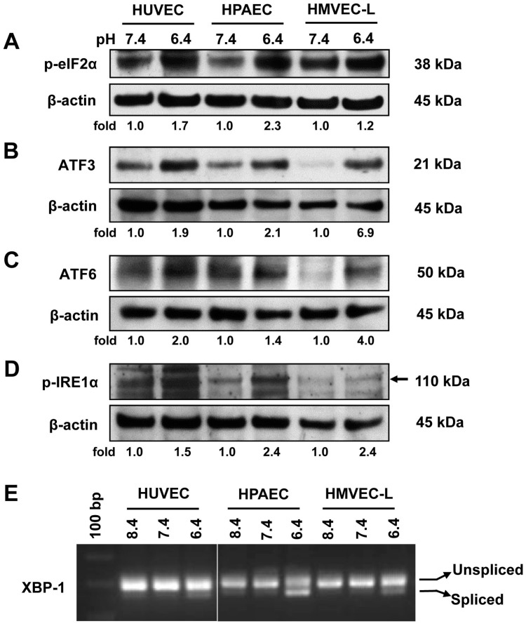
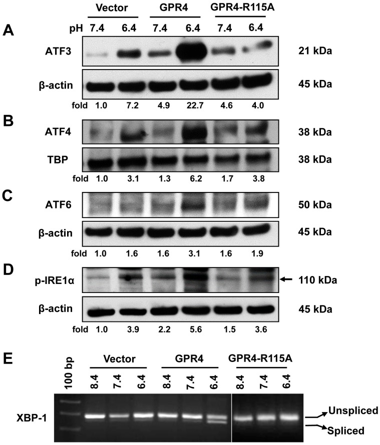
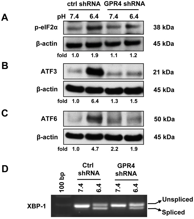
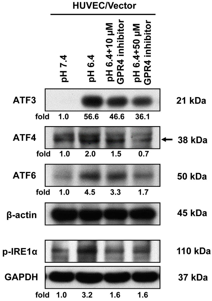
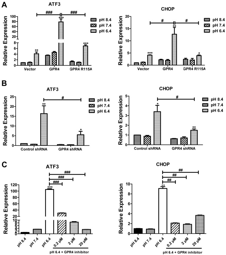
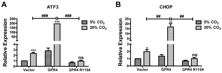
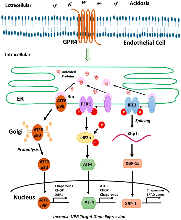
Acidosis commonly exists in the tissue microenvironment of various pathophysiological conditions such as tumors, inflammation, ischemia, metabolic disease, and respiratory disease. For instance, the tumor microenvironment is characterized by acidosis and hypoxia due to tumor heterogeneity, aerobic glycolysis (the "Warburg effect"), and the defective vasculature that cannot efficiently deliver oxygen and nutrients or remove metabolic acid byproduct. How the acidic microenvironment affects the function of blood vessels, however, is not well defined. GPR4 (G protein-coupled receptor 4) is a member of the proton-sensing G protein-coupled receptors and it has high expression in endothelial cells (ECs). We have previously reported that acidosis induces a broad inflammatory response in ECs. Acidosis also increases the expression of several endoplasmic reticulum (ER) stress response genes such as CHOP (C/EBP homologous protein) and ATF3 (activating transcription factor 3). In the current study, we have examined acidosis/GPR4- induced ER stress pathways in human umbilical vein endothelial cells (HUVEC) and other types of ECs. All three arms of the ER stress/unfolded protein response (UPR) pathways were activated by acidosis in ECs as an increased expression of phosphorylated eIF2α (eukaryotic initiation factor 2α), phosphorylated IRE1α (inositol-requiring enzyme 1α), and cleaved ATF6 upon acidic pH treatment was observed. The expression of other downstream mediators of the UPR, such as ATF4, ATF3, and spliced XBP-1 (X box-binding protein 1), was also induced by acidosis. Through genetic and pharmacological approaches to modulate the expression level or activity of GPR4 in HUVEC, we found that GPR4 plays an important role in mediating the ER stress response induced by acidosis. As ER stress/UPR can cause inflammation and cell apoptosis, acidosis/GPR4-induced ER stress pathways in ECs may regulate vascular growth and inflammatory response in the acidic microenvironment.

Keywords: G protein-coupled receptor 4 (GPR4); acidosis; blood vessel; endoplasmic reticulum (ER) stress; endothelial cell (EC); pH; tissue microenvironment; unfolded protein response (UPR).


Conflict of interest statement

The authors declare no conflict of interest.

Figures

Figure 1
Figure 1
Acidic pH activates the endoplasmic reticulum (ER) stress/unfolded protein response (UPR) pathways in vascular endothelial cells (ECs). (AD) Western blot of various protein expression in primary human umbilical vein endothelial cells (HUVEC), human pulmonary artery endothelial cells (HPAEC), and human lung microvascular endothelial cells (HMVEC-L). Cells were treated in EGM-2/HEM or EGM-2-MV/HEM media at physiological (pH 7.4) or acidic (pH 6.4) conditions for 0.5–1 h (p-eIF2α and p-IRE1α), or 5 h (ATF3 and ATF6). Cell lysates were then collected and separated by electrophoresis. Protein expression of (A) phosphorylated-eIF2α, (B) ATF3, (C) active/cleaved ATF6, and (D) phosphorylated-IRE1α was detected using the specific antibodies. β-Actin expression was used as a loading control. The arrow indicates the target band. After being normalized to the loading control, the target bands were quantified by densitometry using the ImageJ software (version 1.51, National Institutes of Health, Bethesda, MD, USA). The relative protein expression level at pH 7.4 of each EC type was set as 1.0-fold; (E) HUVEC, HPAEC, and HMVEC-L cells were treated at pH 8.4, 7.4, or 6.4 for 5 h. Total RNA was isolated and cDNA was synthesized. Unspliced and spliced human XBP-1 mRNA isoforms were examined by reverse transcription polymerase chain reaction (RT-PCR), as described in “Materials and Methods”. The results shown are representative of at least two biological repeats.
Figure 2
Figure 2
Acidosis-induced ER stress response is augmented by overexpression of G protein-coupled receptor 4 (GPR4), but not the signaling defective GPR4 mutant, in HUVEC. (AD) HUVECs transduced with the control vector (Vector), GPR4 expression construct (GPR4), or GPR4 R115A mutant expression vector (GPR4 R115A) were treated in EGM-2/HEM buffered media at physiological (pH 7.4) or acidic (pH 6.4) conditions for 0.5–1 h (p-IRE1α) or 5 h (ATF3, ATF4, and ATF6). Cell lysates were then collected and separated by electrophoresis. Protein expression of (A) ATF3, (B) ATF4, (C) active/cleaved ATF6, and (D) phosphorylated-IRE1α was detected using the specific antibodies. β-Actin or TATA-binding protein (TBP) expression was used as a loading control. The arrow indicates the target band. After being normalized to the loading control, the target bands were quantified by densitometry using the ImageJ software (version 1.51, National Institutes of Health, Bethesda, USA). The relative protein expression level at pH 7.4 of HUVEC/Vector was set as 1.0-fold. (E) HUVECs, as used in (AD), were treated at pH 8.4, 7.4, or 6.4 for 5 h. Total RNA was isolated and cDNA was synthesized. Spliced and unspliced XBP-1 mRNA isoforms were examined by RT-PCR. The results shown are representative of at least two biological repeats.
Figure 3
Figure 3
Acidosis-induced ER stress response is alleviated by knocking down GPR4 expression in HUVEC. HUVECs transduced with the control shRNA (ctrl shRNA) or GPR4 shRNA (GPR4 shRNA) were treated in EGM-2/HEM buffered media at physiological (pH 7.4) or acidic (pH 6.4) conditions for 0.5–1 h (p-eIF2α), 1 h (XBP-1), or 5 h (ATF3 and ATF6). (AC) Cell lysates were collected and separated by electrophoresis. Protein expression of (A) phosphorylated-eIF2α, (B) ATF3, and (C) active/cleaved ATF6 was detected using the specific antibodies. β-Actin expression was used as a loading control. After being normalized to the loading control, the target bands were quantified by densitometry using the ImageJ software (version 1.51, National Institutes of Health, Bethesda, USA). The relative protein expression level at pH 7.4 of HUVEC/Control-shRNA was set as 1.0-fold. (D) Total RNA was isolated from the cells and cDNA was synthesized. Spliced and unspliced XBP-1 isoforms were examined by RT-PCR. The results shown are representative of two or more biological repeats.
Figure 4
Figure 4
Acidosis-induced ER stress response is attenuated by a GPR4 small molecule inhibitor in HUVEC. HUVEC/Vector cells were treated for 0.5–1 h (p-IRE1α) or 5 h (ATF3, ATF4 and ATF6) with EGM-2/HEM pH 7.4 and 6.4 media or with pH 6.4 media containing 10 and 50 µM of the GPR4 inhibitor, for which one-hour pretreatment in EGM-2 medium with the same concentrations of GPR4 inhibitor was performed. Cell lysates were then collected and separated by electrophoresis. Protein expression of ATF3, ATF4, active/cleaved ATF6, and phosphorylated-IRE1α was detected using the specific antibodies. β-actin or GAPDH expression was used as a loading control. The arrow indicates the target band. After being normalized to the loading control, the target bands were quantified by densitometry using the ImageJ software (version 1.51, National Institutes of Health, Bethesda, MD, USA). The relative protein expression level at pH 7.4 of HUVEC/Vector was set as 1.0-fold. The results shown are representative of two or more biological repeats.
Figure 5
Figure 5
GPR4 modulates the ER stress response genes induced by acidic pH at the mRNA level in HUVEC. (A,B) HUVEC transduced with the control vector (Vector), GPR4 expression construct (GPR4), GPR4 R115A mutant expression vector (GPR4 R115A), control shRNA (control shRNA), or GPR4 shRNA expression vector (GPR4 shRNA) were treated in EGM-2/HEM buffered media at basic (pH 8.4), physiological (pH 7.4), or acidic (pH 6.4) conditions for 5 h. (C) HUVEC/GPR4 cells were treated for 5 h with EGM-2/HEM pH 8.4, 7.4, or 6.4 media or with pH 6.4 media containing 0.2, 2, and 20 µM of the GPR4 inhibitor, for which one-hour pretreatment in EGM-2 medium with the same concentrations of GPR4 inhibitor was performed. Total RNA was isolated, and cDNA was synthesized. Real-time qRT-PCR was performed to quantify the mRNA level of ATF3 and CHOP. Ct values were normalized to the housekeeping gene β-actin (ACTB). The expression level of the target genes in (A) HUVEC/Vector, (B) HUVEC/control shRNA or (C) HUVEC/GPR4 cells treated with pH 8.4 was set as 1. Error bars indicate the mean ± SEM. *, p < 0.05; **, p < 0.01; ***, p < 0.001; compared with corresponding pH 8.4 groups. #, p < 0.05; ##, p < 0.01; ###, p < 0.001; comparing the indicated pairs of data. The results shown are the average of at least two biological repeats.
Figure 6
Figure 6
GPR4 is involved in modulating the ER stress response induced by hypercapnic acidosis in HUVEC. HUVECs transduced with the control vector (Vector), GPR4 expression construct (GPR4), or GPR4 R115A mutant expression vector (GPR4 R115A) were treated for 5 h with EGM-2 media buffered with 5% CO2 or 20% CO2. Total RNA was isolated and cDNA was synthesized. Real-time qRT-PCR was performed to quantify the mRNA level of (A) ATF3 and (B) CHOP. Ct values were normalized to the housekeeping gene β-actin (ACTB). The expression level of the target genes in HUVEC/Vector cells treated with 5% CO2-buffered EGM-2 medium was set as 1. Error bars indicate the mean ± SEM. *, p < 0.05; **, p < 0.01; ***, p < 0.001; ns, not significant (p > 0.05); compared with corresponding “5% CO2” groups. #, p < 0.05; ##, p < 0.01; ###, p < 0.001; comparing the indicated pairs of data. The results shown are the average of at least two biological repeats.
Figure 7
Figure 7
A model depicting the acidosis/GPR4-induced ER stress response in vascular ECs. Acidic extracellular pH activates the proton-sensing receptor GPR4 and stimulates all three arms (ATF6, PERK, and IRE1) of the ER stress/UPR pathways in ECs. “P” denotes the phosphate group of a protein.

Similar articles

Cited by

References

    1. Chen A., Dong L., Leffler N.R., Asch A.S., Witte O.N., Yang L.V. Activation of GPR4 by acidosis increases endothelial cell adhesion through the cAMP/Epac pathway. PLoS ONE. 2011;6:e27586. doi: 10.1371/journal.pone.0027586. - DOI - PMC - PubMed
    1. Dong L., Li Z., Leffler N.R., Asch A.S., Chi J.T., Yang L.V. Acidosis activation of the proton-sensing GPR4 receptor stimulates vascular endothelial cell inflammatory responses revealed by transcriptome analysis. PLoS ONE. 2013;8:e61991. doi: 10.1371/journal.pone.0061991. - DOI - PMC - PubMed
    1. Gatenby R.A., Gillies R.J. Why do cancers have high aerobic glycolysis? Nat. Rev. Cancer. 2004;4:891–899. doi: 10.1038/nrc1478. - DOI - PubMed
    1. Justus C.R., Dong L., Yang L.V. Acidic tumor microenvironment and pH-sensing G protein-coupled receptors. Front. Physiol. 2013;4:354. doi: 10.3389/fphys.2013.00354. - DOI - PMC - PubMed
    1. Sanderlin E.J., Justus C.R., Krewson E.A., Yang L.V. Emerging roles for the pH-sensing G protein-coupled receptors in response to acidotic stress. Cell Health Cytoskelet. 2015;7:99–109.

MeSH terms