

Early Healing Events after Periodontal Surgery: Observations on Soft Tissue Healing, Microcirculation, and Wound Fluid Cytokine Levels
- PMID: 28134829
- PMCID: PMC5343819
- DOI: 10.3390/ijms18020283
Early Healing Events after Periodontal Surgery: Observations on Soft Tissue Healing, Microcirculation, and Wound Fluid Cytokine Levels
Abstract
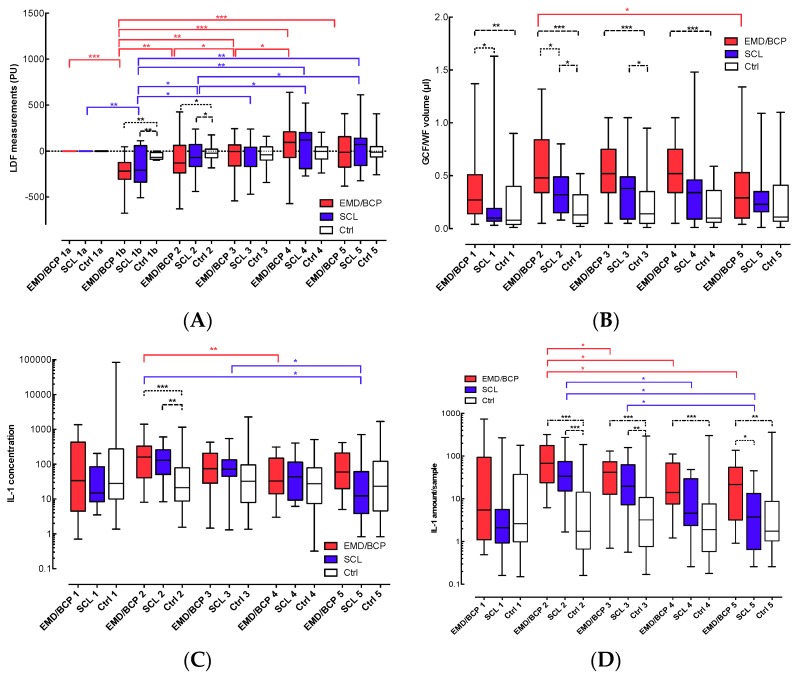
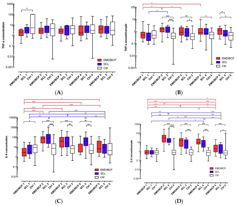
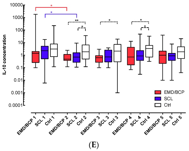
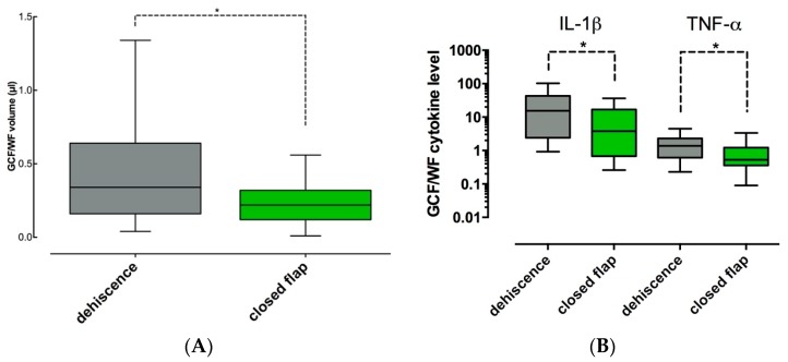
Early wound healing after periodontal surgery with or without enamel matrix derivative/biphasic calcium phosphate (EMD/BCP) was characterized in terms of soft tissue closure, changes of microcirculation, and expression of pro- and anti-inflammatory cytokines in gingival crevicular fluid/wound fluid (GCF/WF). Periodontal surgery was carried out in 30 patients (18 patients: application of EMD/BCP for regeneration of bony defects; 12 patients: surgical crown lengthening (SCL)). Healthy sites were observed as untreated controls. GCF/WF samples were collected during two post-surgical weeks. Flap microcirculation was measured using laser Doppler flowmetry (LDF). Soft tissue healing was evaluated after two weeks. GCF/WF levels of interleukin 1β (IL-1β), tumour necrosis factor (TNF-α), IL-6, and IL-10 were determined using a multiplex immunoassay. Surgery caused similar reductions of flap microcirculation followed by recovery within two weeks in both EMD/BCP and SCL groups. GCF/WF and pro-inflammatory cytokine levels were immediately increased after surgery, and returned only partially to baseline levels within the two-week observation period. Levels of IL-10 were temporarily reduced in all surgical sites. Flap dehiscence caused prolonged elevated levels of GCF/WF, IL-1β, and TNF-α. These findings show that periodontal surgery triggers an immediate inflammatory reaction corresponding to the early inflammatory phase of wound healing, and these inflammation measures are temporary in case of maintained closure of the flap. However, flap dehiscence causes prolonged inflammatory exudation from the periodontal wound. If the biological pre-conditions for periodontal wound healing are considered important for the clinical outcome, care should be taken to maintain primary closure of the flap.
Keywords: cytokines; periodontal regeneration; periodontal surgery; surgical crown lengthening; wound healing.
Conflict of interest statement
The authors declare no conflict of interest. The funding sponsors had no role in the design of the study; in the collection, analyses, or interpretation of data; in the writing of the manuscript, and in the decision to publish the results.
Figures
Similar articles
-
Changes in transforming growth factor-beta1 in gingival crevicular fluid following periodontal surgery.J Clin Periodontol. 2004 Jul;31(7):527-33. doi: 10.1111/j.1600-051x.2004.00521.x. J Clin Periodontol. 2004. PMID: 15191588 Clinical Trial.
-
Expression of inflammatory biomarkers and growth factors in gingival crevicular fluid at different healing intervals following non-surgical periodontal treatment: A systematic review.J Periodontal Res. 2020 Dec;55(6):801-809. doi: 10.1111/jre.12795. Epub 2020 Aug 25. J Periodontal Res. 2020. PMID: 32840888
-
EMD in periodontal regenerative surgery modulates cytokine profiles: A randomised controlled clinical trial.Sci Rep. 2016 Mar 15;6:23060. doi: 10.1038/srep23060. Sci Rep. 2016. PMID: 26976446 Free PMC article. Clinical Trial.
-
Levels of tissue inhibitor of metalloproteinases-1 and matrix metalloproteinases-1 and -8 in gingival crevicular fluid following treatment with enamel matrix derivative (EMDOGAIN).J Periodontal Res. 2001 Oct;36(5):309-16. doi: 10.1034/j.1600-0765.2001.360506.x. J Periodontal Res. 2001. PMID: 11585118 Clinical Trial.
-
Post-Surgical Clinical Monitoring of Soft Tissue Wound Healing in Periodontal and Implant Surgery.Int J Med Sci. 2017 Jul 18;14(8):721-728. doi: 10.7150/ijms.19727. eCollection 2017. Int J Med Sci. 2017. PMID: 28824306 Free PMC article. Review.
Cited by
-
Wound fluid sampling methods for proteomic studies: A scoping review.Wound Repair Regen. 2022 May;30(3):317-333. doi: 10.1111/wrr.13009. Epub 2022 Apr 5. Wound Repair Regen. 2022. PMID: 35381119 Free PMC article.
-
Platelet rich fibrin versus ozone gel for periodontal regeneration in induced rats' intrabony three-wall periodontal defects.J Oral Biol Craniofac Res. 2020 Oct-Dec;10(4):639-649. doi: 10.1016/j.jobcr.2020.09.001. Epub 2020 Sep 4. J Oral Biol Craniofac Res. 2020. PMID: 32983858 Free PMC article.
-
Introduction of "MAPS" wound healing index and its correlation with guided bone regeneration outcome.PLoS One. 2025 Mar 20;20(3):e0319271. doi: 10.1371/journal.pone.0319271. eCollection 2025. PLoS One. 2025. PMID: 40111952 Free PMC article.
-
Soft Tissue Retraction Maneuver in Cone Beam Computed Tomography Prior to Crown-Lengthening Procedure-A Technical Note.J Clin Med. 2024 Jun 24;13(13):3668. doi: 10.3390/jcm13133668. J Clin Med. 2024. PMID: 38999234 Free PMC article.
-
Non-invasive and quantitative methods for assessment of blood flow in periodontal and oral soft tissues: a systematic review.Front Dent Med. 2025 May 22;6:1587821. doi: 10.3389/fdmed.2025.1587821. eCollection 2025. Front Dent Med. 2025. PMID: 40475388 Free PMC article. Review.
References
MeSH terms
Substances
LinkOut - more resources
Full Text Sources
Other Literature Sources
Research Materials
Miscellaneous
