Review
. 2016 Dec 6;7(1):113-170.
doi: 10.1002/cphy.c160006.

Ischemia/Reperfusion

Affiliations
Review

Ischemia/Reperfusion

Theodore Kalogeris et al. Compr Physiol. .

Abstract

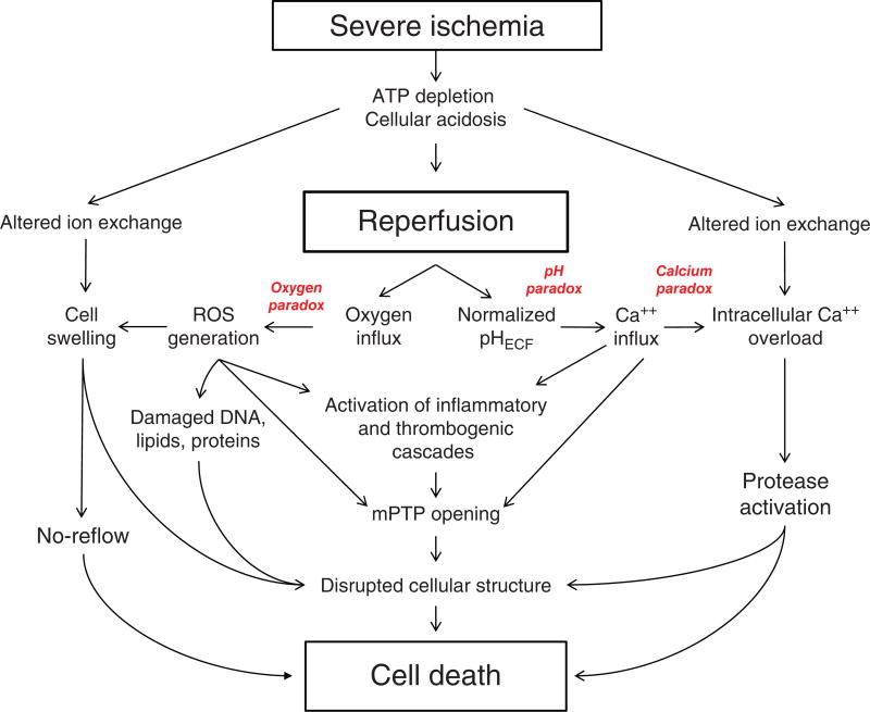
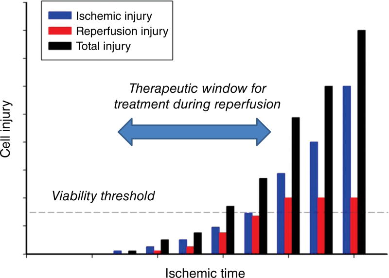
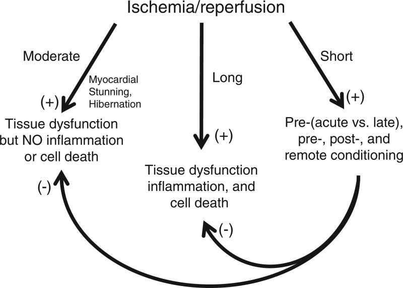
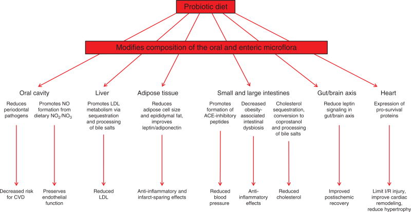
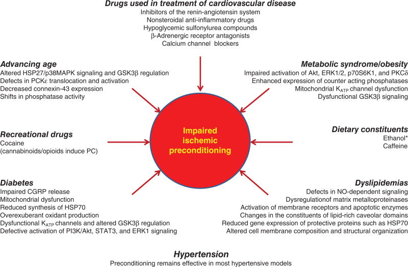
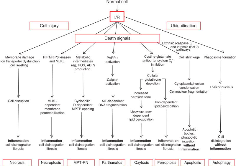
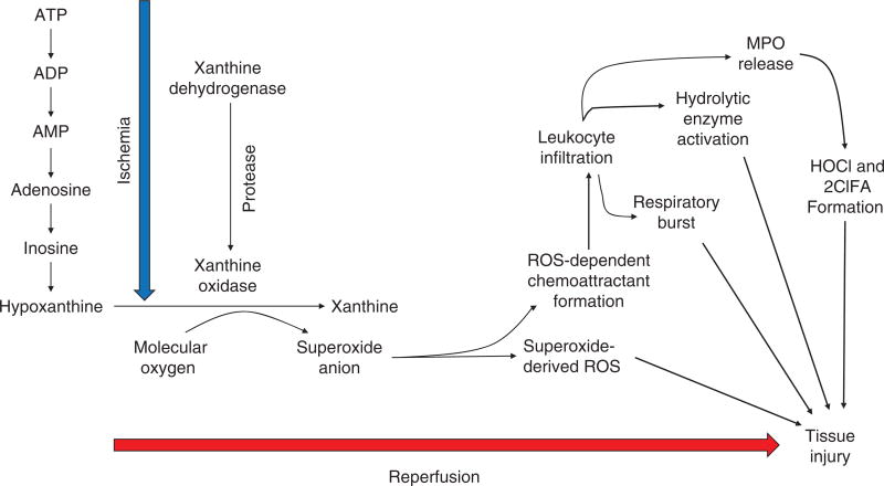
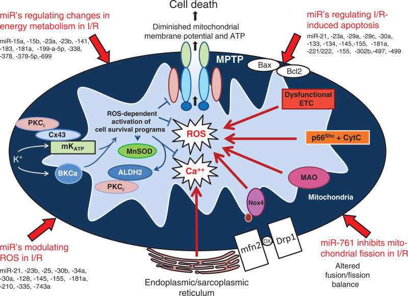
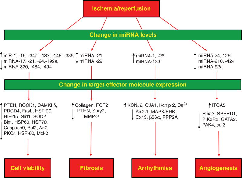
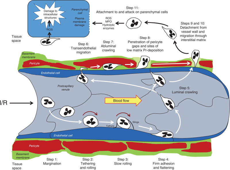
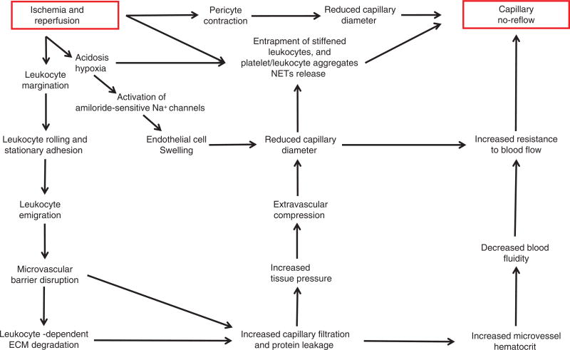
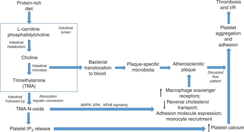
Ischemic disorders, such as myocardial infarction, stroke, and peripheral vascular disease, are the most common causes of debilitating disease and death in westernized cultures. The extent of tissue injury relates directly to the extent of blood flow reduction and to the length of the ischemic period, which influence the levels to which cellular ATP and intracellular pH are reduced. By impairing ATPase-dependent ion transport, ischemia causes intracellular and mitochondrial calcium levels to increase (calcium overload). Cell volume regulatory mechanisms are also disrupted by the lack of ATP, which can induce lysis of organelle and plasma membranes. Reperfusion, although required to salvage oxygen-starved tissues, produces paradoxical tissue responses that fuel the production of reactive oxygen species (oxygen paradox), sequestration of proinflammatory immunocytes in ischemic tissues, endoplasmic reticulum stress, and development of postischemic capillary no-reflow, which amplify tissue injury. These pathologic events culminate in opening of mitochondrial permeability transition pores as a common end-effector of ischemia/reperfusion (I/R)-induced cell lysis and death. Emerging concepts include the influence of the intestinal microbiome, fetal programming, epigenetic changes, and microparticles in the pathogenesis of I/R. The overall goal of this review is to describe these and other mechanisms that contribute to I/R injury. Because so many different deleterious events participate in I/R, it is clear that therapeutic approaches will be effective only when multiple pathologic processes are targeted. In addition, the translational significance of I/R research will be enhanced by much wider use of animal models that incorporate the complicating effects of risk factors for cardiovascular disease. © 2017 American Physiological Society. Compr Physiol 7:113-170, 2017.


Figures

Figure 1
Figure 1
Major pathologic events contributing to ischemia/reperfusion injury. When the blood supply is markedly reduced or absent, ischemic cells switch to anaerobic metabolism to provide ATP. However, this results in cellular acidosis and insufficient ATP production to meet metabolic demand. As a consequence, ATPases are inactivated, while active Ca2+ efflux and Ca2+ reuptake by the endoplasmic reticulum are markedly reduced, with the net effect of this abherent ion transport producing Ca2+ overload in the cell. In addition, xanthine dehydrogenase is converted to XO during ischemia (see Fig. 7), coincident with accumulation of hypoxanthine, one of the substrates required to drive its enzymatic activity. On reperfusion, the delivery of oxygen and substrates required for aerobic ATP generation is restored as is extracellular pH via washout of accumulated H+ (pH paradox). The latter event promotes additional Ca2+ influx (calcium paradox), while the influx of oxygen fuels XO-driven production of ROS (oxygen paradox) (see Fig. 7). ROS produced by this and other mechanisms can damage virtually every biomolecule found in cells, promote opening of mitochondrial PTPs, and activate inflammatory and thrombogenic cascades to exacerbate cell injury. The latter events are further amplified by release of danger signals (e.g., ATP) and other proinflammatory and thrombogenic mediators from damaged cells (see text for further explanation). The ensuing massive influx of immunocytes at previously ischemic sites contribute to cell injury via the NADPH oxidase-driven respiratory burst, release of hydrolytic enzymes, and production of MPO-derived hypochlorous acid and N-chloramines. The development of the capillary no-reflow phenomenon during reperfusion results in nutritive perfusion impairment by mechanisms outlined in Figure 11.
Figure 2
Figure 2
Total injury sustained by a tissue subjected to ischemia followed by reperfusion (I/R) (black bars) is attributable to ischemia per se (blue bars) and a component that is due to reestablishing the blood supply (red bars). At the onset of prolonged ischemia two separate general pathologic processes are initiated. The first are processes of tissue injury that are due to ischemia per se. The second are biochemical changes that occur during ischemia that contribute to the surge in generation of reactive oxygen species and infiltration of proinflammatory neutrophils and other immunocytes when molecular oxygen is reintroduced to the tissues during reperfusion. For a treatment to be effective in reducing cellular dysfunction and/or death when administered at the onset of reperfusion (therapeutic window), reestablishing the blood supply must occur before damage attributable to ischemia per se exceeds the viability threshold for irreversible damage. Concepts from Bulkley, 1987 (100).
Figure 3
Figure 3
Tissue responses to ischemia/reperfusion are bimodal (trimodal in the heart), depending on the duration and magnitude of ischemia. Prolonged and severe ischemia induces cell damage that progresses to infarction, with reperfusion paradoxically exacerbating tissue injury by invoking inflammatory responses. In the heart, shorter bouts of ischemia (5–20 min duration) induce myocardial stunning, wherein contractile function is initially impaired on reperfusion, but slowly improves, without progression to infarction and in the absence of significant inflammation. On the other hand, prolonged exposure to subacute levels of ischemia without reperfusion may induce myocardial hibernation, wherein cardiac cells modify their metabolic phenotype to survive but with a cost of reduced mechanical function. The third mode of response is exemplified by the tissue response to short periods of ischemia (<5 min) followed by reperfusion (ischemic conditioning) that do not produce detectable injury or dysfunction. Far from being innocuous and functionally inert, the response of all organs to such conditioning ischemia is characterized by activation of cell survival programs that confer tolerance to the deleterious effects induced by subsequent exposure to prolonged I/R such that postischemic injury is dramatically reduced. Cardioprotective effects are invoked when tissues are exposed to short bouts of conditioning I/R prior to (ischemic preconditioning) or during (ischemic per-conditioning) prolonged ischemia or at the onset of reperfusion after prolonged cessation of blood flow (ischemic postconditioning). Tolerance to prolonged I/R in one organ can also be activated by subjecting distant organs to conditioning I/R, a remote effect that can also magnify the beneficial actions of local conditioning.
Figure 4
Figure 4
Ingestion of probiotic diets modifies the composition profile of the oral and enteric microbiome to limit I/R via microflora-dependent alterations that decrease risk for cardiovascular disease via reductions in blood pressure, oral pathogens, blood LDL and total cholesterol, preservation of endothelium-dependent vasodilator mechanisms, activation of anti-inflammatory and infarct-sparing cell survival programs, and improved postischemic tissue remodeling.
Figure 5
Figure 5
The presence of coexisting risk factors including metabolic syndrome, obesity, diabetes, advancing age, smoking, and dyslipidemias not only increase the likelihood for cardiovascular disease, but also worsen the outcome for those individuals who do suffer a heart attack or stroke. Interestingly, while ischemic and pharmacologic conditioning strategies are remarkably effective in young, healthy subjects, the presence of the aforementioned comorbid factors reduces their cardioprotective effects. The mechanisms underlying the impaired efficacy of conditioning is listed below each of the italicized co-morbid risk factors in the figure. Surprisingly little attention has been devoted to the effect of cigarette smoking to limit the efficacy of conditioning or with regard to the mechanisms by which this impairment occurs. Caffeine consumption also reduces the effectiveness preconditioning, as does the ingestion of alcoholic beverages at high levels, an effect that disappears as the absorbed ethanol is metabolized and eliminated from the blood. While use of some recreational drugs (eg, cocaine) abolishes ischemic preconditioning, morphine (or other opioids) injections or smoking marijuana may induce preconditioned phenotypes via activation opioid and cannabinoid receptors, respectively. It is also important to note that many of the drugs commonly used in the therapeutic management of patients with cardiovascular disease who are at high risk for myocardial infarction or stroke reduce or abolish the effectiveness of preconditioning stimuli by affecting their underlying signaling mechanisms. Reproduced from Ref. , with permission.
Figure 6
Figure 6
Cell death modalities in ischemia/reperfusion (I/R). I/R-induced necrosis generally occurs as a result of dysfunctional ion transport mechanisms, which causes cells to swell and eventually burst, effects that are exacerbated by plasma membrane damage. Release of proinflammatory mediators and damaged biomolecules initiates the influx of inflammatory cells such as neutrophils, which disrupt the extracellular matrix and cause damage to parenchyal cells by release of cytotoxic oxidants and hydrolytic enzymes. Apopotosis is a regulated form of cell death that causes cell shrinkage and condensation of the cytosol and nucleus, which eventually form apoptotic bodies. Because they are surrounded by cell membranes, apoptotic bodies can be engulfed and digested by phagocytes without evoking an inflammatory response. Autophagy provides a mechanism to remove damaged or senescent protein aggregates and organelles by enclosing them in membrane-lined vesicles called proteasomes which fuse with lysosomes containing enzymes that degrade the ingested material, usually without evoking an inflammatory response. While normally performing this “housekeeping” function, autophagy may also provide cells with a survival mechanism to withstand the deleterious effects of ischemia, by generating amino acids and fatty acids for cell function. However, when uncontrolled, autophagy contributes to ischemic cell death. While necrosis was once believed to occur from non-specific trauma or injury as a result of I/R, it now appears that postischemic infarction may also be attributable to programmed events that require a dedicated molecular circuitry that has been termed programmed necrosis or necroptosis. Necroptosis is initiated by TNF-like cytokines that activate RIP kinases to mediate necrosis via increased production of reactive oxygen species and calcium overload, which in turn modulate the mitochondrial permeability transition pore (MPTP), leading to dissipation of the proton electrochemical gradient, with subsequent ATP depletion, further ROS production, and swelling and rupture of mitochondrial membranes. Recent genetic studies have suggested that the MPTP is predominantly involved in a second form of regulated necrosis that is designated MPT-RN that is critically dependent on cyclophilin D. Parthanotos can be distinguished from other forms of programmed cell death by its requirement for poly-ADP-ribose polymerase activation. Two newly described cell death modalities have been implicated in I/R, ferroptosis and oxytosis. Both involve inhibition of the cytine-glutamate antiporter Xc, but differ in their modes of lipid peroxidation, being iron dependent and lipoxygenase dependent, respectively.
Figure 7
Figure 7
Mechanism for XO-dependent production of ROS at the onset of reperfusion. During the period of ischemia, ATP is step-wise catabolized to hypoxanthine, which accumulates in the tissues because the lack of blood flow does not wash out metabolites from the tissues. Coincident with these changes, xanthine dehydrogenase is converted to XO by a proteolytic mechanism. Thus, a requisite substrate (hypoxanthine) and the activated enzyme (XO) are present in excess in ischemic tissues, but the oxidation of hypoxanthine to xanthine and uric acid cannot proceed, owing to the lack of molecular oxygen that is required to fuel the reaction. On reperfusion, this requisite substrate is suddenly resupplied to the tissue, which fuels the rapid overproduction of ROS. ROS-induced formation of chemoattractants promotes leukocyte infiltration, neutrophils in particular, which in turn exacerbate cellular injury via NADPH oxidase-dependent respiratory burst, MPO-mediated formation of hypochlorous acid, N-chloramines, and 2-chloro fatty acids (2ClFA), and release and activation of hydrolytic enzymes that target every type of biomolecule found in cells and tissues.
Figure 8
Figure 8
Generation of ROS by mitochondria (mitoROS) is a nexus for both activation of cell survival programs that mediate the effect of conditioning stimuli to enhance tolerance to I/R and serves as a focal point for overexuberant ROS-induced ROS release that contributes to the pathogenesis of cell injury in I/R. On the one hand, ROS triggers the activation of cell survival programs in responses to a number of mildly noxious stimuli, such as short bouts of ischemia or antecedent ethanol exposure or pharmacologic agents (activators of mitochondrial ATP-sensitive potassium (mKATP) or large conductance, calcium-activated potassium (BKCa) channels. The enhanced tolerance to ischemia invoked by these mitoROS-dependent conditioning stimuli, which can be delivered before (preconditioning), during (perconditioning) or at the onset of reperfusion (postconditioning), activate protective protein kinases, such as PKCε, the expression of prosurvival genes (e.g., heme oxygenase-1) and mitochondrial antioxidant defenses (e.g., MnSOD, aldehyde dehydrogenase-1, or ALDH2), as well as targeting the MPTP to maintain the channel in a closed state. On the other hand, overexuberant ROS generation at the onset of reperfusion, driven by ROS-induced ROS release that is fueled by electron transport chain dysfunction, especially at complexes I and III, and enhanced activities of p66Shc, MAO, and NADPH oxidase-4 (Nox4) in mitochondria, causes the MPTP to open, leading to swelling, cell disruption, and death. Not depicted is the effect of oxidants to alter the balance of mitochondrial fission and fusion in conditioning and I/R, which emerging evidence has implicated as contributory to both processes. The dual nature of ROS as protective vs damaging species relates to the type of ROS produced, their concentrations, and/or the compartmental localization for their production. MIcroRNAs (miR’s) formed during conditioning and during I/R produce changes in mitochondrial energy metabolism, apoptosis, mitochondrial fission/fusion balance, and ROS production. See text for further explanation. Modified from Refs. and .
Figure 9
Figure 9
Functional roles and target genes for miRNAs implicated in ischemia/reperfusion and preconditioning. See text for further explanation.
Figure 10
Figure 10
Neutrophil trafficking to ischemic sites occurs during reperfusion of ischemic tissues and involves 11 distinct steps. See text for further explanation. Figure modified from Refs. and .
Figure 11
Figure 11
Mechanisms underlying the development of the postischemic capillary no-reflow phenomenon. See text for explanation.
Figure 12
Figure 12
Overnutrition via consumption of a Western diet rich in red meats, lipids, and fructose and lacking fermentable fiber may contribute to a shift in the intestinal microbiome to a profile that favors enhanced risk for myocardial infarction, stroke, and peripheral vascular disease. As one example, consumption of protein-rich diets containing red meats leads to the liberation of carnitine and phosphatidyl choline as the bolus of ingested food is digested in the small bowel, which are converted to TMA by colonic microbes. Upon absorption, TMA is metabolized to the proatherogenic TMAO by the liver to promote the formation of atherosclerotic plaques. The problem is exacerbated by diet-induced dysbiosis, which favors the translocation of bacteria into the bloodstream and promotion of plaque formation. Interestingly, a blood-like microbiome exists in plaques that resembles the patient’s oral and intestinal microbial profiles, with some of the bacterial species correlating with plasma cholesterol levels. TMAO also contributes to a prothrombogenic phenotype via effects to increase platelet IP3 levels, thereby elevating intracellular calcium levels to promote platelet aggregration and adhesion to injured endothelium.

References

    1. Abdellatif M. Differential expression of microRNAs in different disease states. Circ Res. 2012;110:638–650. - PMC - PubMed
    1. Abela CB, Homer-Vanniasinkham S. Clinical implications of ischaemia-reperfusion injury. Pathophysiology. 2003;9:229–240. - PubMed
    1. Abonia JP, Friend DS, Austen WG, Jr, Moore FD, Jr, Carroll MC, Chan R, Afnan J, Humbles A, Gerard C, Knight P, Kanaoka Y, Yasuda S, Morokawa N, Austen KF, Stevens RL, Gurish MF. Mast cell protease 5 mediates ischemia-reperfusion injury of mouse skeletal muscle. J Immunol. 2005;174:7285–7291. - PMC - PubMed
    1. Abramov AY, Scorziello A, Duchon M. Three distinct mechanisms generate oxygen free radicals in neurons and contribute to cell death during anoxia and reoxygenation. J Neurosci. 2007;27:1129–1138. - PMC - PubMed
    1. Adibhatha RM, Hatche JF. Lipid oxidation and peroxidation in CNS health and disease: From molecular mechanisms to therapeutic opportunities. Antiox Redox Signal. 2010;12:125–169. - PubMed

Publication types