

Dendritic Release of Neurotransmitters
- PMID: 28135005
- PMCID: PMC5381730
- DOI: 10.1002/cphy.c160007
Dendritic Release of Neurotransmitters
Abstract
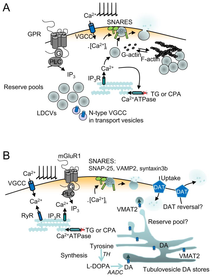
Release of neuroactive substances by exocytosis from dendrites is surprisingly widespread and is not confined to a particular class of transmitters: it occurs in multiple brain regions, and includes a range of neuropeptides, classical neurotransmitters, and signaling molecules, such as nitric oxide, carbon monoxide, ATP, and arachidonic acid. This review is focused on hypothalamic neuroendocrine cells that release vasopressin and oxytocin and midbrain neurons that release dopamine. For these two model systems, the stimuli, mechanisms, and physiological functions of dendritic release have been explored in greater detail than is yet available for other neurons and neuroactive substances. © 2017 American Physiological Society. Compr Physiol 7:235-252, 2017.
Copyright © 2017 John Wiley & Sons, Inc.
Figures
Similar articles
-
Talking back: dendritic neurotransmitter release.Trends Neurosci. 2003 May;26(5):255-61. doi: 10.1016/S0166-2236(03)00072-9. Trends Neurosci. 2003. PMID: 12744842 Review.
-
Dendritic release of vasopressin and oxytocin.J Neuroendocrinol. 1998 Dec;10(12):881-95. doi: 10.1046/j.1365-2826.1998.00279.x. J Neuroendocrinol. 1998. PMID: 9870745 Review.
-
Dendritic transmitter release: a comparison of two model systems.J Neuroendocrinol. 2008 Jun;20(6):677-86. doi: 10.1111/j.1365-2826.2008.01714.x. J Neuroendocrinol. 2008. PMID: 18601689 Review.
-
Oestradiol acutely stimulates exocytosis of oxytocin and vasopressin from dendrites and somata of hypothalamic magnocellular neurons.Neuroscience. 1995 Oct;68(4):1179-88. doi: 10.1016/0306-4522(95)00186-m. Neuroscience. 1995. PMID: 8544991
-
Multiple signalling modalities mediated by dendritic exocytosis of oxytocin and vasopressin.Philos Trans R Soc Lond B Biol Sci. 2015 Jul 5;370(1672):20140182. doi: 10.1098/rstb.2014.0182. Philos Trans R Soc Lond B Biol Sci. 2015. PMID: 26009761 Free PMC article. Review.
Cited by
-
Shielding Effect of Ryanodine Receptor Modulator in Rat Model of Autism.Basic Clin Neurosci. 2023 Mar-Apr;14(2):247-261. doi: 10.32598/bcn.2021.2966.1. Epub 2023 Mar 1. Basic Clin Neurosci. 2023. PMID: 38107532 Free PMC article.
-
The modular organization of the cerebral cortex: Evolutionary significance and possible links to neurodevelopmental conditions.J Comp Neurol. 2019 Jul 1;527(10):1720-1730. doi: 10.1002/cne.24554. Epub 2018 Nov 15. J Comp Neurol. 2019. PMID: 30303529 Free PMC article. Review.
-
Parallel Processing of Two Mechanosensory Modalities by a Single Neuron in C. elegans.Dev Cell. 2019 Dec 2;51(5):617-631.e3. doi: 10.1016/j.devcel.2019.10.008. Epub 2019 Nov 14. Dev Cell. 2019. PMID: 31735664 Free PMC article.
-
Persistent autism-relevant behavioral phenotype and social neuropeptide alterations in female mice offspring induced by maternal transfer of PBDE congeners in the commercial mixture DE-71.Arch Toxicol. 2022 Jan;96(1):335-365. doi: 10.1007/s00204-021-03163-4. Epub 2021 Oct 23. Arch Toxicol. 2022. PMID: 34687351 Free PMC article.
-
Somato-dendritic vasopressin and oxytocin secretion in endocrine and autonomic regulation.J Neuroendocrinol. 2020 Jun;32(6):e12856. doi: 10.1111/jne.12856. Epub 2020 May 14. J Neuroendocrinol. 2020. PMID: 32406599 Free PMC article. Review.
References
-
- Albin RL, Young AB, Penney JB. The functional anatomy of basal ganglia disorders. Trends Neurosci. 1989;12:366–375. - PubMed
-
- An S, Zenisek D. Regulation of exocytosis in neurons and neuroendocrine cells. Cur Opin Neurobiol. 2004;14:522–530. - PubMed
-
- Andersson DR, Nissbrandt H, Bergquist F. Partial depletion of dopamine in substantia nigra impairs motor performance without altering striatal dopamine neurotransmission. Eur J Neurosci. 2006;24:617–624. - PubMed
-
- Armstrong WE. Morphological and electrophysiological classification of hypothalamic supraoptic neurons. Prog Neurobiol. 1995;47:291–339. - PubMed
-
- Bains JS, Ferguson AV. Activation of N-methyl-D-aspartate receptors evokes calcium spikes in the dendrites of rat hypothalamic paraventricular nucleus neurons. Neuroscience. 1999;90:885–891. - PubMed
Publication types
MeSH terms
Substances
Grants and funding
LinkOut - more resources
Full Text Sources
Other Literature Sources
