Review
. 2017 Apr 26;117(8):5457-5520.
doi: 10.1021/acs.chemrev.6b00591. Epub 2017 Jan 30.

Mechanistic Understanding of Lanthipeptide Biosynthetic Enzymes

Affiliations
Review

Mechanistic Understanding of Lanthipeptide Biosynthetic Enzymes

Lindsay M Repka et al. Chem Rev. .

Abstract

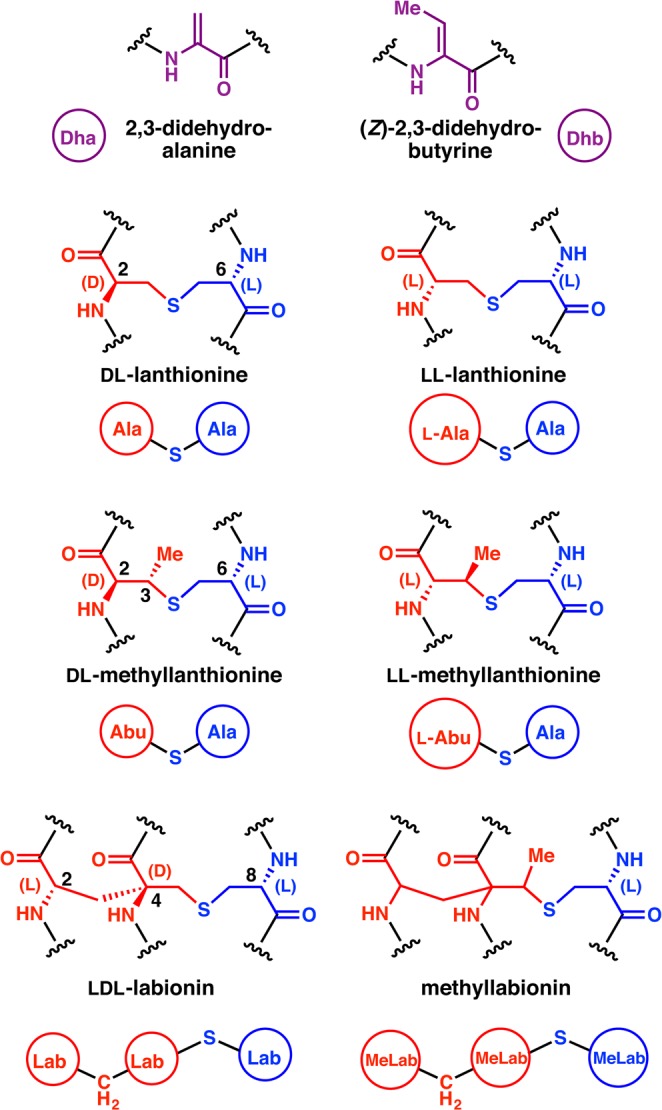
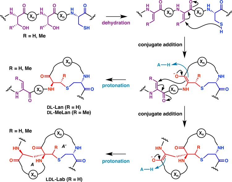
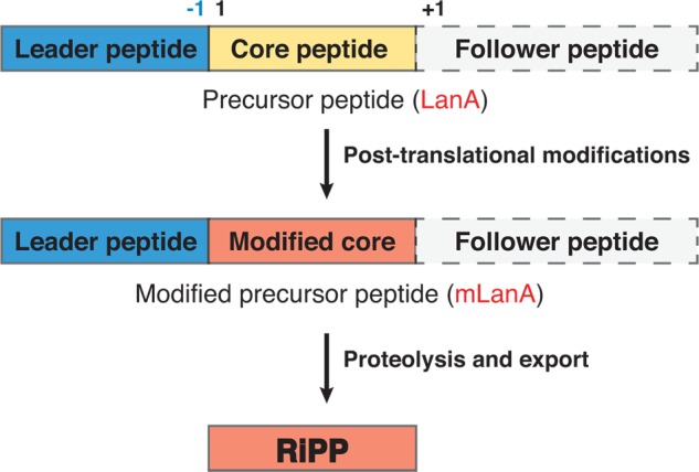
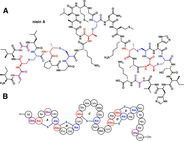
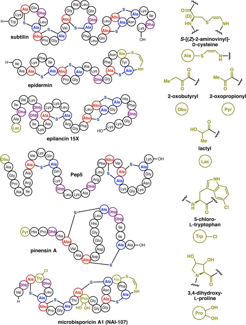
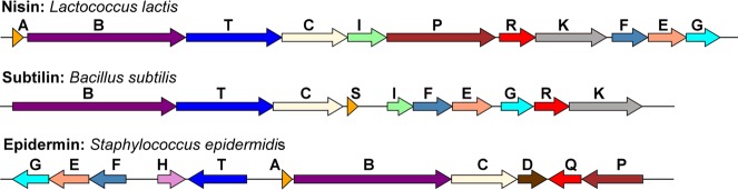
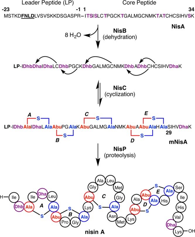
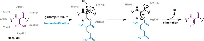
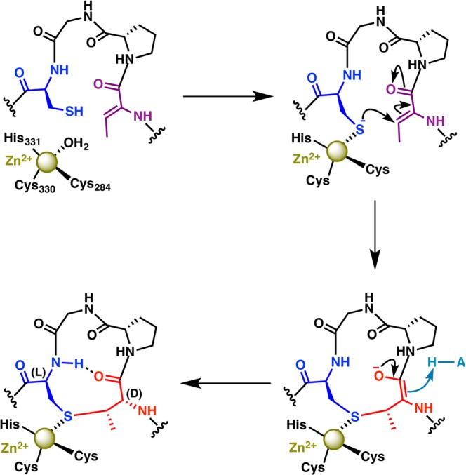
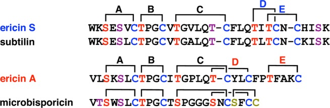
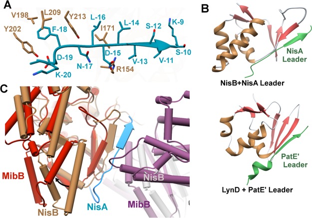
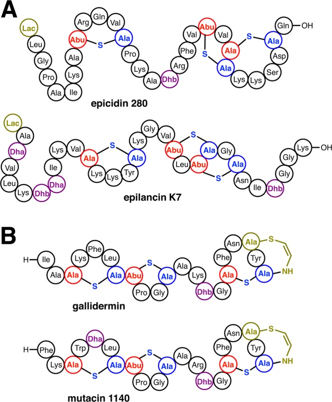
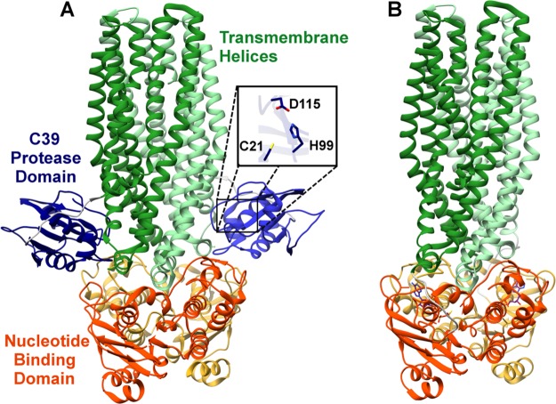
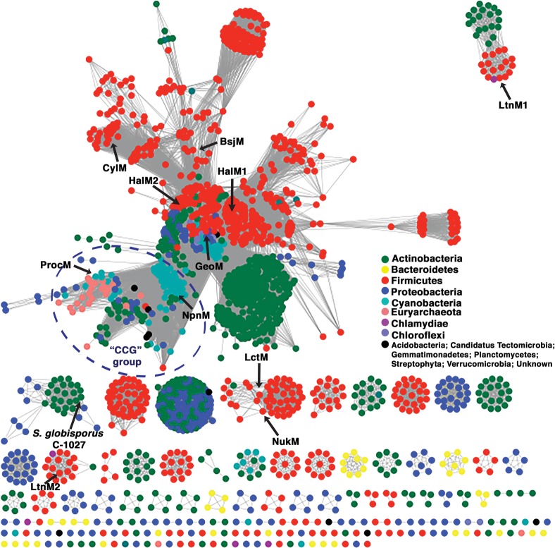
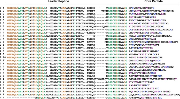
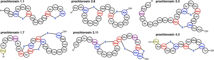
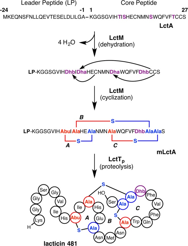
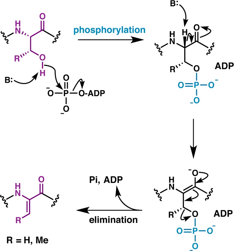
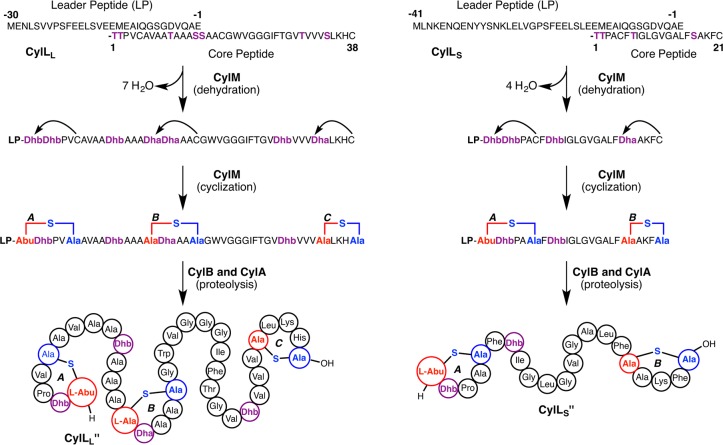
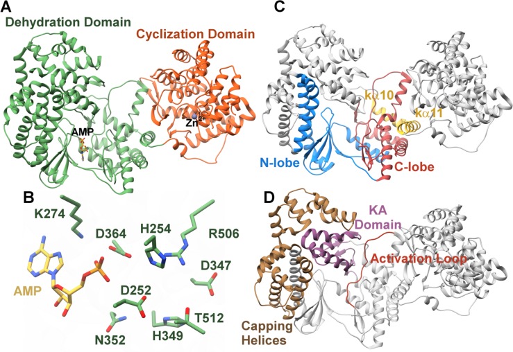
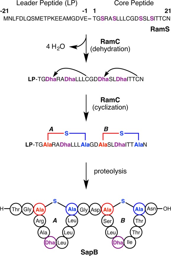
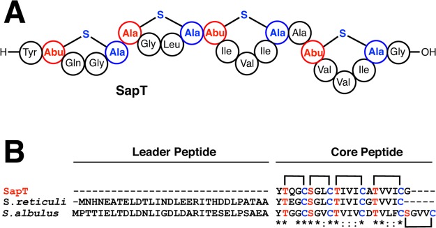
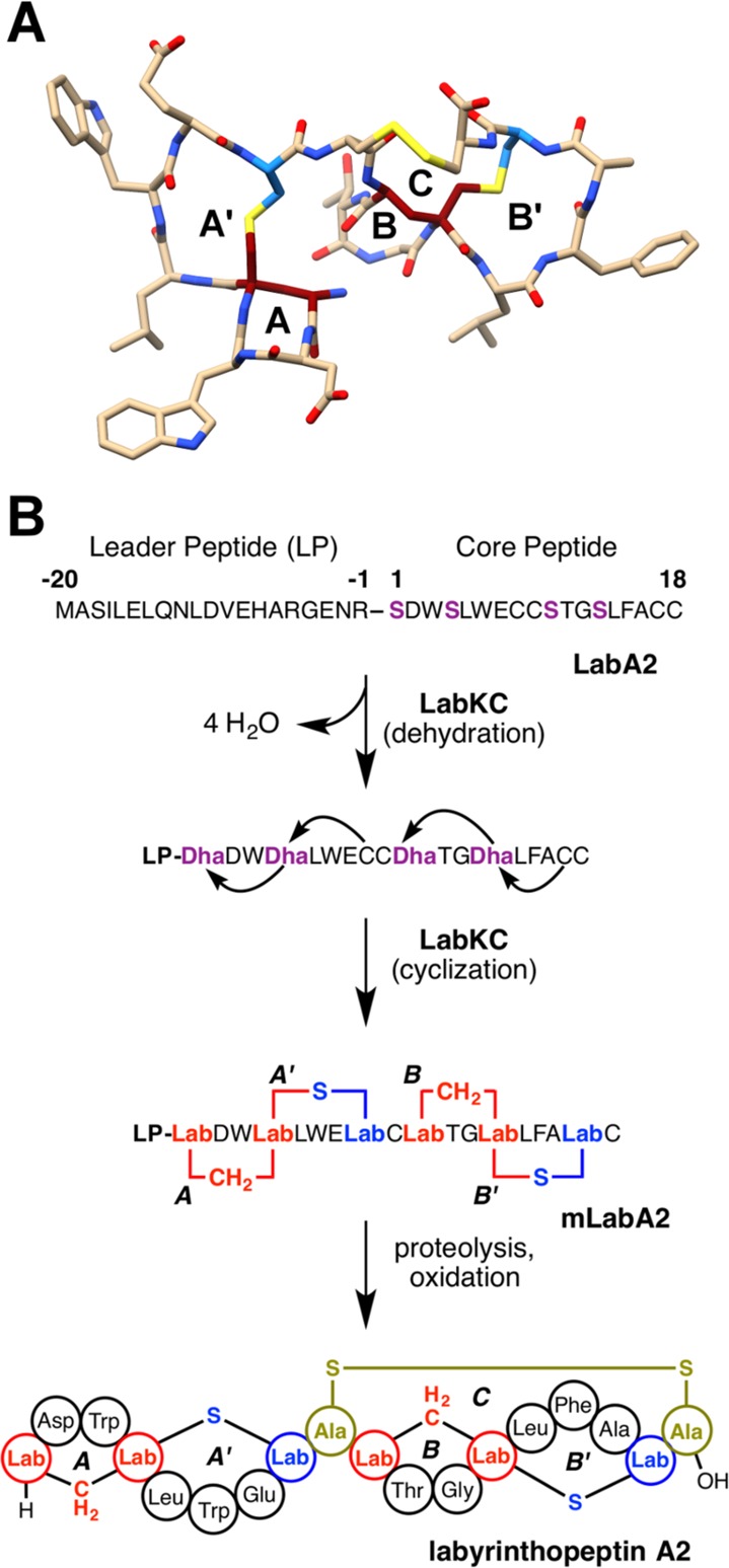
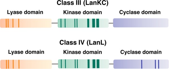
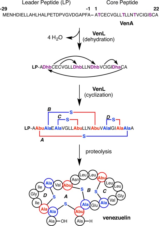
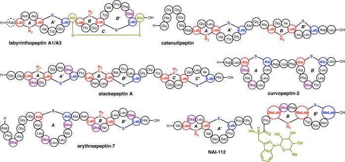
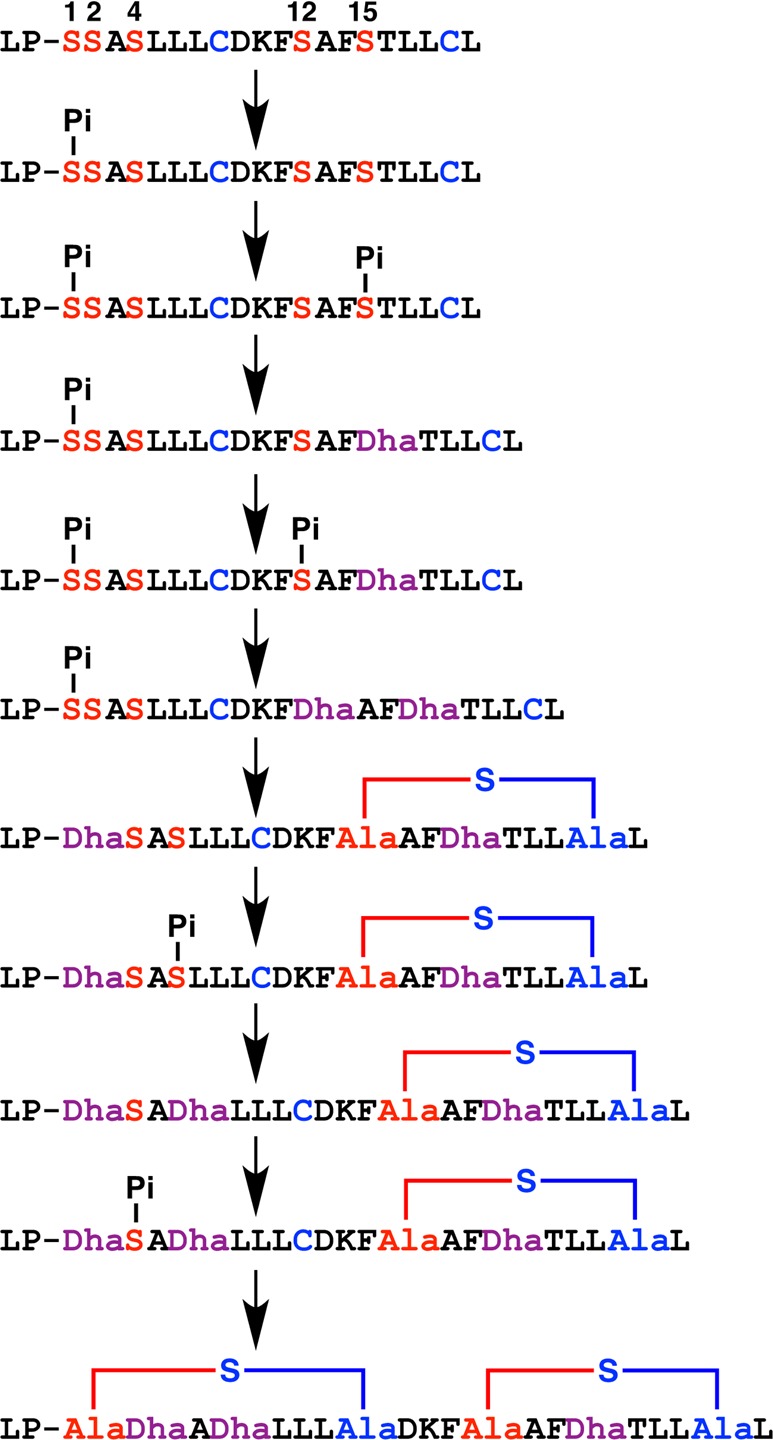
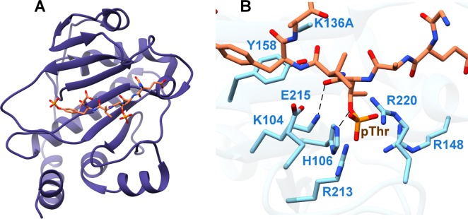
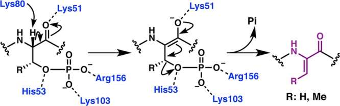
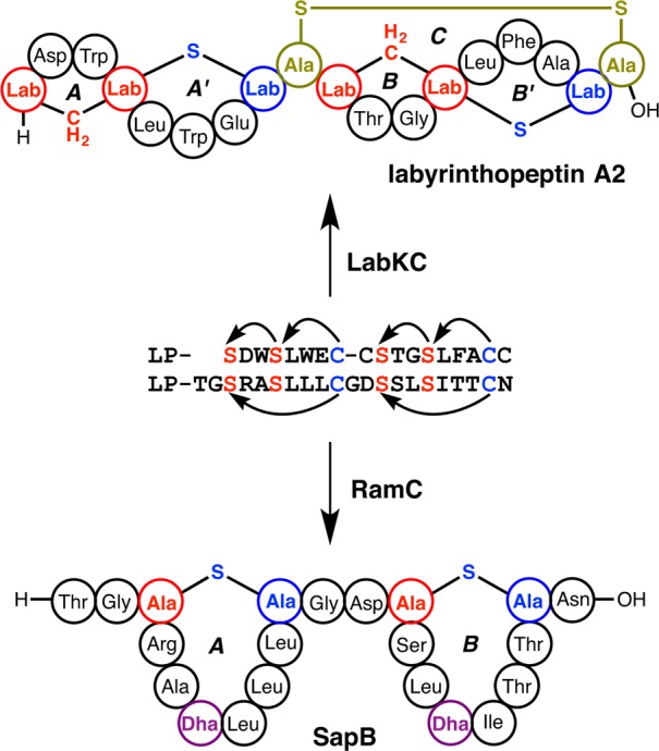
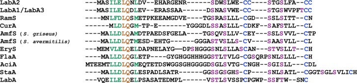
Lanthipeptides are ribosomally synthesized and post-translationally modified peptides (RiPPs) that display a wide variety of biological activities, from antimicrobial to antiallodynic. Lanthipeptides that display antimicrobial activity are called lantibiotics. The post-translational modification reactions of lanthipeptides include dehydration of Ser and Thr residues to dehydroalanine and dehydrobutyrine, a transformation that is carried out in three unique ways in different classes of lanthipeptides. In a cyclization process, Cys residues then attack the dehydrated residues to generate the lanthionine and methyllanthionine thioether cross-linked amino acids from which lanthipeptides derive their name. The resulting polycyclic peptides have constrained conformations that confer their biological activities. After installation of the characteristic thioether cross-links, tailoring enzymes introduce additional post-translational modifications that are unique to each lanthipeptide and that fine-tune their activities and/or stability. This review focuses on studies published over the past decade that have provided much insight into the mechanisms of the enzymes that carry out the post-translational modifications.


Conflict of interest statement

The authors declare no competing financial interest.

Figures

Figure 1
Figure 1
Structures of the thioether cross-links and dehydro amino acids that are characteristic for lanthipeptides. Under each chemical structure is shown a shorthand notation used in this review. At present, two different diastereomers of both Lan and MeLan have been found in natural lanthipeptides, whereas for Dhb and Lab only one diastereomer has been reported thus far. A methyl-substituted Lab (MeLab) has recently been identified and its stereochemistry is unknown. In all drawings of cross-links, atoms originating from Ser/Thr are shown in red, whereas atoms originating from Cys are shown in blue. Abu: α-aminobutyric acid.
Figure 2
Figure 2
Post-translational modification reactions leading to the formation of (Me)Lan or (Me)Lab. Color coding as in Figure 1. The stereochemistry for MeLab has not been determined. Xn = peptide of n amino acids. The stereochemistry of (Me)Lan can be different from DL (see Figure 1).
Figure 3
Figure 3
General biosynthetic pathway for RiPPs. For lanthipeptides, thus far no follower peptides have been reported. The general numbering scheme for the precursor peptides is indicated.
Figure 4
Figure 4
Structure of the class I lanthipeptide nisin A (A) and its shorthand structure representation (B) that is used throughout this review.
Figure 5
Figure 5
Structures of class I lanthipeptides including members containing tailoring modifications (olive green). Shorthand notations used for each post-translational modification (PTM) are indicated. R-stereochemistry has been demonstrated for the lactyl group in epilancin 15X (see section 2.6.1).
Figure 6
Figure 6
Biosynthetic gene clusters of various class I lanthipeptides.,,,− See section 2.1 and Abbreviations section for the general nomenclature of lanthipeptide biosynthetic genes.
Figure 7
Figure 7
Scheme showing the biosynthetic route to nisin A. For clarity, the process is shown as first completion of dehydration and then cyclization, but this is not necessarily the case. The timing of the different PTMs installed on the NisA precursor peptide is discussed in the text. The FNLD motif in the leader peptide (bolded and underlined) is conserved in other class I lanthipeptide leader peptides and appears important in interactions with both NisB and NisC. The cyclization catalyzed by NisC is reversible as shown by experiments involving resubjection of mNisA to the cyclization conditions (section 2.3).
Figure 8
Figure 8
LanB enzymes achieve dehydration by catalyzing a transesterification reaction from glutamyl-tRNAGlu to the side chain of Ser/Thr followed by β-elimination. Chemoselective activation by glutamate via its α-carboxylate is illustrated here as has been demonstrated for the nonlanthipeptide RiPP goadsporin. Conserved LanB residues predicted by mutagenesis studies on NisB to be important for transesterification and elimination are shown in gray. The exact roles of these residues remain to be established.
Figure 9
Figure 9
(A) Structure of the NisB homodimer bound to the NisA leader sequence (cyan). The glutamylation domain (brown) and elimination domain (purple) are indicated for one monomer, and the other protomer is colored in gray. (B) Mapping of the electrostatic potential onto the surface of the NisB monomer identified a basic patch (blue) in the vicinity of the NisA leader (green) predicted to engage glutamyl-tRNAGlu. PDB ID 4WD9.
Figure 10
Figure 10
Residues identified by Ala-scanning mutational analysis that are critical for (A) glutamylation and (B) glutamate elimination map to the two different domains of NisB. The exact roles of these residues remain to be established. PDB ID 4WD9.
Figure 11
Figure 11
Sequence similarity network generated herein using EFI-EST and visualized in Cytoscape with an alignment score threshold of 120 (∼30% sequence identity) showing the distribution of LanB enzymes across phyla. Each node represents protein sequences sharing >95% sequence identity.
Figure 12
Figure 12
(A) Overall structure of the NisC cyclase (PDB ID 2G0D) illustrating the α,α-toroid (orange), and the SH2-like extension (blue and purple). (B) Close-up view of NisC showing the zinc ligands and other residues located in the active site. Residues derived from the SH2-like domain are indicated in blue. (C) Active site of LanCL1 showing coordination of enzyme residues (green) and GSH (red) to zinc (PDB ID 3E73).
Figure 13
Figure 13
Proposed mechanism of cyclization illustrated for the formation of the B-ring of nisin. The active site acid (H-A) that protonates the enolate is likely His212.
Figure 14
Figure 14
Class I cyclase EriC produces the lanthipeptides ericin S and ericin A with different D and E ring topologies resembling those of subtilin and microbisporicin, respectively.
Figure 15
Figure 15
(A) Close-up view of the interaction between the NisA leader sequence (blue) and the NisB dehydratase (brown). (B) Comparison of the winged helix-turn-helix leader-binding motifs in NisB and the cyanobactin heterocyclase LynD, another RiPP biosynthetic enzyme (PDB ID 4V1T). (C) Superimposition of NisB (brown and gray) with bound NisA leader (blue) and unliganded MibB (red and purple, PDB ID 5EHK), focusing on the winged helix-turn-helix domain. The black arrow illustrates the inward tilt of an ampipathic helix in MibB relative to its counterpart in NisB that interacts with the NisA leader peptide.
Figure 16
Figure 16
Tailoring modifications in class I lanthipeptide biosynthesis include installation of an N-terminal lactate (Lac) (A) and installation of a C-terminal aminovinyl Cys (B). The structure shown for epicidin 280 is hypothetical and based on analogy to Pep5 but is known to contain an N-terminal Lac.
Figure 17
Figure 17
(A) Structure of the ElxO oxidoreductase involved in N-terminal Lac installation. (B) Close-up view of the active site showing critical residues and bound NADP(H) (yellow). (C) Electrostatic surface view of the structure illustrating an obvious groove that may be involved in binding to the peptide substrate. PDB ID 4QEC.
Figure 18
Figure 18
(A) Structure of the EpiD flavoprotein homotrimer involved in AviCys installation. (B) Close-up view of the FMN (yellow) cofactor in the vicinity of a bound short peptide substrate (purple). PDB ID 1G5Q.
Figure 19
Figure 19
(A) Solution NMR structures showing the overall folds of the SpaI (PDB ID 2LVL), NisI (PDB ID 2N2E and 2N32) and MlbQ resistance proteins (PDB ID 2MVO). The fold of MlbQ is distinct from those of SpaI and NisI. (B) Crystal structure of the S. agalactiae nisin resistance protein (SaNSR). PDB ID 4Y68.
Figure 20
Figure 20
(A) Overall structure of the NisP catalytic domain. PDB ID 4MZD. (B) Docking model of the unmodified NisA peptide (green) onto the NisP active site (pink) evidencing steric clashes that may explain the substrate preference of NisP for mNisA. (C) Structure of the LicP protease including the prodomain. PDB ID 4ZOQ.
Figure 21
Figure 21
Structures of bovicin HJ50, nukacin ISK-1, and mutacin II. For the first two compounds, the protease activity of the cognate bifunctional LanTps has been reconstituted. Bovicin HJ50 incorporates a disulfide tailoring modification (section 4.6).
Figure 22
Figure 22
Biosynthesis of the two-component lantibiotic lichenicidin. The structures of Licα and Licβ were assigned by NMR and MS analyses. LicTp acts on both peptides to remove the entire leader peptide of Licα and all but six C-terminal residues, NDVNPE (bolded and underlined), of Licβ. The latter are recognized by LicP in the final proteolysis step. The stereochemistry of the Licβ A-ring is unknown but predicted to be LL as it derives from a Dhx-Dhx-Xxx-Xxx-Cys motif (Dhx = Dha/Dhb; section 4.3.3). For discussion of the LanM proteins, see section 4. For clarity, the process is shown as first completion of dehydration and then cyclization, but this is not necessarily the case. For instance, for the related two-component lantibiotic haloduracin, cyclization commences before dehydration is completed.,
Figure 23
Figure 23
Homodimeric structure of the AMS family protease/transporter from C. thermocellum determined in the absence (A) and presence (B) of an inert nucleotide analog. The transmembrane domain is colored in dark and light green and the nucleotide-binding domain is colored in dark and light orange. The protease domain shown in dark and light blue is not ordered in the nucleotide bound structure, and is presumably directed away from the core of the transporter. In the absence of nucleotide, the entrance to the transmembrane tunnel is situated at the interface with the protease domain and positions the active site residues right at the gateway. A close-up view of the active site residues of the protease domain is shown in the inset box. PDB ID 4S0F and 4RY2.
Figure 24
Figure 24
Sequence similarity network of LanMs generated herein using EFI-EST and visualized in Cytoscape with an alignment score threshold of 110 (∼35% sequence identity). Each node represents protein sequences sharing 100% sequence identity. LanMs discussed throughout this review are indicated. The enzymes containing three Cys as Zn2+ ligands (“CCG” group) are mostly found within the dashed circle. Enzymes from clusters that contain both LanM and LanC enzymes (e.g., a cluster from Streptomyces globisporus C-1027) group together.
Figure 25
Figure 25
ProcM has 30 putative substrates, 29 of which contain Cys residues in the core peptide. One substrate, ProcAT.1, has a truncated leader peptide. Fully conserved identical residues in the leader peptide are shown in dark orange and fully conserved similar residues are shown in green. Cys residues in the core peptide are shown in blue and Ser/Thr residues in purple.
Figure 26
Figure 26
Six structurally characterized prochlorosins demonstrate the diverse ring topologies installed by ProcM.
Figure 27
Figure 27
Biosynthesis of lacticin 481. For clarity, the process is shown as first completion of dehydration and then cyclization, but this is not actually the case since cyclization commences before dehydration is completed. See section 4.5.
Figure 28
Figure 28
Dehydration during biosynthesis of class II lanthipeptides involves phosphorylation followed by phosphate elimination with anti stereoselectivity in all cases for which the stereochemistry has been investigated.
Figure 29
Figure 29
Biosynthesis of both peptides that make up cytolysin. For clarity, the process is shown as first completion of dehydration and then cyclization, but the order of the PTMs is currently not known.
Figure 30
Figure 30
(A) Structure of the CylM class II lanthipeptide synthetase showing the dehydration domain (green) and cyclization domain (orange). (B) Close-up view of the dehydration active site showing critical active site residues and bound nucleotide. (C) and (D) Color-coded diagrams highlighting in (C) the N- and C-terminal kinase lobes (blue and red) as well as the kα10 and kα11 helices (yellow), and in (D) the LanM specific KA-domain (purple), activation loop (red), and capping helices (brown). PDB ID 5DZT.
Figure 31
Figure 31
Proposed mechanism of phosphorylation and phosphate elimination by CylM based on mutagenesis studies. In the absence of a cocrystal structure, many details are still missing. For instance, the interactions of Asp252/His254 with the nucleotide phosphate could be mediated via a Mg2+. Not shown are Asn352, Asp364, and Glu366, which also appear to engage the nucleotide phosphates.
Figure 32
Figure 32
Structure of the CylM cyclization domain shown in the same orientation as NisC in Figure 12A. The antiparallel β-sheet that may constitute a substrate-binding platform is shown in purple.
Figure 33
Figure 33
Biosynthesis of haloduracin β. BH1491 is an uncharacterized protease that has been suggested to remove residues −1 through −6 after cleavage by HalTp at the GS motif. Although for clarity the process is drawn as dehydration and then cyclization, the cyclization process commences before dehydration is completed., See section 4.4.
Figure 34
Figure 34
Cyclization catalyzed by both HalM2 and NisC is reversible, as shown with α-D-labeled mHalA2 and mNisA. D/H exchange in α-D-labeled mHalA2 occurs in the presence of HalM2 but could occur either by a deprotonation/reprotonation mechanism or by a reversible cyclization mechanism. The reversibility of cyclization was confirmed for α-D-labeled mHalA2 and mNisA by trapping of the ring-opened species by conjugate addition to NEM.
Figure 35
Figure 35
Modification of LctA(−24–20, S11A/Q20A) by LctM and by a cyclization-deficient mutant showed that LctM enforces the chemoselectivity of cyclization. The major products are shown but the native ring topology was observed as a minor product using the cyclization-deficient mutant.
Figure 36
Figure 36
Nonenzymatic and enzymatic cyclization of ProcA substrates containing a single ring installed by ProcM-catalyzed modification of a linear substrate with one Cys protected. In all cases, nonenzymatic cyclization was much slower (A and B). When B ring formation was blocked by Cys protection of ProcA3.3, an incorrect ring topology was favored as characterized for the enzymatic cyclization (B). LP = leader peptide.
Figure 37
Figure 37
Class II two-component natural product bicereucin comprises a lanthipeptide and a linear peptide. The Lan ring was produced as a mixture of stereoisomers in the in vitro reconstitution of bicereucin biosynthesis.
Figure 38
Figure 38
Residue-specific incorporation of deuterium at the α-carbon of Ser/Thr enables monitoring of the directionality of dehydration. Dehydration of unlabeled Ser/Thr involves a loss of 18 Da (A), whereas dehydration of α-deuterated Ser/Thr involves a loss of 19 Da (B).
Figure 39
Figure 39
Conformational selection model whereby leader peptide binding shifts an equilibrium to an active enzyme state that promotes core peptide binding to the enzyme and facilitates catalysis. The enzyme, leader peptide, and core peptide are shown in blue, pink, and green, respectively. The leader peptide in class II LanAs is predicted to form a helix but this conformation has not been directly proven in the context of interactions with the modifying enzymes and is not generalizable to class I lanthipeptide biosynthesis, as the precursor NisA does not adopt a helical conformation in binding to its modifying enzyme NisB (section 2.5).
Figure 40
Figure 40
Examples of class II lanthipeptides containing d-Ala residues and a 2-oxobutyryl (Obu) or 2-oxopropionyl (pyruvyl, Pyr) tailoring modification at the N-terminus.
Figure 41
Figure 41
(A) Cinnamycin and duramycin peptides share identical post-translational modifications including Lal formation and Asp hydroxylation, for which the structures are shown in (B).
Figure 42
Figure 42
Structures of actagardine and Ala(0)-actagardine.
Figure 43
Figure 43
Biosynthesis of SapB. For clarity the process is drawn as first complete dehydration and then cyclization, but the order of the PTMs for SapB is not actually known.
Figure 44
Figure 44
SapT (A) is predicted to be a class I lanthipeptide based on sequence alignment (B) with putative LanAs from S. reticuli and S. albulus that cluster with genes encoding putative LanB enzymes. Asterisks indicate fully conserved identical residues whereas colons indicate conserved similar residues.
Figure 45
Figure 45
(A) X-ray crystal structure of labyrinthopeptin A2 (CCDC number 721326). The color coding uses standard atom-type colors (red, oxygen; blue, nitrogen; yellow, sulfur). For Lab, the carbons originating from Cys are shown in light blue and those originating from Ser are in magenta. (B) Biosynthesis of labyrinthopeptin A2. For clarity the process is drawn as complete dehydration and then cyclization, but the order of the PTMs can be more complicated; e.g. see text and Figure 49 for curvopeptin.
Figure 46
Figure 46
Class III and class IV lanthipeptide synthetases are distinguished by their cyclase domains. LanL (class IV) contains zinc-binding ligands conserved in class I and II but these ligands are absent in the class III LanKC.
Figure 47
Figure 47
Biosynthesis of venezuelin. For clarity the process is drawn as completion of dehydration before cyclization, but the order of the PTMs for venezuelin is not currently known.
Figure 48
Figure 48
Structures of class III lanthipeptides. Labyrinthopeptin A3 differs from labyrinthopeptin A1 by a single additional Asp residue at the N-terminus thought to derive from incomplete leader removal (section 5.4).
Figure 49
Figure 49
CurKC catalyzes phosphorylation, phosphate elimination, and cyclization of CurA in a distributive fashion and unusual order. Only the main pathway is shown, with minor parallel pathways also observed. Pi = phosphate; LP = leader peptide.
Figure 50
Figure 50
(A) Structure of SpvC type III effector protein K136A mutant (purple) bound to a model pThr-bearing substrate (brown). (B) Close-up view of the SpvC active site. PDB ID 2Z8P.
Figure 51
Figure 51
Proposed mechanism of VenL-catalyzed phosphate elimination to afford Dhx.
Figure 52
Figure 52
Class III lanthipeptides labyrinthopeptin A2 and SapB contain different PTMs (Lab and Lan, respectively) despite high similarity of their precursor sequences.
Figure 53
Figure 53
Alignment of class III lanthipeptide precursors. Lanthipeptides used: labyrinthopeptins A1-A3 (LabA1-A3), SapB (RamS), curvopeptin (CurA), griseopeptin (Streptomyces griseus AmfS), avermipeptin (Streptomyces avermitilis AmfS), erythreapeptin (EryS), flavipeptin (FlaA), catenulipeptin (AciA), stackepeptin (StaA), and NAI-112 (LabA). Fully conserved identical residues in the leader region are shown in dark orange and conserved similar residues are shown in green. Ser/Thr residues that undergo dehydration are shown in purple, and Cys residues are shown in blue.

Similar articles

Cited by

References

    1. Arnison P. G.; Bibb M. J.; Bierbaum G.; Bowers A. A.; Bugni T. S.; Bulaj G.; Camarero J. A.; Campopiano D. J.; Challis G. L.; Clardy J.; et al. Ribosomally synthesized and post-translationally modified peptide natural products: overview and recommendations for a universal nomenclature. Nat. Prod. Rep. 2013, 30 (1), 108–160. 10.1039/C2NP20085F. - DOI - PMC - PubMed
    1. Schnell N.; Entian K. D.; Schneider U.; Götz F.; Zahner H.; Kellner R.; Jung G. Prepeptide sequence of epidermin, a ribosomally synthesized antibiotic with four sulphide-rings. Nature 1988, 333 (6170), 276–278. 10.1038/333276a0. - DOI - PubMed
    1. Mayer H.; Bauer H.; Breuss J.; Ziegler S.; Prohaska R. Characterization of rat LANCL1, a novel member of the lanthionine synthetase C-like protein family, highly expressed in testis and brain. Gene 2001, 269 (1–2), 73–80. 10.1016/S0378-1119(01)00463-2. - DOI - PubMed
    1. Park S.; James C. D. Lanthionine synthetase components C-like 2 increases cellular sensitivity to adriamycin by decreasing the expression of P-glycoprotein through a transcription-mediated mechanism. Cancer Res. 2003, 63 (3), 723–727. - PubMed
    1. Mohr K. I.; Volz C.; Jansen R.; Wray V.; Hoffmann J.; Bernecker S.; Wink J.; Gerth K.; Stadler M.; Müller R. Pinensins: the first antifungal lantibiotics. Angew. Chem., Int. Ed. 2015, 54 (38), 11254–11258. 10.1002/anie.201500927. - DOI - PubMed

MeSH terms