. 2017 Jan 30;13(1):e1006131.
doi: 10.1371/journal.ppat.1006131. eCollection 2017 Jan.

Blocking two-component signalling enhances Candida albicans virulence and reveals adaptive mechanisms that counteract sustained SAPK activation

Affiliations

Blocking two-component signalling enhances Candida albicans virulence and reveals adaptive mechanisms that counteract sustained SAPK activation

Alison M Day et al. PLoS Pathog. .

Abstract

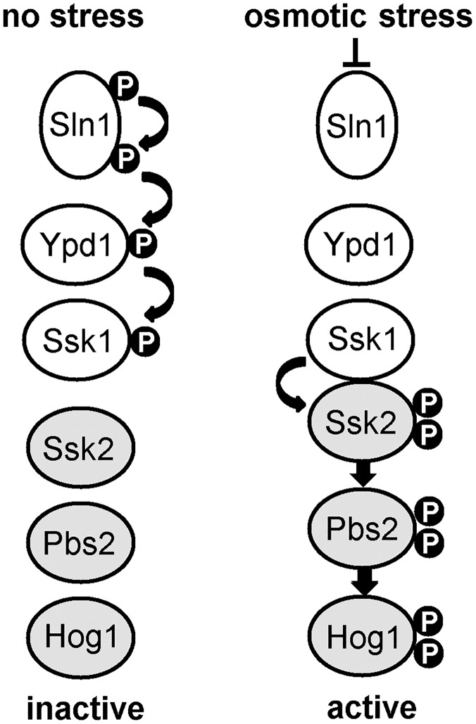
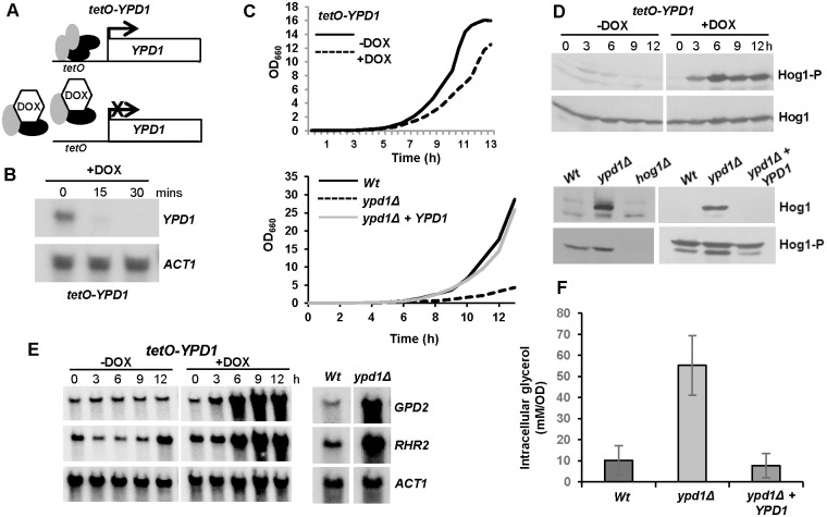
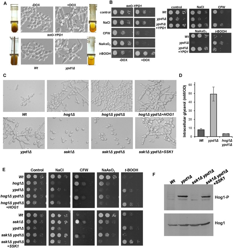
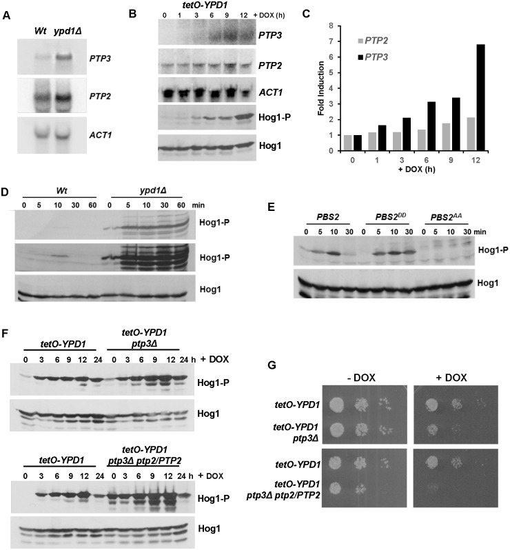
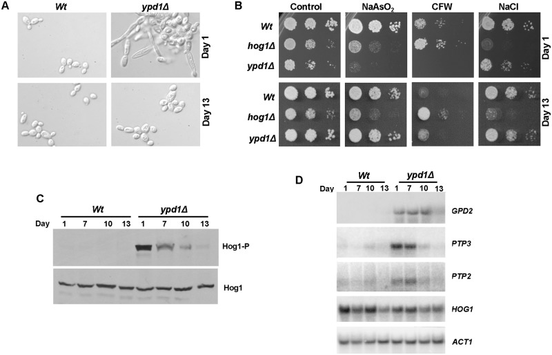
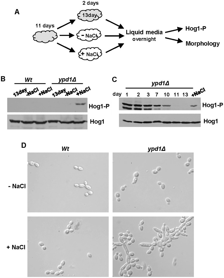
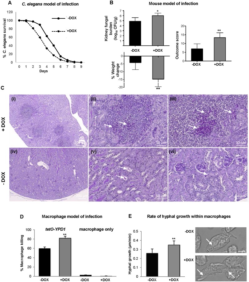
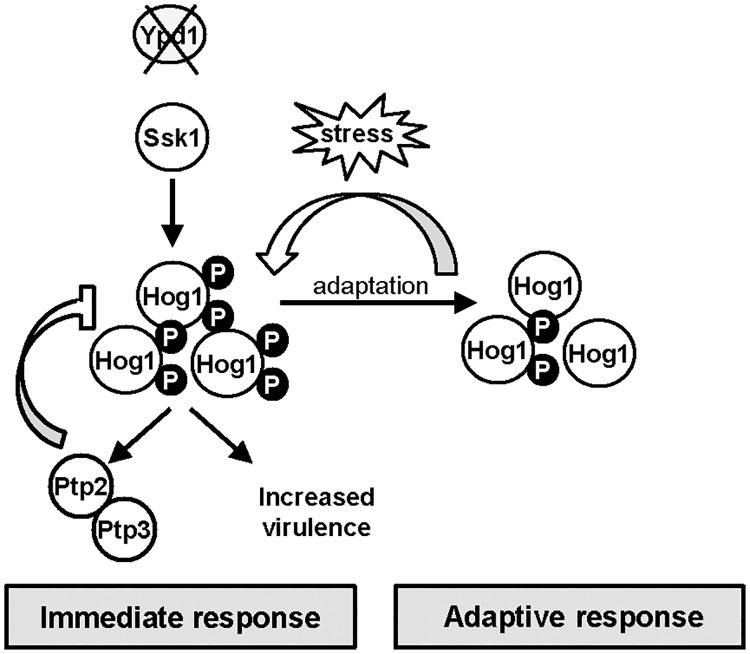
The Ypd1 phosphorelay protein is a central constituent of fungal two-component signal transduction pathways. Inhibition of Ypd1 in Saccharomyces cerevisiae and Cryptococcus neoformans is lethal due to the sustained activation of the 'p38-related' Hog1 stress-activated protein kinase (SAPK). As two-component signalling proteins are not found in animals, Ypd1 is considered to be a prime antifungal target. However, a major fungal pathogen of humans, Candida albicans, can survive the concomitant sustained activation of Hog1 that occurs in cells lacking YPD1. Here we show that the sustained activation of Hog1 upon Ypd1 loss is mediated through the Ssk1 response regulator. Moreover, we present evidence that C. albicans survives SAPK activation in the short-term, following Ypd1 loss, by triggering the induction of protein tyrosine phosphatase-encoding genes which prevent the accumulation of lethal levels of phosphorylated Hog1. In addition, our studies reveal an unpredicted, reversible, mechanism that acts to substantially reduce the levels of phosphorylated Hog1 in ypd1Δ cells following long-term sustained SAPK activation. Indeed, over time, ypd1Δ cells become phenotypically indistinguishable from wild-type cells. Importantly, we also find that drug-induced down-regulation of YPD1 expression actually enhances the virulence of C. albicans in two distinct animal infection models. Investigating the underlying causes of this increased virulence, revealed that drug-mediated repression of YPD1 expression promotes hyphal growth both within murine kidneys, and following phagocytosis, thus increasing the efficacy by which C. albicans kills macrophages. Taken together, these findings challenge the targeting of Ypd1 proteins as a general antifungal strategy and reveal novel cellular adaptation mechanisms to sustained SAPK activation.


Conflict of interest statement

The authors have declared that no competing interests exist.

Figures

Fig 1
Fig 1. Overview of two component-mediated regulation of the Hog1 SAPK in S. cerevisiae.
Under non-stress conditions the Sln1 histidine kinase autophosphorylates, and this phosphate is transferred via the Ypd1 phosphorelay protein to the Ssk1 response regulator protein. Following osmotic stress Sln1 is inactivated, thus halting the phosphorelay which culminates in unphosphorylated Ssk1 which is a potent activator of the Ssk2 MAPKKK. This results in the phosphorylation and activation of the downstream Hog1 SAPK.
Fig 2
Fig 2. C. albicans cells lacking YPD1 exhibit hyperactivation of Hog1 but are viable.
(A) Strategy to control YPD1 expression. One YPD1 allele was deleted, and the remaining allele placed under the control of the E. coli tet operator (tetO) in strain THE1 to generate strain tetO-YPD1 (JC1586). THE1 cells express an E. coli tet repressor–S. cerevisiae Hap4 activation domain fusion protein. In the absence of doxycycline (DOX), this fusion protein binds as a dimer to the tet operator resulting in transcriptional activation. However, doxycycline prevents dimerisation of the fusion protein and blocks transcription. (B) Doxycycline treatment inhibits YPD1 expression. Northern analysis of YPD1 and ACT1 (control) transcript levels in tetO-YPD1 cells treated with doxycycline for the indicated times. (C) Repression or deletion of YPD1 results in a slow growth phenotype. Growth analysis of tetO-YPD1 cells, untreated or treated with doxycycline (top panel), and wild-type (Wt, JC21), ypd1Δ (JC2001) and ypd1Δ+YPD1 (JC2002) cells (bottom panel). (D) Repression or deletion of YPD1 results in constitutive phosphorylation of Hog1. Western blot analysis of whole cell extracts isolated from tetO-YPD1 cells following treatment with doxycycline for the indicated times, or from exponentially growing wild-type (Wt), ypd1Δ and ypd1Δ+YPD1 cells. Blots were probed for phosphorylated Hog1 (Hog1-P), stripped and reprobed for total Hog1 (Hog1). (E) Repression or deletion of YPD1 results in high levels of GPD2 and RHR2 expression. RNA was isolated from tetO-YPD1 cells, treated with or without doxycycline for the indicated times, or wild-type and ypd1Δ cells, and analyzed using gene-specific probes with ACT1 as a loading control. (F) Deletion of YPD1 results in increased intracellular glycerol levels. The mean ± SD is shown for 3 biological replicates.
Fig 3
Fig 3. Phenotypes associated with loss of Ypd1 are dependent on Hog1 and Ssk1.
(A) Repression or deletion of YPD1 triggers flocculation and a swollen pseudohyphal filamentous phenotype. Micrographs of Wt, ypd1Δ, and tetO-YPD1 cells plus or minus doxycycline (DOX) grown overnight in rich media. Images of culture tubes demonstrate the rapid sedimentation rate of cells lacking YPD1. (B) Repression or deletion of YPD1 results in pleiotropic stress phenotypes. 104 cells, and 10-fold dilutions thereof, of exponentially growing tetO-YPD1 cells, or wild-type (Wt), ypd1Δ and ypd1Δ+YPD1 cells, were spotted onto rich media plates (plus or minus DOX for tetO-YPD1 cells) containing NaCl (1.0 M), calcofluor white (CFW, 30 μg/ml), NaAsO2 (1.5 mM) and t-BOOH (2 mM), and incubated at 30°C for 24h. (C) The morphological defects exhibited by ypd1Δ cells are dependent on Hog1 and Ssk1. Micrographs of wild-type (Wt), ypd1Δ, hog1Δ (JC50), ssk1Δ (JC1552), hog1Δ ypd1Δ (JC1475) hog1Δypd1Δ+HOG1 (JC1478), ssk1Δ ypd1Δ (JC1683), and ssk1Δ ypd1Δ+SSK1 (JC1704) cells. (D) The high glycerol levels in ypd1Δ cells are dependent on Hog1. The mean ± SD is shown for 3 biological replicates. (E) The stress phenotypes exhibited by ypd1Δ cells are dependent on Hog1 and Ssk1. Exponentially growing strains were spotted onto rich media plates containing the additives detailed in B above, and incubated at 30°C for 24h. (F) The sustained Hog1 activation in ypd1Δ cells is dependent on Ssk1. Western blots depicting basal levels of Hog1 phosphorylation in the indicated strains. Blots were probed for phosphorylated Hog1 (Hog1-P), stripped and reprobed for total Hog1 (Hog1).
Fig 4
Fig 4. C. albicans cells adapt to loss of Ypd1 function by inducing negative regulators of Hog1.
(A) PTP2 and PTP3 are induced in ypd1Δ cells. Northern blot analysis of PTP2 and PTP3 expression in exponentially growing Wt (JC21 and ypd1Δ (JC2001) cells. ACT1 was used as a loading control. (B) The kinetics of PTP3 and PTP2 are similar to that of Hog1 activation following doxycycline treatment of tetO-YPD1 cells. Northern blot analyses of PTP2 and PTP3 expression, and western blot analysis of Hog1 phosphorylation in tetO-YPD1 cells (JC1586) following treatment with doxycycline for the indicated times. (C) Quantification of PTP3 and PTP2 induction following doxycycline treatment of tetO-YPD1 cells. (D) The tyrosine phosphatase inhibitor, arsenite, further activates Hog1 in ypd1Δ cells. Western blot analysis of whole cell extracts isolated from exponentially growing Wt and ypd1Δ cells treated with 5mM NaAsO2 for the specified times. Duplicate blots were probed for phosphorylated Hog1 (Hog1-P) or total Hog1 (Hog1) levels. A darker exposure of the Hog1-P blot is included (middle panel) to show the level of Hog1-P observed in Wt cells following arsenite treatment. (E) Hog1 is activated by arsenite in PBS2DD cells. Western blot analysis of whole cell extracts isolated from exponentially growing PBS2 (JC112), PBS2AA (JC126) and PBS2DD (JC124) cells after treatment with 5mM NaAsO2 for the specified times. Blots were probed for phosphorylated Hog1 (Hog1-P), stripped, and reprobed for total Hog1 (Hog1) levels. (F) Deletion of PTP genes trigger greater activation of Hog1 following repression of YPD1. Western blot analysis of whole cell extracts isolated from tetO-YPD1, tetO-YPD1 ptp3Δ (JC2188) and tetO-YPD1 ptp3Δ PTP2/ptp2 (JC2195) cells following treatment with doxycycline for the indicated times. Duplicate blots were probed for phosphorylated Hog1 (Hog1-P) or total Hog1 (Hog1) levels. (G) Deletion of PTP genes impairs cell growth following repression of YPD1. 104 cells, and 10-fold dilutions thereof, of the indicated strains were spotted onto rich media plates plus or minus DOX, and incubated at 30°C for 24h.
Fig 5
Fig 5. C. albicans adapts to long term Ypd1 loss by lowering Hog1 activity.
(A) ypd1Δ cells become morphologically similar to wild-type cells over time. Micrographs of exponentially growing Wt (JC21) and ypd1Δ (JC2001) cells taken from rich media plates after 1 or 13 days. (B) ypd1Δ cells gradually accumulate stress phenotypes characteristic of wild-type cells. Approximately 104 cells, and 10-fold dilutions thereof, of exponentially growing Wt, hog1Δ (JC50) and ypd1Δ cells taken from rich media plates after 1 or 13 days were spotted onto plates containing; NaAsO2 (1.5 mM), calcofluor white (CFW 30 μg/ml) and NaCl (0.5 M). Plates were incubated at 30°C for 24 hrs. (C) Hog1 phosphorylation is not sustained in ypd1Δ cells over time. Western blot analysis of whole cell extracts isolated from exponentially growing Wt and ypd1Δ cells taken from rich media plates after the number of days indicated. Blots were probed for phosphorylated Hog1 (Hog1-P), stripped and reprobed for total Hog1 (Hog1). (D) PTP2, PTP3, and GPD2 expression is not sustained in ypd1Δ cells. Northern blot analysis of the indicated genes in exponentially growing Wt and ypd1Δ cells taken from plates after the number of days indicated. ACT1 was used as a loading control.
Fig 6
Fig 6. The reduction of Hog1 phosphorylation following loss of Ypd1 function can be reversed by stress exposure.
(A) Experimental overview. Freshly isolated cells were incubated on rich solid media for 11 days (depicted in grey) and then either re-streaked onto fresh rich media with (+NaCl) or without (-NaCl) 0.3M NaCl (depicted in white) and incubated for a further 2 days, or maintained on the original plate for a further 2 days (13 day). Cells were then cultured overnight in liquid rich media lacking NaCl, and Hog1 phosphorylation and cellular morphology examined. (B) Western blot analysis of both phosphorylated and total Hog1 levels in Wt (JC21) and ypd1Δ (JC2001) cells treated as described in A. (C) Western blot analysis of whole cell extracts isolated from exponentially-growing ypd1Δ cells taken either from rich media plates after the number of days indicated, or after being re-streaked on rich media with NaCl (+NaCl) as described in A. Duplicate blots were probed for phosphorylated Hog1 (Hog1-P) or total Hog1 (Hog1) levels. (D) Micrographs of exponentially-growing Wt and ypd1Δ strains treated as described in A.
Fig 7
Fig 7. Repression of YPD1 expression during infection potentiates C. albicans virulence.
(A) C. elegans model of infection. Nematodes were infected with the conditional tetO-YPD1 strain (JC1586) and transferred to liquid medium either with (+DOX) or without (-DOX) doxycycline. Doxycycline treatment consistently increased the rate of killing of the nematodes infected with the tetO-YPD1 strain (P<0.001). These data are from a single experiment representative of three independent biological replicates. (B) Mouse model of infection. Kidney fungal burden measurements, percentage weight loss, and outcome score measurements of mice infected with tetO-YPD1 cells and administered doxycycline (+DOX) or not (-DOX). Comparison of +DOX and -DOX treated groups by Kruskal-Wallis statistical analysis demonstrates significant differences for all three parameters with doxycycline treated mice giving a significantly greater outcome score (*P<0.05, ** P<0.01). (C) Increased virulence of cells with repressed YPD1 expression is associated with increased numbers of fungi and inflammation in the infected kidneys. Representative images from kidneys of mice infected with tetO-YPD1 cells and treated with (+DOX; i-iii) or without (-DOX; iv-vi) doxycycline. Sections (5 μm) were Periodic Acid-Schiff stained and post-stained with Hematoxylin. Low magnification images in (i) and (iv) (scale bar = 200 μm) show the difference in lesion number and extent of inflammatory response. Higher magnification (scale bar = 50μm) shows the presence of clusters of filamentous fungal cells (white arrows) in the lesions of doxycycline-treated mouse kidneys (ii & iii) and isolated single pseudohyphal fungal cells (white arrows) in placebo-treated mouse kidneys (v & vi). (D) Macrophage model of infection. C. albicans mediated killing of macrophages was determined by detecting the number of ruptured macrophages following co-culture with or without tetO-YPD1 cells in the presence (+DOX) or absence (-DOX) of doxycycline. ANOVA was used to determine statistical significance (** P ≤ 0.01). (E) Assessment of hyphal growth following phagocytosis by tetO-YPD1 cells in the presence (+DOX) or absence (-DOX) of doxycycline. The left panel shows that +DOX treatment resulted in a faster rate of hyphal growth. ANOVA was used to determine statistical significance (** P ≤ 0.01). The right panel shows images taken 61 mins post engulfment of yeast cells. The scale bar is 9 μm, and the white arrows indicate hyphal cells within the macrophage.
Fig 8
Fig 8. Ypd1 inactivation in C. albicans triggers Hog1 hyperactivation, increased virulence, and in the long term a reduction in Hog1 activity.
Model depicting outcomes following YPD1 loss in C. albicans. Loss of Ypd1 results in the accumulation of the unphosphorylated Ssk1 response regulator, which drives the activation of Hog1 under non-stressed conditions. The levels of Hog1 phosphorylation are modulated by the induction of the negative regulators Ptp2 and Ptp3 which allows cells to adapt and survive Ypd1 loss. Loss of Ypd1 function during infection increases the virulence of C. albicans, by possibly enhancing Hog1 activity promoting stress resistance and/or filamentation. Furthermore, C. albicans adapts to long-term activation of Hog1 by reducing the levels of the phosphorylated Hog1 kinase. This adaptation process prevents phenotypes associated with sustained SAPK activation and ypd1Δ cells now phenotypically resemble wild-type cells. Notably, however, this adaptation mechanism to circumvent Hog1 phosphorylation can be over-ridden following transient stress exposure and thus sustained Hog1 activation is restored.

Similar articles

Cited by

References

    1. Pfaller MA, Diekema DJ. Epidemiology of invasive candidiasis: a persistent public health problem. Clin Microbiol Rev. 2007;20:133–63. 10.1128/CMR.00029-06 - DOI - PMC - PubMed
    1. Brown AJ, Budge S, Kaloriti D, Tillmann A, Jacobsen MD, Yin Z, et al. Stress adaptation in a pathogenic fungus. J Exp Biol. 2014;21:144–55. - PMC - PubMed
    1. Kyriakis JM, Avruch J. Mammalian MAPK signal transduction pathways activated by stress and inflammation: a 10-year update. Physiol Rev. 2012;92:689–737. 10.1152/physrev.00028.2011 - DOI - PubMed
    1. Saito H, Posas F. Response to hyperosmotic stress. Genetics. 2012;192:289–318. 10.1534/genetics.112.140863 - DOI - PMC - PubMed
    1. Alonso-Monge R, Navarro-Garcia F, Roman E, Negredo AI, Eisman B, Nombela C, et al. The Hog1 mitogen-activated protein kinase is essential in the oxidative stress response and chlamydospore formation in Candida albicans. Eukaryot Cell. 2003;2:351–61. 10.1128/EC.2.2.351-361.2003 - DOI - PMC - PubMed

MeSH terms

Substances