Comment
. 2017 Mar 6;216(3):571-581.
doi: 10.1083/jcb.201610093. Epub 2017 Jan 30.

Arabidopsis HAP2/GCS1 is a gamete fusion protein homologous to somatic and viral fusogens

Affiliations
Comment

Arabidopsis HAP2/GCS1 is a gamete fusion protein homologous to somatic and viral fusogens

Clari Valansi et al. J Cell Biol. .

Abstract

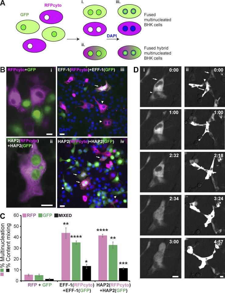
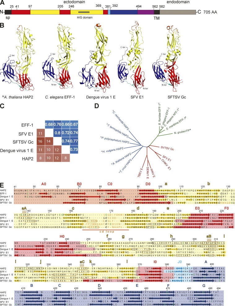
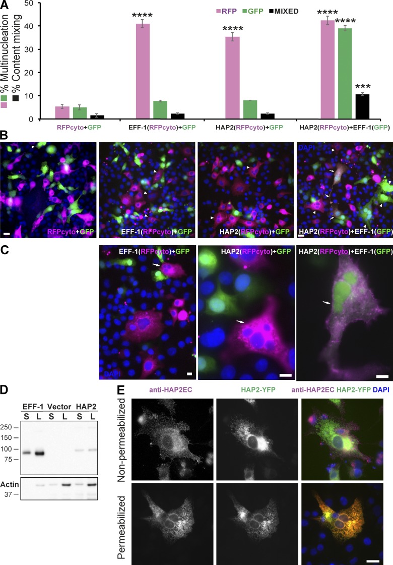
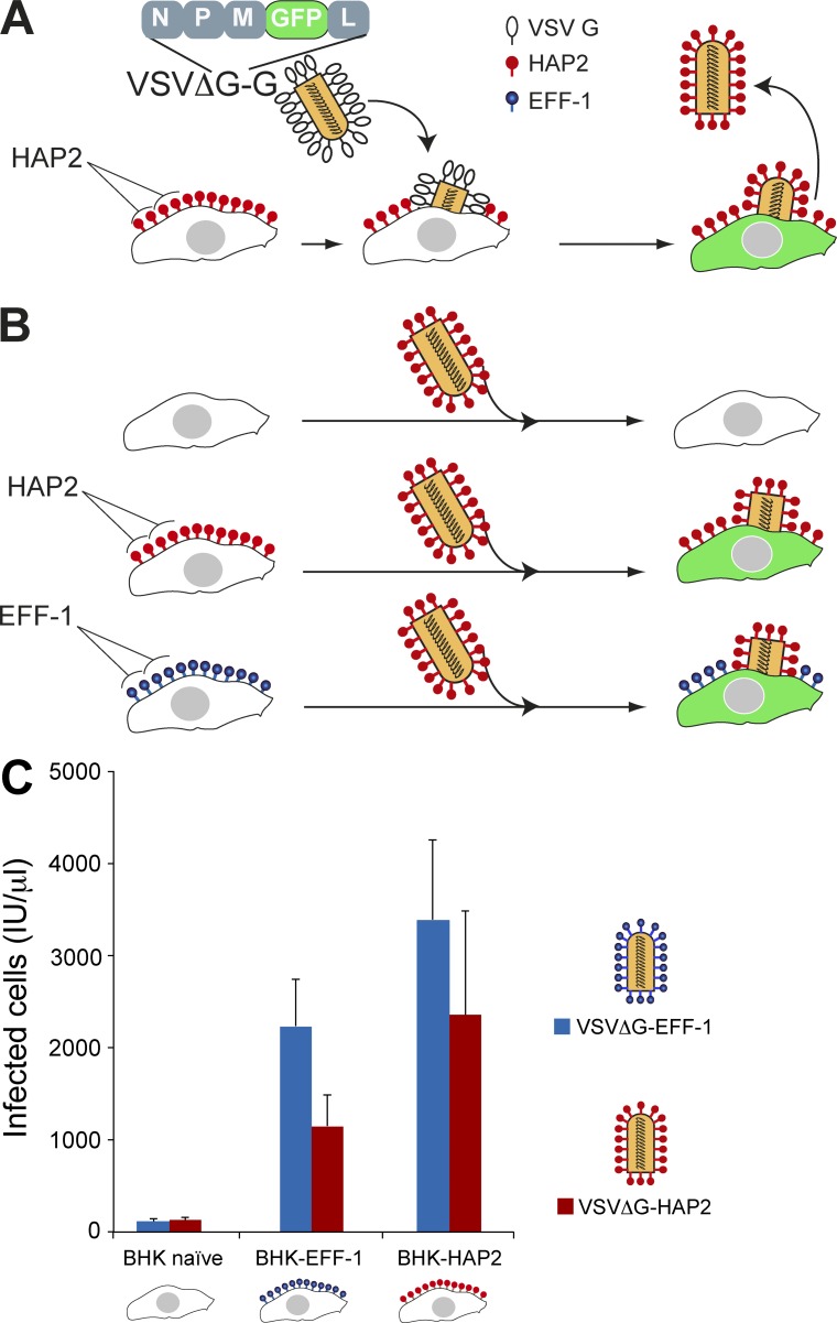
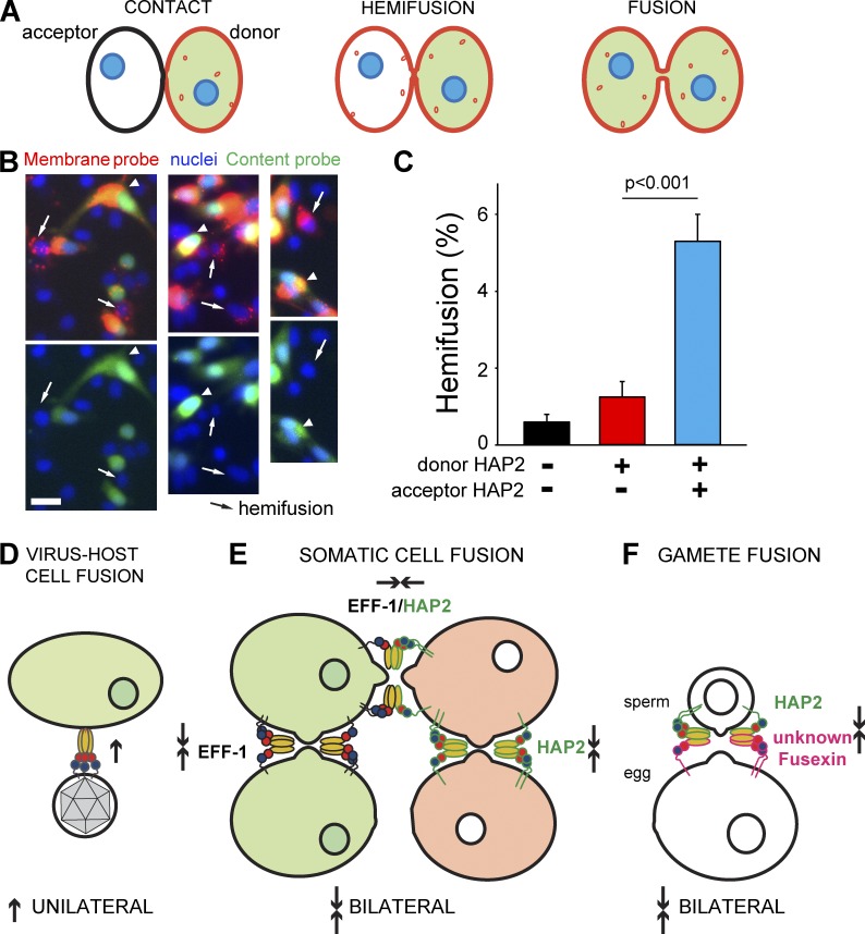
Cell-cell fusion is inherent to sexual reproduction. Loss of HAPLESS 2/GENERATIVE CELL SPECIFIC 1 (HAP2/GCS1) proteins results in gamete fusion failure in diverse organisms, but their exact role is unclear. In this study, we show that Arabidopsis thaliana HAP2/GCS1 is sufficient to promote mammalian cell-cell fusion. Hemifusion and complete fusion depend on HAP2/GCS1 presence in both fusing cells. Furthermore, expression of HAP2 on the surface of pseudotyped vesicular stomatitis virus results in homotypic virus-cell fusion. We demonstrate that the Caenorhabditis elegans Epithelial Fusion Failure 1 (EFF-1) somatic cell fusogen can replace HAP2/GCS1 in one of the fusing membranes, indicating that HAP2/GCS1 and EFF-1 share a similar fusion mechanism. Structural modeling of the HAP2/GCS1 protein family predicts that they are homologous to EFF-1 and viral class II fusion proteins (e.g., Zika virus). We name this superfamily Fusexins: fusion proteins essential for sexual reproduction and exoplasmic merger of plasma membranes. We suggest a common origin and evolution of sexual reproduction, enveloped virus entry into cells, and somatic cell fusion.


Figures

Figure 1.
Figure 1.
A. thaliana HAP2 is sufficient to fuse mammalian BHK cells. (A) BHK cell–cell fusion assay: after discarding a possible failure in cell division (Table S1), cell–cell fusion is measured by the appearance of multinucleated cells labeled with either RFPcyto (magenta) or nuclear and cytoplasmic GFP (green; i). (ii) Fusion is also indicated by the appearance of multinucleated cells containing nuclear GFP and fluorescence from both RFPcyto and GFP in the cytoplasm. (iii) Nuclei are labeled with DAPI (blue) after fixation and permeabilization of the cells. (B, i) RFPcyto + GFP: negative control shows mononucleated cells expressing RFPcyto (magenta) or nuclear and cytoplasmic GFP (green). (ii) HAP2(RFPcyto) + HAP2(GFP): BHK cells were transfected with AtHAP2 and GFP (green) or RFPcyto (magenta); merged image of hybrid cell that contains mixed cytoplasm and three nuclei. (iii) EFF-1(RFPcyto) + EFF-1(GFP): hybrid binucleate cell emerged after EFF-1 expression and mixing of magenta and green cells (arrow). EFF-1(RFPcyto) binucleate cells (arrowheads). (iv) HAP2(RFPcyto) + HAP2(GFP): heterokaryons (hybrids) express magenta cytoplasm and green nuclei and cytoplasm (arrows). Multinucleate green cells (arrowheads). Bars: (B, i and ii) 10 µm; (B, iii and iv) 20 µm. (C) Quantification of multinucleation and content-mixing experiments. Magenta and green bars represent the fraction of multinucleated cells (two nuclei or higher) out of all the cells in contact (magenta or green, respectively). Black bars represent the RFPcyto and GFP content-mixing index. The fusion and mixing indexes are presented as means ± SEM of three independent experiments. Total number of nuclei counted in multinucleated cells and in cells in contact n ≥ 1,000 for each experimental condition. Used unpaired t test comparing each color (RFPcyto, GFP, or mixed) for EFF-1 and HAP2 to the negative control (RFPcyto+GFP). *, P < 0.01; **, P < 0.005; ***, P < 0.001; ****, P < 0.0005. (D) Still images from time-lapse experiments reveal merging of two mononucleated (i) and three cells (ii) expressing RFPcyto and HAP2 (arrows and arrowheads, respectively). Time indicated in hours:minutes (see Videos 1 and 2 for panels i and ii in D, respectively). Note that the top nucleus (arrow in D, i) disappears because of defocus at 2:34. Two nuclei are out of focus at 4:57 (D, ii, bottom; see Fig. S1 A). Bars, 20 µm.
Figure 2.
Figure 2.
Model of the 3D structure of A. thaliana HAP2 ectodomain indicates it is a class II fusogen. (A) Diagram of A. thaliana HAP2 protein colored by domains according to the ectodomain modeled structure: signal peptide (sp) and transmembrane domain (TM), black; domain I, red; domain II, yellow; domain III, blue; domain I–III linker, cyan; stem, magenta; and intracellular domain, gray. (B) Cartoon of AtHAP2 ectodomain modeled structure (residues 41–494), alongside with C. elegans EFF-1 (PDB 4OJC), dengue virus E glycoprotein (PDB 4GSX), Semliki Forest virus E1 glycoprotein (SFV E1; PDB 1RER), and severe fever with thrombocytopenia syndrome virus glycoprotein Gc (SFTSV Gc; PDB 5G47) class II fusion proteins. Structures are colored according to domains as in A and E; cd loops are shown in orange (facing up). (C) Structural similarity scores between experimental and modeled fusogens computed after flexible structural alignment. Blue matrix: TM-scores (values >0.5 are indicative of significant structural similarity); orange matrix: z-scores (values >2 are considered as indicative of significant structural similarity). (D) Unrooted tree inferred using a distance matrix. Superscript gray M indicates modeled structure; colors are HAP2, blue; EFF-1/AFF-1/FF, green; class II viral fusogens, red (Fig. S3 A). (E) Structure-based alignment of AtHAP2 ectodomain with fusion proteins shown in B. Background colors indicate the domains organization (as in A and B); arrows in color and rounded rectangles denote β sheets and α helices, respectively. The black box in AtHAP2 sequence marks the HAP2/GCS1 domain; the cd loops are marked in orange. Cysteines involved in conserved disulfide bonds stabilizing the cd loop are denoted in bold italics.
Figure 3.
Figure 3.
HAP2 is a bilateral fusogen. (A) Quantification of content mixing experiments showing that HAP2 and EFF-1 are required in both interacting cells to form hybrids. Control RFPcyto + GFP is the same as in Fig. 1 B. The fusion and mixing indexes are presented as means ± SEM of three independent experiments. Total number of nuclei counted in multinucleated cells and in cells in contact, n ≥ 1,000 for each experimental condition. ***, P < 0.001; ****, P < 0.0005. P > 0.05 is not significant. (B) Representative fields used to determine percentages of multinucleation and content mixing. RFPcyto + GFP: mixed control cells; nuclear staining DAPI (blue). Dividing green cell with two nuclei is marked by arrowhead. EFF-1(RFPcyto) + GFP: BHK–EFF-1 (magenta) do not mix with GFP-expressing cells, revealing that EFF-1–mediated fusion is bilateral (homotypic). Multinucleate cells expressing RFPcyto (arrowheads). HAP2(RFPcyto) + GFP: BHK-HAP2 multinucleation (arrowheads) and failure to mix with GFP-expressing cells revealing HAP2-mediated fusion is bilateral. HAP2(RFPcyto) + EFF-1(GFP): hybrids (arrows) between BHK–EFF-1 (green) and BHK-HAP2 (magenta) reveal heterotypic merger of cells. Multinucleated green or red cells (arrowheads). Some hybrids are mononucleate probably because of cell division or nuclear fusion after merger. Bars, 20 µm. (C) Examples of multinucleate cells containing two to four nuclei are marked with arrows. The cells are able to divide after fusion; therefore, the number of nuclei per cells is usually smaller than six. EFF-1(RFPcyto) + GFP and HAP2(RFPcyto) + GFP images show no mixing indicating that HAP2-mediated fusion is bilateral (homotypic) in BHK cells. Bars, 10 µm. (D) Immunoblot of vector (negative control) and EFF-1 and HAP2 proteins carrying a V5 epitope fused to the cytoplasmic tail. Surface biotinylation of BHK cells expressing the different proteins. S indicates surface expression after affinity purification using neutravidin agarose beads; L is lysate. The amount of sample of HAP2 “S” is 600 times higher than EFF-1. The amount of lysate for HAP2 “L” is 12 times higher than for EFF-1. (E) Immunofluorescence of HAP2-YFP shows surface expression in cells revealed by anti-HAP2Extracellular (HAP2EC) polyclonal antibody. Without permeabilization, 20% of the cells expressing HAP2-YFP showed punctate expression on the surface; merged image (top, magenta). Untransfected BHK cells showed no immunoreactivity. After permeabilization with detergent, there is colocalization between immunostaining with anti-HAP2EC and the YFP signal revealing the specificity of the antibody that recognizes HAP2 in the reticular ER and perinuclear localization (merged image; bottom, yellow). Immunofluorescence in permeabilized BHK-HAP2-V5 cells using anti-V5 antibody shows similar localization (Fig. S1, B and C). Bar, 20 µm.
Figure 4.
Figure 4.
HAP2 can fuse the VSVΔG pseudovirus to target cells. (A) Cartoon illustrates the generation of VSVΔG-HAP2 pseudoviruses. Transfected BHK cells express HAP2 protein on the surface and were infected with VSVΔG-G. The viral genome encodes GFP, replacing VSVG. Infection results in viral-induced expression of GFP by target cells (green cytoplasm). VSVΔG-HAP2 pseudoviruses were collected from the supernatant. (B) The activity of VSVΔG-HAP2 was tested on BHK (untransfected, naive), BHK-HAP2, and BHK-EFF-1 cells. Figure adapted with permission from Avinoam et al. (2011). (C) Titers of VSVΔG pseudoviruses. The type of protein on the viral membrane (EFF-1 or HAP2) and on the BHK target cells (naive, EFF-1, or HAP2) is indicated. Titers in infectious units (IU) represent the number of cells expressing GFP per microliter 24 h after virus inoculation. Data are mean ± SEM (n = 3 experiments). We found no significant difference between infection with VSVΔG-HAP2 and VSVΔG–EFF-1 of the different BHK target cells (two-tailed paired t test).
Figure 5.
Figure 5.
Hemifusion in HAP2-mediated fusion is bilateral, and Fusexins use divergent mechanisms. (A) Cartoon illustrates the hemifusion assay in which one cell type, labeled with two fluorescent probes, acts as the “donor” cell and the unlabeled cell acts as “acceptor.” Because of internalization of fluorescent lipid from the plasma membrane, by the time we score fusion, this probe mostly labels intracellular membranes. (B) Fluorescence microscopy images of AtHAP2-transfected cells labeled with both cell tracker (green, content probe) and Vybrant DiI (red, membrane probe) coplated with unlabeled AtHAP2-transfected cells. (top) Green cell tracker, DiI (red), and Hoechst 33342 (blue). (bottom) Green cell tracker and Hoechst 33342 (blue). Hemifusion event is detected as an appearance of a cell (marked by arrows) that acquired only membrane probe apparently from an adjacent double-labeled cell (arrowheads). Bar, 20 µm. (C) Hemifusion extents quantified as the ratio between numbers of cells labeled only with membrane probe and numbers of cells labeled with both membrane and content probes. The results are means ± SEM (n = 3). (D) Viral class II trimeric fusion proteins (viral Fusexins) have a unilateral fusion mechanism, and the Fusexin is present only in the virus’ envelope or in one cell during cell–cell fusion. (E) Somatic cellular Fusexins (e.g., EFF-1 and AFF-1; on green cells) use a bilateral homotypic mechanism, and the model proposes Fusexin has to be in both cells, forming trans-trimeric complexes (Podbilewicz et al., 2006; Pérez-Vargas et al., 2014). Our results suggest a mechanistic model in which sperm AtHAP2 fuses animal cells using a homotypic design (bilateral; pink cells). HAP2 and EFF-1 can fuse cells in trans using a heterotypic mechanism. (F) We hypothesize that the egg of A. thaliana (the central cell too) expresses an unidentified Fusexin that interacts with sperm HAP2.

Comment on

Similar articles

Cited by

References

    1. Aguilar P.S., Baylies M.K., Fleissner A., Helming L., Inoue N., Podbilewicz B., Wang H., and Wong M.. 2013. Genetic basis of cell-cell fusion mechanisms. Trends Genet. 29:427–437. 10.1016/j.tig.2013.01.011 - DOI - PMC - PubMed
    1. Avinoam O., Fridman K., Valansi C., Abutbul I., Zeev-Ben-Mordehai T., Maurer U.E., Sapir A., Danino D., Grünewald K., White J.M., and Podbilewicz B.. 2011. Conserved eukaryotic fusogens can fuse viral envelopes to cells. Science. 332:589–592. 10.1126/science.1202333 - DOI - PMC - PubMed
    1. Baker N.A., Sept D., Joseph S., Holst M.J., and McCammon J.A.. 2001. Electrostatics of nanosystems: Application to microtubules and the ribosome. Proc. Natl. Acad. Sci. USA. 98:10037–10041. 10.1073/pnas.181342398 - DOI - PMC - PubMed
    1. Bianchi E., Doe B., Goulding D., and Wright G.J.. 2014. Juno is the egg Izumo receptor and is essential for mammalian fertilization. Nature. 508:483–487. 10.1038/nature13203 - DOI - PMC - PubMed
    1. Chernomordik L.V., and Kozlov M.M.. 2005. Membrane hemifusion: crossing a chasm in two leaps. Cell. 123:375–382. 10.1016/j.cell.2005.10.015 - DOI - PubMed

Publication types

MeSH terms