. 2017 Feb 14;114(7):1702-1707.
doi: 10.1073/pnas.1618606114. Epub 2017 Jan 30.

DSCAM promotes axon fasciculation and growth in the developing optic pathway

Affiliations

DSCAM promotes axon fasciculation and growth in the developing optic pathway

Freyja M Bruce et al. Proc Natl Acad Sci U S A. .

Abstract

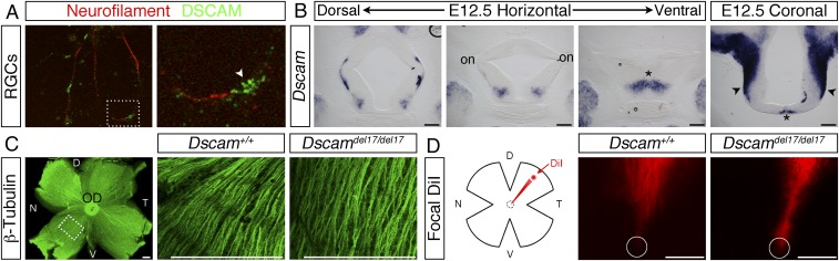
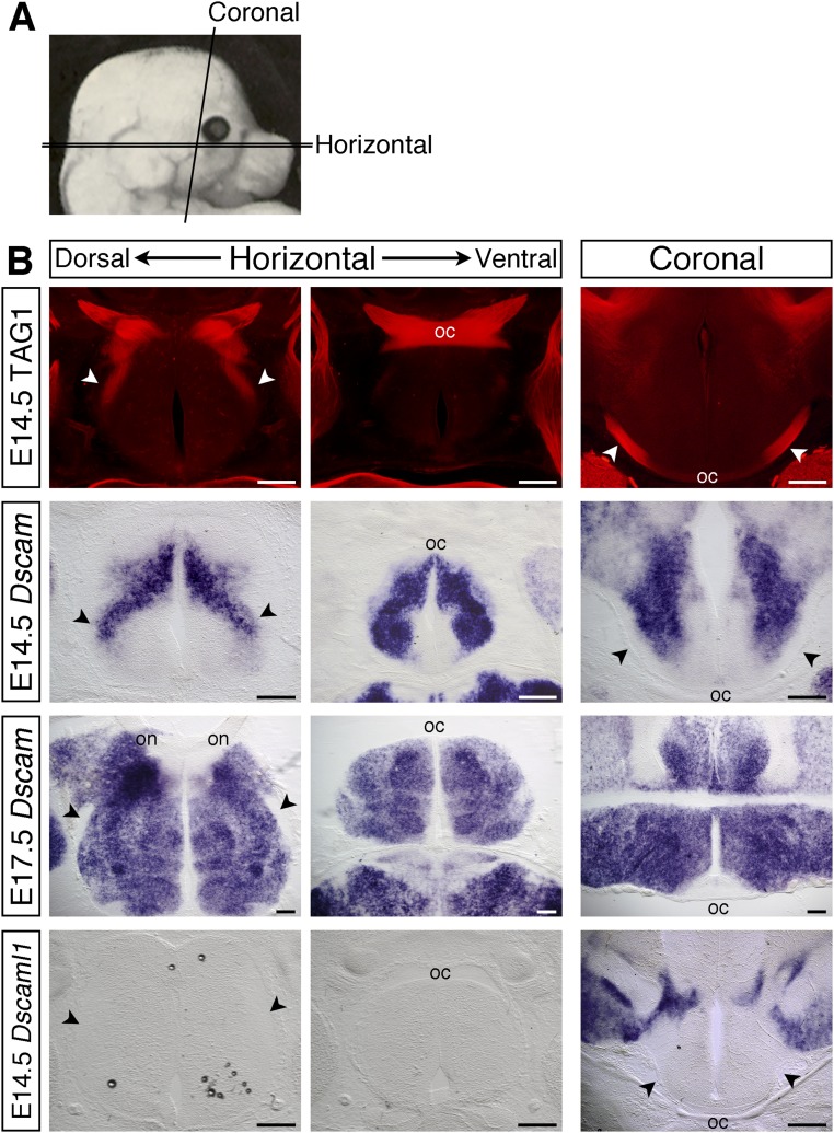
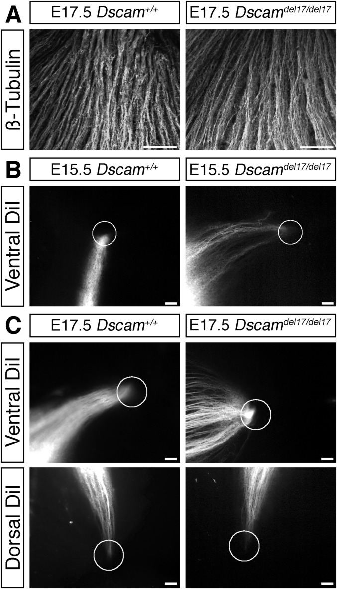
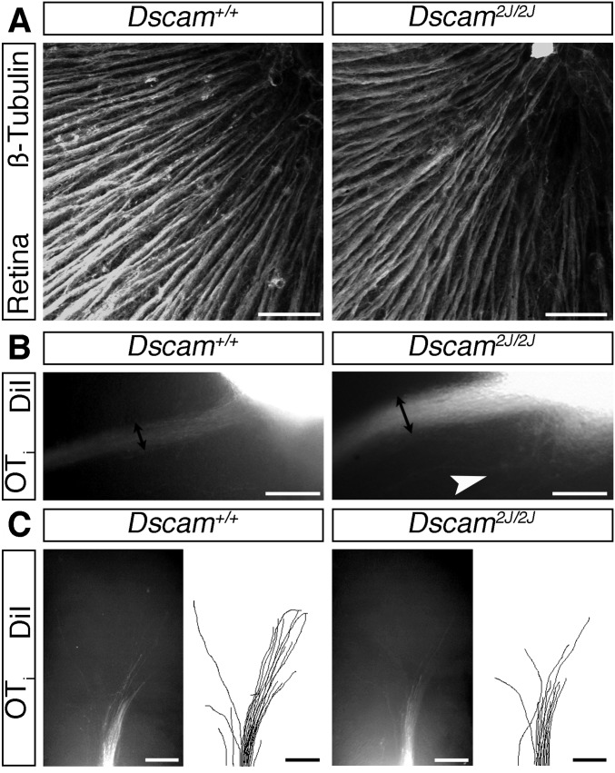
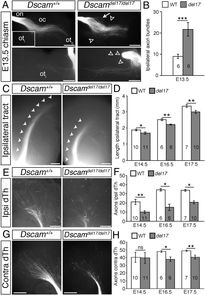
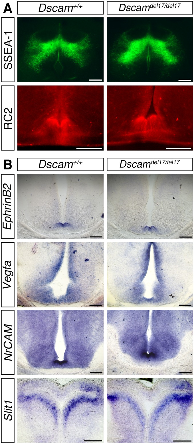
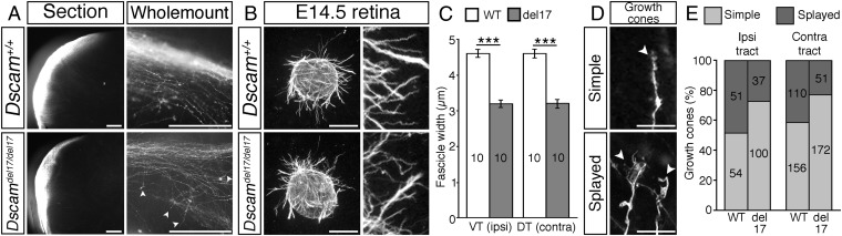
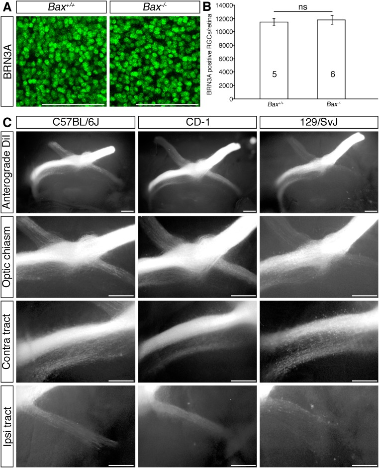
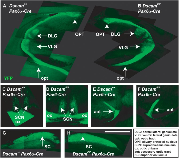
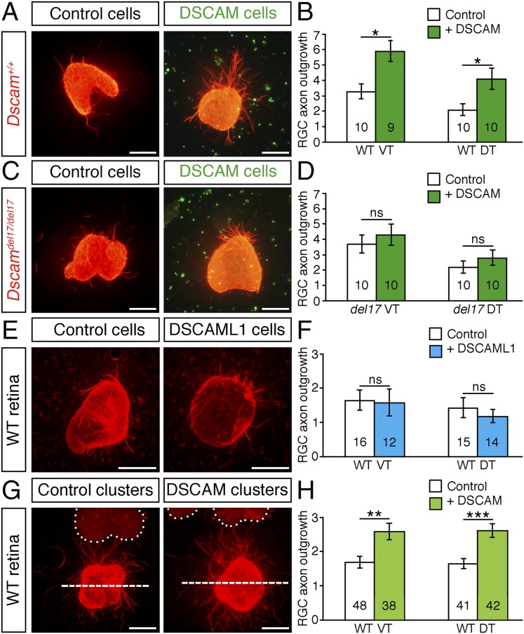
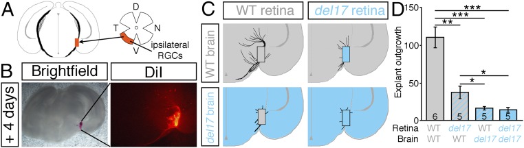
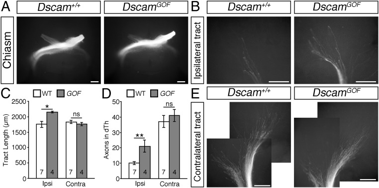
Although many aspects of optic pathway development are beginning to be understood, the mechanisms promoting the growth of retinal ganglion cell (RGC) axons toward visual targets remain largely unknown. Down syndrome cell adhesion molecule (Dscam) is expressed by mouse RGCs shortly after they differentiate at embryonic day 12 and is essential for multiple aspects of postnatal visual system development. Here we show that Dscam is also required during embryonic development for the fasciculation and growth of RGC axons. Dscam is expressed along the developing optic pathway in a pattern consistent with a role in regulating RGC axon outgrowth. In mice carrying spontaneous mutations in Dscam (Dscamdel17 ; Dscam2J), RGC axons pathfind normally, but growth from the chiasm toward their targets is impaired, resulting in a delay in RGC axons reaching the dorsal thalamus compared with that seen in wild-type littermates. Conversely, Dscam gain of function results in exuberant growth into the dorsal thalamus. The growth of ipsilaterally projecting axons is particularly affected. Axon organization in the optic chiasm and tract and RGC growth cone morphologies are also altered in Dscam mutants. In vitro DSCAM promotes RGC axon growth and fasciculation, and can act independently of cell contact. In vitro and in situ DSCAM is required both in the RGC axons and in their environment for the promotion of axon outgrowth, consistent with a homotypic mode of action. These findings identify DSCAM as a permissive signal that promotes the growth and fasciculation of RGC axons, controlling the timing of when RGC axons reach their targets.

Keywords: axon guidance; development; growth cone; optic chiasm; visual system.


Conflict of interest statement

The authors declare no conflict of interest.

Figures

Fig. 1.
Fig. 1.
DSCAM is expressed by retinal axons and along the developing optic pathway, but is not essential for intraretinal axon pathfinding. (A) Double immunofluorescent staining of cultured P0 retinal neurons with antibodies against DSCAM (green) or neurofilaments (red). (Right) Boxed region shown at higher power. The white arrowhead indicates the growth cone. (B) In situ hybridization for Dscam on horizontal and coronal sections through the ventral diencephalon of E12.5 WT embryos. on, optic nerve; asterisk, presumptive chiasm; black arrowheads, presumptive optic tracts. (C) Flat-mounted E15.5 WT retina stained with antibodies against neuron-specific β-tubulin to label all retinal axons. The box indicates the region in which confocal images through the optic fiber layer were captured in E15.5 Dscam+/+ and Dscamdel17/del17 retinas (Right). (D) Schematic illustration of the method used to label small groups of RGC axons and images of labeled RGC axons in dorsal retina of E15.5 Dscamdel17/del17 WT and mutants. Circles indicate the position of the optic disk. D, dorsal; N, nasal; OD, optic disk; T, temporal; V, ventral. (Scale bars: 200 µm.)
Fig. S1.
Fig. S1.
Expression of Dscam along the developing optic pathway. (A) Schematic illustration of the plains of sections used. (B) Serial horizontal sections through the ventral diencephalon and coronal sections at the level of the optic tracts of E14.5 and E17.5 C57BL/6J embryos stained with antibodies against TAG-1 to label the RGC axons (Top), or by in situ hybridization with probes specific for Dscam or Dscaml1. Horizontal sections, anterior up; coronal sections, dorsal up. oc, optic chiasm; on, optic nerve; arrowheads, optic tracts. (Scale bars: 200 µm.)
Fig. S2.
Fig. S2.
DSCAM is not required for intraretinal RGC axon guidance. (A) Confocal images of RGC axons labeled with antibodies against β-tubulin in the optic fiber layer of E17.5 Dscamdel17 WT and mutant retinas. The direction of the optic disk is up. (B and C) Small groups of DiI-labeled RGC axons originating in ventral or dorsal retina in E15.5 (B) and E175 (C) WT and Dscamdel17 mutant littermates. Circles indicate the positions of the optic disk. (Scale bars: 50 µm.)
Fig. S3.
Fig. S3.
Dscam2J mutants display similar RGC axon guidance phenotypes to Dscamdel17 mutants. (A) Confocal images of RGC axons in the optic fiber layer of E16.5 Dscam2J WT and mutant retinas stained with antibodies against β-tubulin. The direction of the optic disk is up. (B and C) Whole-mount views of anterograde DiI-labeled RGC axons in the ipsilateral optic tract adjacent to the chiasm (B) and dorsal thalamus (C) of E16.5 Dscam2J WT and mutant littermates. In C, the cortex has been removed, and the brains are viewed from the side. Double-headed black arrows indicate the width of the optic tract; white arrowhead, axons straying away from their normal pathway. (Scale bars: 50 µm in A; 250 µm in B and C.)
Fig. 2.
Fig. 2.
RGC axon growth through the optic tracts is impaired in Dscam mutants. (A, C, E, and G) Whole-mount views of anterograde DiI-labeled RGC axons in the optic nerve (on), optic chiasm (oc), and proximal contralateral (otc) and ipsilateral (oti) optic tracts of E13.5 Dscamdel17 WT and mutant littermates (A) and in the ipsilateral optic tracts (C) and ipsilateral and contralateral dorsal thalamus (E and G) of E16.5 Dscamdel17 WT and mutant littermates. The boxed region in A indicates the region of the ipsilateral optic tract shown at higher power in the lower panels. White arrows indicate defasciculated axons in the optic nerve; open arrowheads, straying axons. (A) Ventral views. (C, E, and G) Side views following removal of the cortex. (B, D, F, and H) Mean ± SEM number of axon bundles in the ipsilateral optic tract of E13.5 Dscamdel17 WT and mutant (del17) littermates (B), and ipsilateral optic tract length (D) and number of axon bundles in the ipsilateral (F) and contralateral (H) dorsal thalamus (dTh) of E14.5–E17.5 Dscamdel17 WT and mutant littermates. Analyses were performed blinded to genotype. Numbers on bars indicate the numbers analyzed. *P < 0.05; **P < 0.01; ***P < 0.001; ns, not significant. (Scale bars: 250 µm.)
Fig. S4.
Fig. S4.
RGC genesis is increased in Dscamdel17 mutants, but specification of ipsilaterally projecting RGCs occurs normally. (A) Schematic illustration of the method used to quantify RGC, mitotic, and apoptotic cell numbers. Retinas were stained by double immunofluorescence with antibodies against BRN3A to label RGCs, with phosphohistone H to label mitotic cells, or with cleaved caspase-3 to label apoptotic cells. Confocal images were captured through the RGC, and neuroblastic layers from four peripheral (p) and four central (c) regions in each retina of BRN3A- and phosphohistone H3-positive cells, and the number of labeled cells in each image was quantified and used to calculate the total number of labeled cells in each retina. Caspase-3–positive cells were quantified by counting all labeled cells. D, dorsal; N, nasal; NL, neuroblastic layer; RGCL, RGC layer; T, temporal; V, ventral. (B) Confocal images of BRN3A-positive RGCs and phosphohistone H3-positive mitotic cells in the RGC layer and neuroblastic layer respectively, and caspase-3–positive cells in E14.5 Dscamdel17 WT and mutant retinas. (C–E) Mean ± SEM number of BRN3A-positive RGCs (C), phosphohistone-H3–positive mitotic cells (D), and caspase-3–positive apoptotic cells (E) in retinas from E14.5 and E15.5 Dscamdel17 WT and mutant (del17) retinas. (F) Sections of E15.5 Dscamdel17 WT and mutant retinas stained by in situ hybridization with a probe specific for Zic2, a marker of ipsilaterally specified RGCs. (G) Peripheral ventrotemporal retina from E16.5 Dscamdel17 WT and mutant littermates stained with an antibody against ZIC-2. (H) Mean ± SEM number of ZIC2-positive cells in retinas from E16.5 Dscamdel17 mutant (del17) and WT littermates. *P < 0.05; ns, not significant. The numbers analyzed at each age and genotype are indicated on the bars. (Scale bars: 100 µm.)
Fig. S5.
Fig. S5.
RGC axon projections in Dscamdel17 mutant and WT littermates. (A) Whole-mount views of anterograde DiI-labeled RGC axons at the optic chiasm (Top) and proximal ipsilateral optic tract (Bottom) of E17.5 Dscamdel17 WT and mutant littermates. Double-headed black arrows indicate the width of the ipsilateral optic tract. (B) Mean ± SEM width of the ipsilateral optic tract in E15.5 and E17.5 Dscamdel17 WT and mutant (del17) littermates. (C) Schematic diagram illustrating the method used to quantify RGC axons projecting between the two eyes. RGC axons from one eye were labeled with DiI, and after allowing time for the dye to diffuse, the contralateral retina was removed and flat-mounted, and the number of labeled cells was quantified. (D) Labeled cells in the contralateral retina of E16.5 Dscamdel17 WT, heterozygous, and homozygous mutant embryos. (E) Mean ± SEM number of labeled cells in the contralateral retina of E16.5 Dscamdel17 WT, heterozygous, and homozygous mutant embryos. (F) Whole-mount views of anterograde DiI-labeled RGC axons in the contralateral optic tracts of E16.5 Dscamdel17 WT and mutant littermates. Here the cortex has been removed, and the diencephalon is viewed from the side in each image. (G) Mean ± SEM contralateral optic tract length in E14.5–E17.5 Dscamdel17 WT and mutant littermates. Analyses were performed blinded to genotype. Numbers on the bars indicate the numbers analyzed at each age and genotype. **P < 0.01; ***P < 0.001; ns, not significant. (Scale bars: 250 µm in A and F; 200 µm in D.)
Fig. S6.
Fig. S6.
The number of RGC axons reaching the dorsal thalamus is lower in Dscamdel17 mutant embryos compared with WT littermates. (A) Schematic illustration of the method used to retrogradely label RGCs from the dorsal thalamus (dTh). Crystals of DiI (asterisks) were placed in the dorsal thalamus on one side of the brain to label all RGC axons that had extended into that region. After allowing time for diffusion back to the eyes, the ipsilateral and contralateral retinas were flat-mounted, and the number of labeled cells was quantified. (B) Flat-mounted ipsilateral and contralateral retinas from E16.5 Dscamdel17 WT and mutant littermates after retrograde labeling from the dorsal thalamus. (C and D) Mean ± SEM numbers of retrogradely labeled RGCs in the ipsilateral (C) and contralateral (D) retinas of E15.5 and E16.5 Dscamdel17 WT and mutant (del17) littermates. D, dorsal; dTh, dorsal thalamus; N, nasal; oc, optic chiasm; OT, optic tract; T, temporal; V, ventral; ns, not significant. Numbers on the bars indicate the numbers analyzed at each age and genotype. *P < 0.05. (Scale bars: 250 µm.)
Fig. S7.
Fig. S7.
Cellular organization and guidance cue expression are similar in the ventral diencephalon of Dscamdel17 WT and mutant littermates. (A) Immunostaining of the SSEA-1–positive neurons located posterior to the developing optic chiasm and RC2-positive radial glia in horizontal (SSEA-1) and coronal (RC2) sections through the optic chiasm of E14.5 Dscamdel17 WT and mutant embryos. (B) In situ hybridization for ephrinB2, Vegfa, NrCAM, and Slit1 in coronal (ephrinB2, Vegfa, and NrCAM) and horizontal (Slit1) sections through the optic chiasm of E14.5 Dscamdel17 WT and mutant littermates. (Scale bars: 200 µm.)
Fig. 3.
Fig. 3.
DSCAM modulates RGC axon fasciculation and growth cone morphology. (A) Coronal sections at the level of the optic tracts and whole-mount views of the ipsilateral optic tract of anterograde DiI-labeled Dscamdel17 WT and mutant littermates. Sections, E17.5 embryos; whole mounts, E14.5 embryos. White arrowheads indicate straying axons. (B) E14.5 retinal explants from Dscamdel17 mutant and WT littermates cultured in collagen gels for 24 h and stained with antibodies against neuron-specific β-tubulin. (Right) Higher-magnification images of the axon bundles. (C) Mean ± SEM axon bundle width of Dscam+/+ and Dscamdel17/del17 retinal axons. Results are the mean from four independent experiments analyzed blinded to genotype. (D) DiI-labeled simple and splayed growth cones (white arrowheads) in the ipsilateral optic tract of E14.5 WT embryos. (E) Percentage of simple and splayed growth cones in the ipsilateral and contralateral optic tracts of E14.5 Dscamdel17 mutants and WT littermates. Analyses were performed blinded to genotype. Numbers on the bars indicate the numbers analyzed. ***P < 0.001. (Scale bars: 250 µm in A and B; 25 µm in D.)
Fig. S8.
Fig. S8.
Quantitation of RGCs in E17.5 Bax-deficient mice and RGC axon organization in mice on different genetic backgrounds. (A) Confocal images of BRN3A-positive RGCs in flat-mounted E17.5 WT and Bax−/− retinas. (B) Mean ± SEM number of BRN3A-positive RGCs in E17.5 WT and Bax−/− retinas. The numbers on the bars indicate the number of each genotype analyzed. ns, not significant. (C) Whole-mount views following anterograde DiI labeling of all axons from one eye of E15.5 C57BL/6J, CD-1, and 129/SvJ mice. Higher-magnification views of the optic chiasm, contralateral (contra), and ipsilateral (ipsi) optic tract are shown. (Scale bars: 100 µm in A; 250 µm in C.)
Fig. S9.
Fig. S9.
Dscam mutant RGCs target brain loci normally. A floxed allele of Dscam (DscamFF) was targeted with Pax6a-Cre, which induces recombination before RGC differentiation and activates a YFP reporter under control of the Thy1 promoter. No differences were observed when comparing targeting of WT and Dscam−/− RGC axons (n = 4 mice). Targeting of brain loci by WT (A, C, E, and G) and Dscam mutant (B, D, F, and H) RGCs. A, G, and H are composite pictures generated by overlying in Adobe Illustrator images captured at overlapping positions of the optic tract (A) or superior colliculus (G and H). Abbreviations in Inset: DLG, dorsal lateral geniculate; VLG, ventral lateral geniculate; opt, optic tract; OPT, olivary prectectal nucleus; SCN, suprachiasmatic nucleus; ox, optic chiasm; aot, accessory optic tract; SC, superior colliculus. (The scale bar in G is equivalent to 1.92 mm in A and B, 514 mm in C and D, 1.41 mm in E and F, and 880 mm in G and H.)
Fig. 4.
Fig. 4.
DSCAM promotes RGC axon outgrowth in vitro. (A, C, E, and G) Retinal explants from E14.5 WT (A, E, and G) and Dscamdel17 mutants (C) cultured for 24 h in collagen gels seeded with control, Dscam-expressing cells (A and C), Dscaml1-expressing cells (E), or 100–300 µm from clusters of control or Dscam-expressing cells (G). Explants were fixed and stained with antibodies against β-tubulin (red) or DSCAM (green). Dotted lines in G indicate the outlines of the cell clusters. Outgrowth was quantified in the area above the dashed line. (B, D, F, and H) Mean ± SEM axon outgrowth of presumptive ipsilateral (VT) and contralateral (DT) RGCs from WT (B, F, and H) and Dscamdel17/del17 (D) retinal explants in the presence of control, DSCAM-producing, or DSCAML1-producing cells. The number of explants analyzed is indicated on the bars. Data are the mean of at least four independent experiments analyzed blinded to genotype and condition. ns, not significant; *P < 0.05; **P < 0.01; ***P < 0.001. (Scale bars: 250 µm.)
Fig. 5.
Fig. 5.
DSCAM promotes RGC axon outgrowth in situ. (A) Schematic diagram of the culture approach. Ventrotemporal retinal explants, containing predominately ipsilaterally projecting RGCs, and brain slices at the level of the optic tract were prepared from E16.5 Dscamdel17 mutant and WT littermates. Retinal explants were cultured on top of the brain slices in different genetic combinations. (B) Bright-field and fluorescent (DiI) images of an E16.5 WT retinal explant cultured on a WT brain slice. (C) Manual tracing of RGC axons from Dscamdel17 WT (gray) or mutant (blue) retinal explants cultured on WT (gray) or mutant (blue) brain slices. (D) Mean ± SEM RGC axon outgrowth from E16.5 Dscamdel17 WT and mutant retinal explants cultured on brain slices from WT and mutant embryos. Numbers on the bars indicate the numbers analyzed. Results are from five independent experiments analyzed blinded to genotype. *P < 0.05; **P < 0.01; ***P < 0.001.
Fig. 6.
Fig. 6.
DSCAM gain of function promotes RGC axon outgrowth in vivo. (A, B, and E) Whole-mount views of anterograde DiI-labeled RGC axons at the optic chiasm (A) and in the ipsilateral (B) and contralateral (E) optic tracts of E14.5 DscamGOF and WT littermates. Panels in E are composite pictures generated by overlying in Adobe Photoshop images captured at overlapping positions along the optic tract. (C and D) Mean ± SEM optic tract length (C) and number of axon bundles in the ipsilateral (ipsi) and contralateral (contra) dorsal thalamus (D) of E14.5 DscamGOF (GOF) and WT littermates. Analyses were performed blinded to genotype. Numbers on the bars indicate the numbers analyzed for each genotype. ns, not significant; *P < 0.05; **P < 0.01. (Scale bars: 250 µm.)

References

    1. Erskine L, Herrera E. Connecting the retina to the brain. ASN Neuro. 2014;6(6):1759091414562107. - PMC - PubMed
    1. Hattori D, Millard SS, Wojtowicz WM, Zipursky SL. Dscam-mediated cell recognition regulates neural circuit formation. Annu Rev Cell Dev Biol. 2008;24:597–620. - PMC - PubMed
    1. Petrovic M, Schmucker D. Axonal wiring in neural development: Target-independent mechanisms help to establish precision and complexity. BioEssays. 2015;37(9):996–1004. - PubMed
    1. Schmucker D, Chen B. Dscam and DSCAM: Complex genes in simple animals, complex animals yet simple genes. Genes Dev. 2009;23(2):147–156. - PubMed
    1. Fuerst PG, et al. DSCAM and DSCAML1 function in self-avoidance in multiple cell types in the developing mouse retina. Neuron. 2009;64(4):484–497. - PMC - PubMed

Publication types

MeSH terms

Substances

LinkOut - more resources