. 2017 Feb 14;114(7):E1062-E1071.
doi: 10.1073/pnas.1617309114. Epub 2017 Jan 30.

Binding, sliding, and function of cohesin during transcriptional activation

Affiliations

Binding, sliding, and function of cohesin during transcriptional activation

Melinda S Borrie et al. Proc Natl Acad Sci U S A. .

Abstract

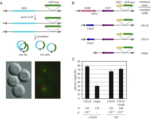
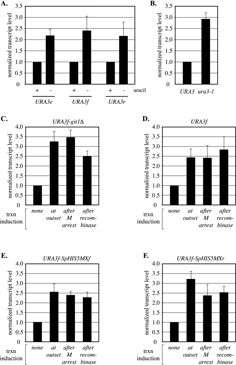
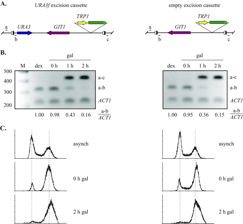
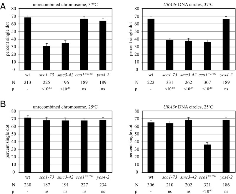
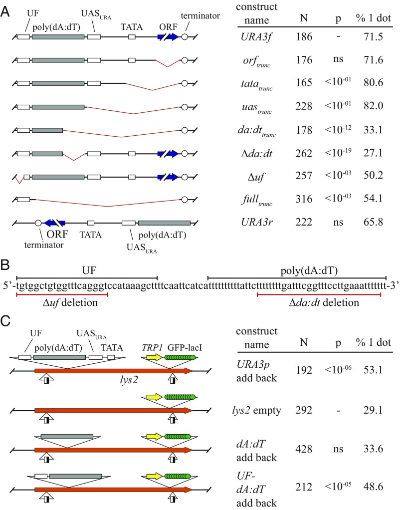
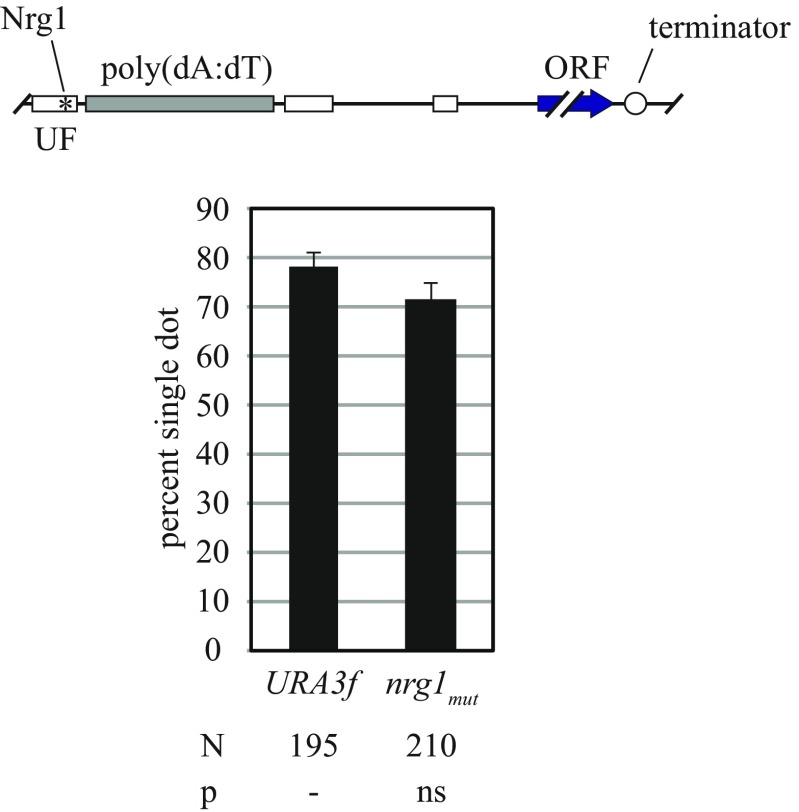
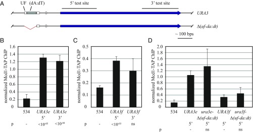
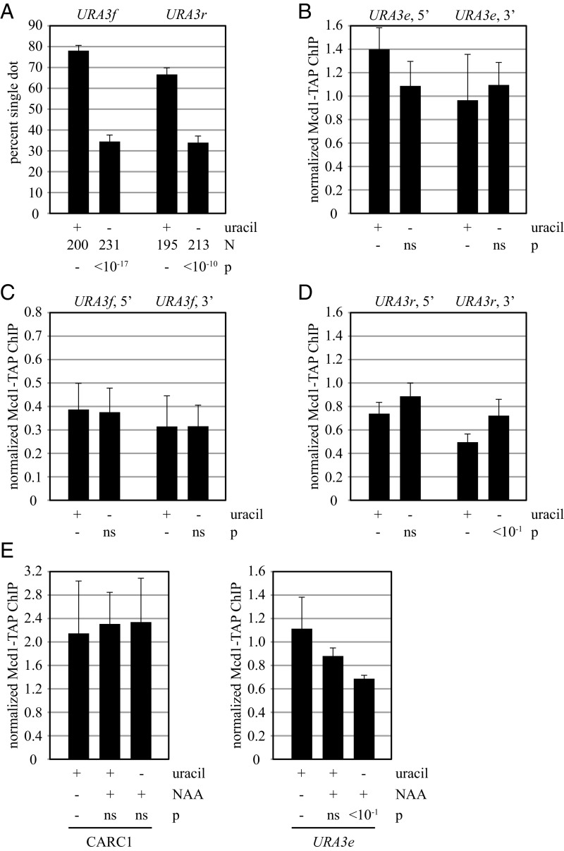
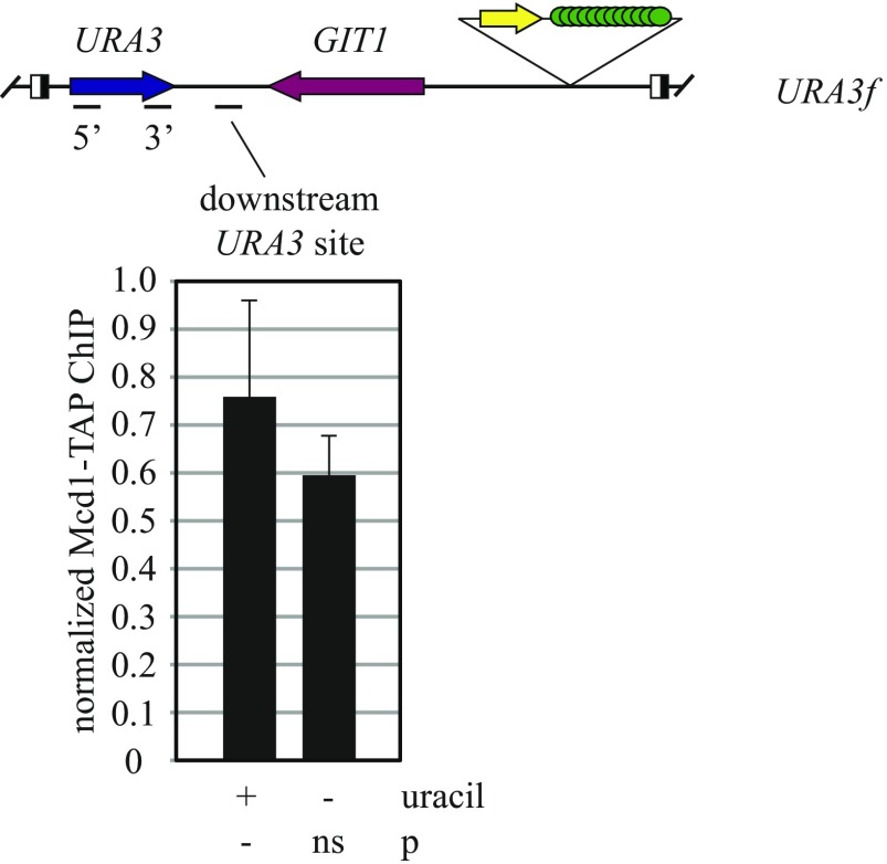
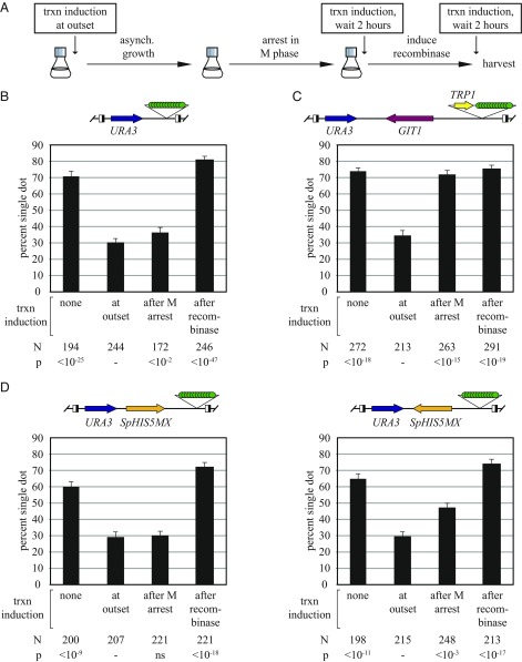
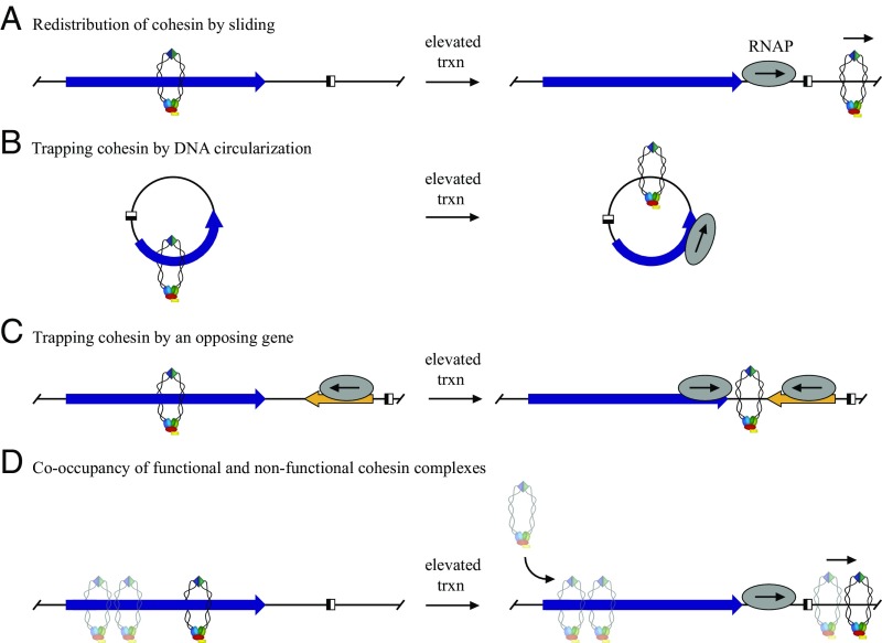
The ring-shaped cohesin complex orchestrates long-range DNA interactions to mediate sister chromatid cohesion and other aspects of chromosome structure and function. In the yeast Saccharomyces cerevisiae, the complex binds discrete sites along chromosomes, including positions within and around genes. Transcriptional activity redistributes the complex to the 3' ends of convergently oriented gene pairs. Despite the wealth of information about where cohesin binds, little is known about cohesion at individual chromosomal binding sites and how transcription affects cohesion when cohesin complexes redistribute. In this study, we generated extrachromosomal DNA circles to study cohesion in response to transcriptional induction of a model gene, URA3. Functional cohesin complexes loaded onto the locus via a poly(dA:dT) tract in the gene promoter and mediated cohesion before induction. Upon transcription, the fate of these complexes depended on whether the DNA was circular or not. When gene activation occurred before DNA circularization, cohesion was lost. When activation occurred after DNA circularization, cohesion persisted. The presence of a convergently oriented gene also prevented transcription-driven loss of functional cohesin complexes, at least in M phase-arrested cells. The results are consistent with cohesin binding chromatin in a topological embrace and with transcription mobilizing functional complexes by sliding them along DNA.

Keywords: URA3; cohesin; poly(dA:dT); sister chromatid cohesion; transcription.


Conflict of interest statement

The authors declare no conflict of interest.

Figures

Fig. 1.
Fig. 1.
URA3 supports cohesion. (A) Schematic of experimental protocol. ROI, region of interest. Half-filled boxes represent RS sites for the R site-specific recombinase. Fluorescence micrographs of strain JSC97 are provided. Two large-budded, M phase-arrested cells are shown, one with a single focus of GFP-lacI fluorescence and the other with two foci of GFP-lacI fluorescence (red arrows). (B) Schematic of the constructs used. (C) Cohesion measurements. Strains MSB7 (URA3f) and MSB13 (empty) were arrested in M phase by Cdc20 depletion, whereas JSC97 (URA3r) was arrested with nocodazole (+NZ). Nicotinamide (NAM; final concentration, 2 mM) was added to half of the JSC97 culture 2 h after DNA circularization, and cells were harvested 2 h later. N denotes the number of cells examined. P values for pairwise χ2 tests are presented relative to a benchmark strain designated with a dash.
Fig. S1.
Fig. S1.
Transcript analysis of URA3. (A) Relative induction levels of ring-borne URA3 genes and the endogenous locus. Control strain PJ1 with URA3 at its normal location (labeled URA3e) was grown in SC media either containing or lacking uracil. Strains MSB14 (URA3f) and MSB20 (URA3r) were grown similarly, but methionine was withheld until midlog phase, when methionine was added to induce M phase arrest by Cdc20 depletion. DNA circles were formed by the addition of galactose and RNA extracts were prepared two hours later. URA3 transcript levels were normalized first to an internal control (ACT1 mRNA) and then to the uninduced condition for each strain (+uracil). The data show that ring-borne URA3 genes were induced at endogenous levels. (B) Effect of Ura3 activity on transcription. The ura3-1 mutation of strain W303-1A (glycine to glutamate at amino acid 234) causes uracil auxotrophy by disrupting the Ura3 active site (51). Isogenic strains PJ1 (URA3) and W303-1A (ura3-1) were grown in YPDA media. After normalization to ACT1 mRNA, samples were normalized to strain PJ1. The data show that ura3 transcription elevated in the absence of Ura3 activity. (CF) Effect of induction timing on URA3 transcriptional output. Induction (uracil depletion) was initiated at each of the following stages: (i) at the outset of the experiment, (ii) following M phase arrest but before DNA circle formation, or (iii) following DNA circle formation (Fig. 6A). Strains MSB105 (URA3f-Δgit1), MSB14 (URA3f), MSB110 (URA3f-SpHISMXf), and MSB112 (URA3f-SpHISMXr) were used. The data show that transcriptional output of URA3 was not affected by the time of induction during the cohesion assay. Note that URA3r DNA circles did not induce fully after circularization and were therefore not used for this set of experiments.
Fig. S2.
Fig. S2.
Validation of DNA excision and cell cycle arrest. (A) Diagram of the binding sites for the primers used to monitor excision (Table S2). (B) Time course of excision. Strains MSB7 (URA3f) and MSB13 (empty) were first grown asynchronously in dextrose, subcultured in raffinose, and then arrested in M phase by Cdc20 depletion. At 0, 1, and 2 h after the addition of galactose, aliquots of cell culture were harvested. One part was used to make DNA extracts, and the other was fixed with ethanol and stained with SYTOX Green (Molecular Probes). qPCR of the extracts was used to determine the extent of excision (primers a and b) using ACT1 as an internal control. The values reported under each gel lane are the average of two independent trials. The data indicate that excision reached 85% completion in the 2-h time frame of a typical cohesion assay. Ethidium bromide-stained agarose gels show the progression of excision. Each lane contains three pooled PCR reactions (a–c, a–b, and ACT1) after 24 amplification cycles. Lane M, 100 bp ladder. (C) DNA histograms during the cell cycle arrest. Flow cytometry was performed at the Rutgers Environmental and Occupational Health Sciences Institute core facility. The results show that a significant M phase arrest was maintained over the 2-h time frame of a typical cohesion assay.
Fig. 2.
Fig. 2.
URA3 cohesion requires activated cohesin but not condensin. Strains JSC97 (wt), JSC96 (scc1-73), MSB147 (smc3-42), MSB157 (eco1W216G), and JSC164 (ycs4-2) bearing the URA3r excision cassette were grown at 25 °C and arrested in M phase with nocodazole. To examine the unrecombined chromosome, dextrose was added, and cells were fixed 2 h later. To examine DNA circles, galactose was added. After 2 h, the cultures were split, and half was shifted to 37 °C for 2 additional hours (A). The other half was left at 25 °C for 2 h (B). ns, nonsignificant.
Fig. S3.
Fig. S3.
Effect of the eco1W216G mutation on cohesion. (A) Analysis of a CEN/ARS minichromosome. A GFP-tagged, CEN/ARS minichromosome (pJSC2) was constructed in yeast by joining plasmids pRS416 and pSR10 via in vivo recombination, according to the method of ref. . pJSC2 is comparable in size to DNA circles bearing either HMR or URA3. Stains JSC209 (wt), JSC81 (eco1W216G), and JSC208 (smc3-42) containing pJSC2 were grown overnight in SC-trp plus dextrose at 25 °C. An equal volume of YPAD was added when the cultures reached midlog, and 20 min later, nocodazole was added. After 3 h, half of the culture was shifted to 37 °C. Cohesion was evaluated after 2 additional hours. The data show that cohesion of a circular, CEN/ARS minichromosome was not diminished by eco1W216G. (B) Analysis of HMR. DNA circles bearing HMR were examined in nocodazole-arrested strains RDY215 (wt) and JSC52 (eco1W216G) at 25 °C per the cohesion assay. To examine the unrecombined chromosome, cells were grown to midlog in dextrose at 25 °C and arrested with nocodazole. The data show that cohesion of HMR DNA circles was abolished in the eco1W216G mutant, whereas cohesion of the unrecombined chromosomal arm was not.
Fig. 3.
Fig. 3.
Sequence elements in the URA3 promoter are necessary and sufficient for cohesion. (A) The poly(dA:dT) tract is necessary for cohesion. Strains MSB7 (URA3f), HJ5 (orftrunc), HJ6 (tatatrunc), HJ7 (uastrunc), HJ19 (da:dttrunc), HJ4 (fulltrunc), and HJ24 (Δuf) were arrested in M phase by Cdc20 depletion. Strains HJ6388 (Δda:dt) and JSC97 (URA3r) were arrested with nocodazole. (B) DNA sequence surrounding the poly(dA:dT) element in the URA3 promoter. Underlines highlight the portions of the element that were deleted in the Δda:dt and Δuf constructs. (C) The poly(dA:dT) and UF elements generate cohesion in the lys2 context. Strains CSW91 (lys2 cassette alone), HJ22 (URA3 promoter add back), HJ23 (UF-dA:dT add back), and MSB155 (dA:dT alone add back) were arrested in M phase with nocodazole.
Fig. S4.
Fig. S4.
Effect of the Nrg1 site in the URA3 promoter on cohesion. DNA circles in strains MSB7 (URA3f) and HJ25 (nrg1mut) were compared in the cohesion assay after M phase arrest by Cdc20 depletion. In strain HJ25, the Nrg1 binding site of the URA3f excision cassette was mutated from 5′-AGGGTCC-3′ to 5′-AaaaTCC-3′. The data show that the Nrg1 site did not contribute to URA3 cohesion.
Fig. 4.
Fig. 4.
Binding of cohesin to URA3. (A) Maps of the constructs used with regions tested by ChIP highlighted. (B and C) ChIP of TAP-tagged Mcd1 cohesin subunit. Binding was measured in strains MSB37 (URA3e, where e denotes the endogenous locus) and MSB19 (URA3f). Primers used to evaluate the 5′ and 3′ test sites are listed in Table S2. P values were obtained by Student’s t tests and are presented relative to a negative control site designated with a dash. ns, not significant. (D) Effect of the Δ(uf-da:dt) deletion on cohesin binding. Strains MSB37, MSB19, MSB153 [URA3e Δ(uf-da:dt)], and MSB156 [URA3f Δ(uf-da:dt)] were used as in B and C.
Fig. 5.
Fig. 5.
Transcriptional induction of URA3 abolishes cohesion but not cohesin binding. (A) Cohesion of DNA circles bearing the URA3 gene. Strains MSB7 (URA3f) and MSB20 (URA3r) were grown in media containing or lacking uracil from the outset of the experiment. (B) ChIP of Mcd1-TAP at chromosomal URA3 genes. Strain MSB37 (URA3e) was grown in media containing or lacking uracil from the outset and cross-linked with formaldehyde following M phase arrest by Cdc20 depletion. (C and D) Strains MSB19 (URA3f) and JSC230 (URA3r) were evaluated as in B. (E) Transcription-dependent loss of cohesin from URA3 in the absence of Scc2. Strain MSB128 (SCC2-AID-9myc ADH1p-OsTIR1) was arrested in M phase by Cdc20 depletion. Three hours later the culture was split into thirds. DMSO was added to one third, and 500 μM NAA was added to another. Uracil was removed from the final third when 500 μM NAA was added. Cohesin levels were assessed 2 h later at the 5′ end of the endogenous URA3 gene, as well as at a site on chromosome III (CARC1) that binds cohesin stably (42).
Fig. S5.
Fig. S5.
Cohesin binding between URA3 and the convergent GIT1 gene. ChIP was performed with strain MSB19 (URA3f) as in Fig. 5C. Primers used are listed in Table S2. The data show that transcriptional induction (uracil depletion) does not affect cohesin binding levels downstream of URA3.
Fig. S6.
Fig. S6.
Conditional depletion of Scc2-AID. (A) Effect of Scc2 depletion on viability. Fivefold serial dilutions of strains MSB37 (SCC2), MSB123 (SCC2-AID-9xmyc), and MSB128 (SCC2-AID-9xmyc OsTIR1) were plated on SC-met media containing or lacking 500 μM NAA. The data show that cell growth ceased when Scc2 was depleted. (B) Time course of Scc2-AID-9xmyc depletion. Strain MSB128 was arrested in M phase by Cdc20 depletion for 3 h. Thereafter, Scc2-AID-9xmyc levels were measured at intervals following the addition of 500 μM NAA. Immunoblotting was performed as described in ref. using anti-myc (9E10; Roche) and anti-Pgk1 (ab-113687; Abcam) antibodies. The data show that Scc2 levels were greatly reduced by 60 min.
Fig. 6.
Fig. 6.
DNA circularization or an opposing transcribed gene prevents transcription-dependent loss of URA3 cohesion. (A) Experimental flowchart. Media containing uracil was replaced with media lacking uracil to induce URA3 transcription at the various times indicated during cell growth for the cohesion assay. (B) DNA circularization before URA3 induction prevents cohesion loss. Strain MSB35 (URA3f-git1Δ) was used. (C) A convergently oriented GIT1 gene prevents cohesion loss if URA3 transcription is induced after M phase. Strain MSB7 (URA3f) was used. (D) Orientation dependence of the SpHIS5MX gene in preventing transcription-dependent loss of URA3 cohesion. Strains MSB30 (URA3f-SpHIS5MXf) and MSB31 (URA3f-SpHIS5MXr) were used.
Fig. 7.
Fig. 7.
Model for binding and sliding of cohesin at URA3. Only one sister chromatid is shown for clarity. (A) Before transcriptional activation, functional cohesin binds the gene, but the complex is displaced by sliding upon collision with RNA polymerase. (B) Circularization of DNA prevents escape of functional cohesin complexes that are mobilized by transcription. (C) An opposing gene prevents escape of functional cohesin complexes. (D) Cooccupancy of functional and nonfunctional cohesin. Nonfunctional complexes are depicted as semitranslucent because their true nature of binding is not known. Upon URA3 induction, cohesin slides downstream, but additional cohesin is loaded onto the gene without acquiring cohesive function in M phase-arrested cells.

References

    1. Michaelis C, Ciosk R, Nasmyth K. Cohesins: Chromosomal proteins that prevent premature separation of sister chromatids. Cell. 1997;91(1):35–45. - PubMed
    1. Guacci V, Koshland D, Strunnikov A. A direct link between sister chromatid cohesion and chromosome condensation revealed through the analysis of MCD1 in S. cerevisiae. Cell. 1997;91(1):47–57. - PMC - PubMed
    1. Bose T, Gerton JL. Cohesinopathies, gene expression, and chromatin organization. J Cell Biol. 2010;189(2):201–210. - PMC - PubMed
    1. Cucco F, Musio A. Genome stability: What we have learned from cohesinopathies. Am J Med Genet C Semin Med Genet. 2016;172(2):171–178. - PubMed
    1. Onn I, Heidinger-Pauli JM, Guacci V, Ünal E, Koshland DE. Sister chromatid cohesion: A simple concept with a complex reality. Annu Rev Cell Dev Biol. 2008;24:105–129. - PubMed

Publication types

MeSH terms

LinkOut - more resources