. 2017 Feb 28;114(9):E1688-E1697.
doi: 10.1073/pnas.1620729114. Epub 2017 Jan 30.

AMH/MIS as a contraceptive that protects the ovarian reserve during chemotherapy

Affiliations

AMH/MIS as a contraceptive that protects the ovarian reserve during chemotherapy

Motohiro Kano et al. Proc Natl Acad Sci U S A. .

Abstract

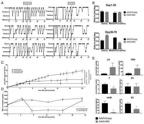
The ovarian reserve represents the stock of quiescent primordial follicles in the ovary which is gradually depleted during a woman's reproductive lifespan, resulting in menopause. Müllerian inhibiting substance (MIS) (or anti-Müllerian hormone/AMH), which is produced by granulosa cells of growing follicles, has been proposed as a negative regulator of primordial follicle activation. Here we show that long-term parenteral administration of superphysiological doses of MIS, using either an adeno-associated virus serotype 9 (AAV9) gene therapy vector or recombinant protein, resulted in a complete arrest of folliculogenesis in mice. The ovaries of MIS-treated mice were smaller than those in controls and did not contain growing follicles but retained a normal ovarian reserve. When mice treated with AAV9/MIS were paired with male breeders, they exhibited complete and permanent contraception for their entire reproductive lifespan, disrupted vaginal cycling, and hypergonadotropic hypogonadism. However, when ovaries from AAV9-MIS-treated mice were transplanted orthotopically into normal recipient mice, or when treatment with the protein was discontinued, folliculogenesis resumed, suggesting reversibility. One of the important causes of primary ovarian insufficiency is chemotherapy-induced primordial follicle depletion, which has been proposed to be mediated in part by increased activation. To test the hypothesis that MIS could prevent chemotherapy-induced overactivation, mice were given carboplatin, doxorubicin, or cyclophosphamide and were cotreated with AAV9-MIS, recombinant MIS protein, or vehicle controls. We found significantly more primordial follicles in MIS-treated animals than in controls. Thus treatment with MIS may provide a method of contraception with the unique characteristic of blocking primordial follicle activation that could be exploited to prevent the primary ovarian insufficiency often associated with chemotherapy.

Keywords: AAV9; AMH; MIS; contraceptive; oncofertility.


Conflict of interest statement

The authors declare no conflict of interest.

Figures

Fig. 1.
Fig. 1.
Comparative analysis of multiple modes of MIS administration. (A) Rat fetal (E14.5) urogenital ridges were incubated ex vivo with recombinant protein (Right), and the contralateral ridge was mock-treated (Left) for 72 h. Shown are representative sections from ridges treated with fresh rhMIS (LR-MIS) protein at 5 μg/mL (Top), rhMIS recovered from a pump implanted for 1 wk in a mouse at 5 μg/mL (Middle), or commercially obtained (R&D Systems) C-terminal MIS protein produced in Escherichia coli at 5 μg/mL (Bottom). Regressed (Top and Middle) or intact (Bottom) Müllerian ducts are indicated by arrows. (BF) Pharmacokinetics of MIS administered by different delivery routes with serum levels of MIS measured serially with a human-specific ELISA. (B) Bolus injection, 3 mg/kg s.c. (n = 3). (C) Bolus injection, 3 mg/kg i.v. (n = 3). (D) Bolus injection, 1.5 mg/kg i.p. (n = 3). (E) Implanted i.p. osmotic pump delivering rhMIS at a rate of 0.6 μg/h. (F) AAV9-MIS 3e11 particles per mouse (n = 5). (G) A representative Western blot of MIS protein showing the prohormone (70 kDa) and cleaved C-terminal (12.5 kDa) peptide from tissue protein lysates of a mouse injected with 3e11 particles of AAV9-MIS. Controls include CHO-K1–LR-MIS–conditioned medium as a positive control and lysate from uninjected (WT) muscle protein as a negative control.
Fig. S1.
Fig. S1.
Expression of MIS in the ovary following AAV9 treatment and chemotherapy-induced changes in endogenous murine MIS. (A) Immunoblot of tissue protein lysates from animals treated with AAV9-empty vector or AAV9-MIS probed for the expression of MIS (murine and human) and β-actin. (B) Average change in endogenous murine MIS levels, as measured by ELISA, comparing pretreatment and posttreatment serum concentrations (14 d after two weekly doses of saline, DOX, CBP, or CPA) in individual mice (n = 3 per group). *P < 0.05 by Student’s t test compared with saline control.
Fig. S2.
Fig. S2.
Validation of the treatment modality. (A) Neonatal (postnatal day 7) mouse ovaries were sectioned and stained for Misr2 (green), Mvh (red), P63 (purple), and DAPI (blue). (Inset) A representative primordial follicle (DAPI excluded for clarity). (B) Total follicle counts 60 d after administration of 1e10–1e12 viral particles of AAV9-MIS or 3e11 particles of GFP (control). (C) Viral titrations with serial serum analysis for circulating rhMIS by a human-specific ELISA. (D) Average number of pups produced per female during a 1-mo mating period at 12 mo of age in n = 10 females treated with either AAV9-MIS or AAV9-empty control at 7 wk of age. **P < 0.01 by Student's t test. (E and F) Representative (n = 3) vaginal cytology consisting of cornified epithelium typical of estrus in females treated with (E) AAV9-empty vector controls or (F) AAV9-MIS at day 70 of treatment. (G) A representative middle section from an AAV9-GFP CBP-treated ovary at end point stained by H&E. (Magnification: 40×.) (H) Representative cleaved caspase-3 IHC-stained follicle from the same ovary. (Magnification: 100×.)
Fig. 2.
Fig. 2.
AAV9-MIS treatment results in reversible ovarian quiescence. Mice were treated with a single dose of AAV9-MIS at 3e11 particles per mouse, and ovaries were examined after 39 d. (A) Representative gross morphology (ovary circled with dashed line). (B) A representative middle section compared with an AAV9-GFP control ovary. (C) Total follicle counts in AAV9-MIS and AAV9-GFP mice 39 d after treatment with a single dose of 3e11 viral particles (n = 5 mice per group). ***P < 0.001 by Student’s t test. (D) Schematic for the transplantation of ovaries from mice treated with AAV9-MIS for 60 d into AAV9-GFP or AAV9-MIS recipients. (E) Representative intact contralateral ovaries (Top) or middle sections from transplanted ovaries (n = 5) (Middle) from two donor mice pretreated with AAV9-MIS, transplanted 60 d later into AAV9-GFP or AAV9-MIS recipients, and recovered 12 d after transplantation. (Bottom, Left) Higher-magnification image of growing follicles indicated by arrows. (Bottom, Right) Quiescent primordial follicles indicated by arrowhead. (Inset magnification: 400×.)
Fig. 3.
Fig. 3.
Treatment with rhMIS protein results in reversible ovarian quiescence. (A) Mice were treated twice daily (every 12 h) with an s.c. injection of 750 μg/kg of rhMIS for 40 consecutive days and were euthanized at days 0, 5, 10, and 15 posttreatment. Shown are representative middle sections from ovaries of mice at days 0, 5, 10, and 15 posttreatment (Upper Row, Left to Right) and higher-magnification examples of quiescent primordial (Lower Row, Left Image) or growing (Lower Row, Three Right Images) follicles. (B) Total follicle counts from ovaries of mice treated for 40 d with rhMIS protein and released for 0, 5, 10, or 15 d and controls consisting of AAV9-MIS as a positive control of complete quiescence (Left), or saline as a negative control with normal folliculogenesis (Right, control). Different letters (a–f) above the bars indicate significance (P < 0.05) within each category of follicle by one-way ANOVA. (C) Serum MIS levels as measured by ELISA during the 40 d of s.c. treatment with 1.5 mg/kg of rhMIS protein, taken at the trough (12 h after injection). (D) Simulation of the pharmacokinetics of s.c. administration of 1.5 mg/kg of rhMIS protein every 12 h over a 24-h interval.
Fig. S3.
Fig. S3.
Model of induced reversible follicle quiescence and reawakening following temporary treatment with MIS protein. Treatment with MIS protein inhibits primordial follicle activation, but already recruited primary, secondary, and antral follicles are committed to continue their irreversible development and are progressively depleted from the ovary. At the conclusion of the treatment period (day 40), the ovary consists entirely of primordial follicles. Cessation of treatment at this step can allow some of the quiescent primordial follicles gradually to resume their progress to the primary, secondary, and antral stages.
Fig. 4.
Fig. 4.
Treatment with AAV9-MIS results in a progressive loss of cycling and fertility and the induction of a hypergonadotropic hypogonadic hormonal profile. (A) Mice were treated with a single injection of 3e11 particles of AAV9-MIS or AAV9-empty vector control, and cycling was monitored by daily vaginal swabs over 70 d. (B) The relative amount of time spent in estrus was compared for the first half (day 1–day 35) and the second half (day 36–day 70) of the observation period in both groups. (C) Mating trios (n = 10 trios) consisting of a proven male breeder with an AAV9-MIS and an AAV9-empty vector control female were housed together continuously for a 6-mo interval, and the average cumulative number of pups per female was compared by ANOVA with a Holm–Sidak post hoc test (P values are indicated). (D) Females treated with AAV9-MIS were split into two groups, AAV9-MIS high and low, based whether their serum levels of MIS, as measured by ELISA, were above or below the 0.25 μg/mL threshold for the duration of the experiment. (E) Following the 6-mo breeding experiment, serum samples were collected, and the levels of LH, FSH, testosterone (T), P4, InhB, and E2 were measured by ELISA. **P < 0.01 and ***P < 0.001 by Student’s t test.
Fig. 5.
Fig. 5.
Treatment with MIS protects the ovarian reserve from the primordial follicle depletion induced by chemotherapy. (A) Mice were treated with a single injection of 3e11 particles of AAV9-MIS or AAV9-GFP control, and weekly chemotherapy started 1 d later. Mice were euthanized either 3 d (DOX) or 5 d (CBP) after the second chemotherapeutic injection. (B and C) Total follicle counts were performed for AAV9-MIS (n = 5) and AAV9-GFP (n = 5) mice treated with 80 mg/kg CBP i.p. (B) or 3 mg/kg DOX i.v. (C) and were analyzed by Student’s t test. (D) Mice were implanted i.p. with pumps delivering either rhMIS protein at 0.6 μg/h or saline as a negative control. Pumps were replaced every 5 or 7 d. Weekly rounds of chemotherapy were started 1 d after implantation of the pump. Mice were euthanized 1 wk after the last dose of chemotherapy. (EG) Total follicle counts were performed for saline pump + saline i.p. controls, rhMIS protein pump + saline i.p. controls, saline pump + chemotherapy i.p., and rhMIS protein pump + chemotherapy i.p. (n = 5 per group) for (E) DOX 7.5 mg/kg i.p., (F) CBP 60 mg/kg i.p., and (G) CPA 60 mg/kg i.p. Average follicle counts were analyzed by two-way ANOVA with a Holm–Sidak post hoc test; *P < 0.05, ***P < 0.001, and ****P < 0.0001.
Fig. 6.
Fig. 6.
Model of hormone-replacement treatment with exogenous MIS to protect the ovarian reserve from primordial follicle depletion induced by chemotherapy. Chemotherapeutic agents (CBP, DOX, CPA), which are toxic to the dividing cells of growing follicles, disrupt the negative feedback that such follicles normally provide to inhibit primordial follicle recruitment, leading to a self-amplifying depletion of the ovarian reserve. By restoring negative feedback with exogenous MIS, primordial follicle activation was inhibited, thus lessening the depletion of the ovarian reserve.

Comment in

References

    1. Baker ML, Metcalfe SA, Hutson JM. Serum levels of Müllerian inhibiting substance in boys from birth to 18 years, as determined by enzyme immunoassay. J Clin Endocrinol Metab. 1990;70(1):11–15. - PubMed
    1. Hudson PL, et al. An immunoassay to detect human müllerian inhibiting substance in males and females during normal development. J Clin Endocrinol Metab. 1990;70(1):16–22. - PubMed
    1. Josso N, Legeai L, Forest MG, Chaussain J-L, Brauner R. An enzyme linked immunoassay for anti-müllerian hormone: A new tool for the evaluation of testicular function in infants and children. J Clin Endocrinol Metab. 1990;70(1):23–27. - PubMed
    1. Seifer DB, et al. Gonadotropin-releasing hormone agonist-induced differences in granulosa cell cycle kinetics are associated with alterations in follicular fluid müllerian-inhibiting substance and androgen content. J Clin Endocrinol Metab. 1993;76(3):711–714. - PubMed
    1. Josso N, Picard JY, Rey R, di Clemente N. Testicular anti-Müllerian hormone: History, genetics, regulation and clinical applications. Pediatr Endocrinol Rev. 2006;3(4):347–358. - PubMed

Publication types