. 2017 Feb 14;114(7):1637-1642.
doi: 10.1073/pnas.1621363114. Epub 2017 Jan 30.

cGAS is essential for the antitumor effect of immune checkpoint blockade

Affiliations

cGAS is essential for the antitumor effect of immune checkpoint blockade

Hua Wang et al. Proc Natl Acad Sci U S A. .

Abstract

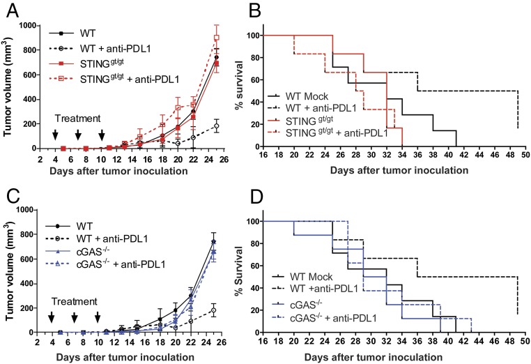
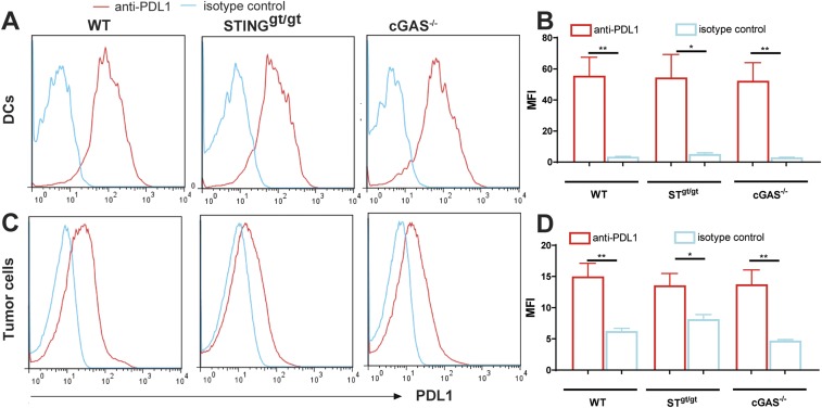
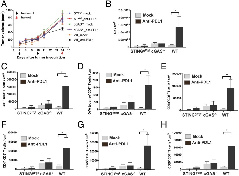
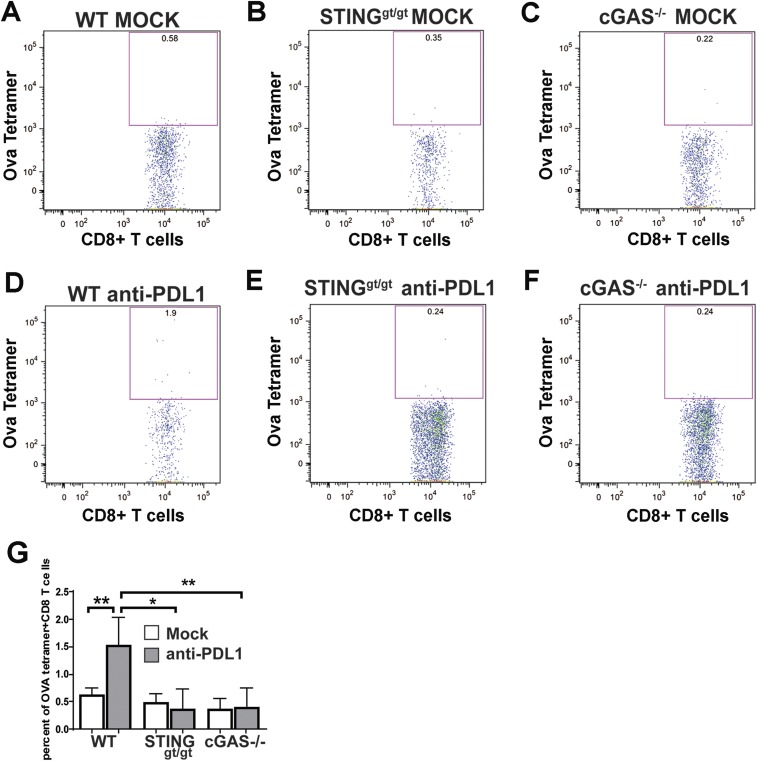
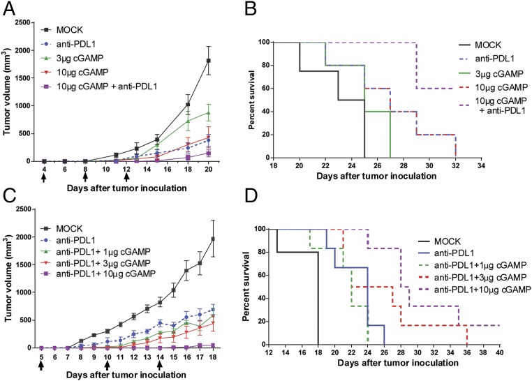
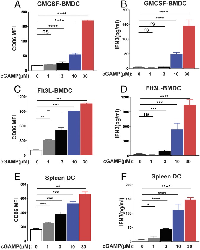
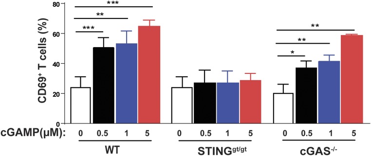
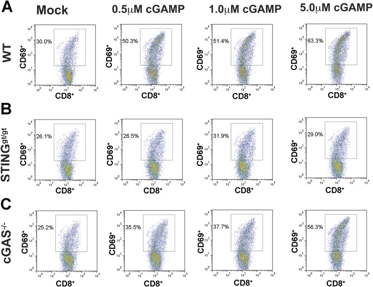
cGMP-AMP (cGAMP) synthase (cGAS) is a cytosolic DNA sensor that activates innate immune responses. cGAS catalyzes the synthesis of cGAMP, which functions as a second messenger that binds and activates the adaptor protein STING to induce type I interferons (IFNs) and other immune modulatory molecules. Here we show that cGAS is indispensable for the antitumor effect of immune checkpoint blockade in mice. Wild-type, but not cGAS-deficient, mice exhibited slower growth of B16 melanomas in response to a PD-L1 antibody treatment. Consistently, intramuscular delivery of cGAMP inhibited melanoma growth and prolonged the survival of the tumor-bearing mice. The combination of cGAMP and PD-L1 antibody exerted stronger antitumor effects than did either treatment alone. cGAMP treatment activated dendritic cells and enhanced cross-presentation of tumor-associated antigens to CD8 T cells. These results indicate that activation of the cGAS pathway is important for intrinsic antitumor immunity and that cGAMP may be used directly for cancer immunotherapy.

Keywords: PD-L1; STING; cGAMP; cGAS; cancer.


Conflict of interest statement

The authors declare no conflict of interest.

Figures

Fig. 1.
Fig. 1.
cGAS and STING are essential for the antitumor effect of PD-L1 blockade. WT, cGAS−/−, and STINGgt/gt mice (n = 6–8 per group) were injected s.c. with 1 × 10⁵ B16F10 melanoma cells, followed by three treatments with 200 μg of PD-L1 antibody at indicated time points. Tumor volumes were measured on the indicated dates and calculated according to the following formula: π/6 × length × width × height. Data are shown as mean ± SEM (A and C). Loss of survival was defined as death or when tumor diameter reached or exceeded 2 cm in any dimension (B and D).
Fig. S1.
Fig. S1.
Normal expression of PD-L1 on DCs and tumor cells in cGAS−/− and STINGgt/gt mice. WT, cGAS−/−, and STINGgt/gt mice (n = 3–4 per group) were injected s.c. with 1 × 10⁶ B16F10 melanoma cells, and tumors were harvested on day 14. Homogenous tumor suspension was prepared and analyzed by FACS using antibodies against CD45, MHCII, CD11c, and PD-L1. Dendritic cells are defined as MHCII+ CD11c+ (A and B) and tumor cells as CD45 population (C and D). Representative FACS plots are shown in A and C, and mean fluorescence intensity (MFI) of PD-L1–expressing cells in indicated experimental groups is shown in B and D. Data are shown as mean ± SEM. Statistical analysis was performed with a one-tailed, unpaired Student’s t test. *P < 0.05 and **P < 0.01.
Fig. 2.
Fig. 2.
cGAS and STING are required for generation of tumor-infiltrating CD8 T cells. (A) WT, cGAS−/−, and STINGgt/gt mice (n = 5 each group) were injected s.c. with 1 × 10⁶ B16F10-OVA cells. PD-L1 antibody was administered on days 7 and 10 after tumor inoculation. Tumor volume was measured on indicated dates (A). Tumors were harvested on day 14 to isolate and analyze tumor-infiltrating leukocytes (TILs). TILs are defined as CD45+ cells and normalized with tumor volume (B). CD8+ T cells are defined as CD8+CD3+ CD45+ TILs (C). CD4+ T cells are defined as CD4+CD3+ CD45+ TILs (F). Ova-specific CD8+ T cells are those bearing the T-cell receptor specific for an Ova-H2Kb tetramer (D). Regulatory T cells are defined as CD25+ CD4+ T cells (G). Activated CD8 (E) and CD4 (H) T cells are defined as CD69+ CD8+ and CD69+ CD4+ T cells. Data are shown as mean ± SEM. Statistical analysis was performed with a one-tailed, unpaired Student’s t test. *P < 0.05 and **P < 0.01.
Fig. S2.
Fig. S2.
cGAS and STING are required for tumor-specific CD8 T-cell response to PD-L1 antibody treatment. WT, cGAS−/−, and STINGgt/gt mice (n = 5 per group) were injected s.c. with 1 × 10⁶ B16F10-Ova melanoma cells, followed by two treatments with 200 μg of PD-L1 antibody as described in Fig. 2. Tumors were harvested on day 14, and tumor-infiltrating leukocytes were prepared and analyzed by FACS using an Ova-H2Kb tetramer and antibodies against CD8 and CD3. Representative FACS plots are shown in AF, and data from all experimental groups are shown in G as mean ± SEM. *P < 0.05 and **P < 0.01.
Fig. 3.
Fig. 3.
Antitumor effects of cGAMP and PD-L1 antibody. C57BL/6J mice (n = 4–5 per group) were injected with 1 × 10⁵ B16F10 melanoma cells, followed by treatments with cGAMP at indicated doses or 200 μg of PD-L1 antibody on days 4, 8, and 12 after tumor inoculation. Tumor volumes were measured on indicated dates (A). Survival of the tumor-bearing mice is shown in B. (C and D) Similar to A and B, except that mice (n = 5–6 per group) were treated with 200 μg of PD-L1 antibody in combination with different amounts of cGAMP as indicated on days 5, 10, and 14 after tumor inoculation. Data are shown as mean ± SEM in A and C.
Fig. 4.
Fig. 4.
cGAMP activates dendritic cells. GM-CSF DCs (A and B), Flt3 DCs (C and D), and splenic DCs (E and F) were cultured in the presence of cGAMP at different concentrations (1, 3, 10, and 30 µM). Eighteen hours after incubation, CD86 expression on MHCII+CD11c+ DCs was analyzed by FACS (A, C, and E). IFNβ in the cell culture media was analyzed by ELISA (B, D, and F). Data are shown as mean ± SEM. Statistical analysis was performed with a one-tailed, unpaired Student’s t test. *P < 0.05, **P < 0.01, ***P < 0.001, and ****P < 0.0001.
Fig. 5.
Fig. 5.
cGAMP stimulates cross-presentation of a tumor-associated antigen. BMDCs from WT, cGas−/−, and Stinggt/gt mice were incubated with irradiated B16F10-OVA cells in the presence of indicated concentrations of cGAMP. CD11c+ DC cells were then purified and cocultured with CD8+ T cells isolated from OT-I T-cell receptor transgenic mice. Cross-presentation efficiency of DCs was analyzed by CD69 expression of OT-I T cells and the average of four independent experiments is shown (representative FACS plots are shown in Fig. S3). Statistical analysis was performed with a one-tailed, unpaired Student’s t test. Data are shown as mean ± SEM. *P < 0.05, **P < 0.01, and ***P < 0.001.
Fig. S3.
Fig. S3.
cGAMP enhances tumor antigen cross-presentation in dendritic cells in a STING-dependent manner. BMDCs from WT (A), STINGgt/gt (B), and cGAS−/− (C) mice were incubated with irradiated B16F10-Ova cells and indicated concentrations of cGAMP. CD11c+ DCs were then isolated and cocultured with CD8 T cells from transgenic mice expressing the Ova-specific T-cell receptor (OT-I). Activated T cells were analyzed by FACS using antibodies specific for CD69 and CD8. Representative FACS plots from four independent experiments are shown, and data from all experimental mouse groups are shown in Fig. 5.

References

    1. Chen Q, Sun L, Chen ZJ. Regulation and function of the cGAS-STING pathway of cytosolic DNA sensing. Nat Immunol. 2016;17(10):1142–1149. - PubMed
    1. Ablasser A, Gulen MF. The role of cGAS in innate immunity and beyond. J Mol Med (Berl) 2016;94(10):1085–1093. - PubMed
    1. Sun L, Wu J, Du F, Chen X, Chen ZJ. Cyclic GMP-AMP synthase is a cytosolic DNA sensor that activates the type I interferon pathway. Science. 2013;339(6121):786–791. - PMC - PubMed
    1. Wu J, et al. Cyclic GMP-AMP is an endogenous second messenger in innate immune signaling by cytosolic DNA. Science. 2013;339(6121):826–830. - PMC - PubMed
    1. Civril F, et al. Structural mechanism of cytosolic DNA sensing by cGAS. Nature. 2013;498(7454):332–337. - PMC - PubMed

Publication types

MeSH terms