. 2017 Feb;140(2):287-305.
doi: 10.1093/brain/aww307.

Loss-of-function mutations in the ATP13A2/PARK9 gene cause complicated hereditary spastic paraplegia (SPG78)

Affiliations

Loss-of-function mutations in the ATP13A2/PARK9 gene cause complicated hereditary spastic paraplegia (SPG78)

Alejandro Estrada-Cuzcano et al. Brain. 2017 Feb.

Abstract

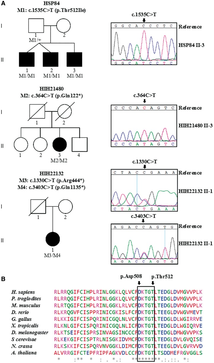
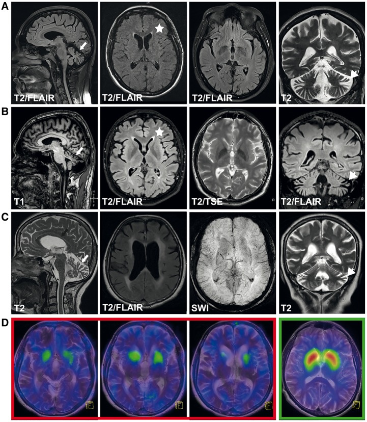
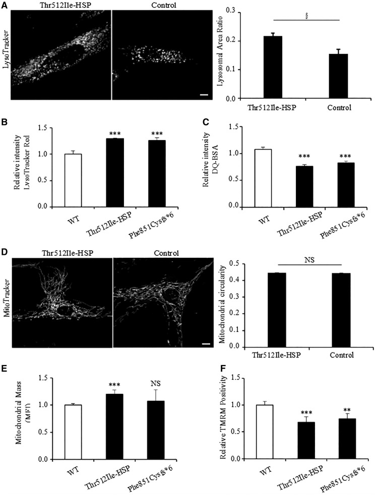
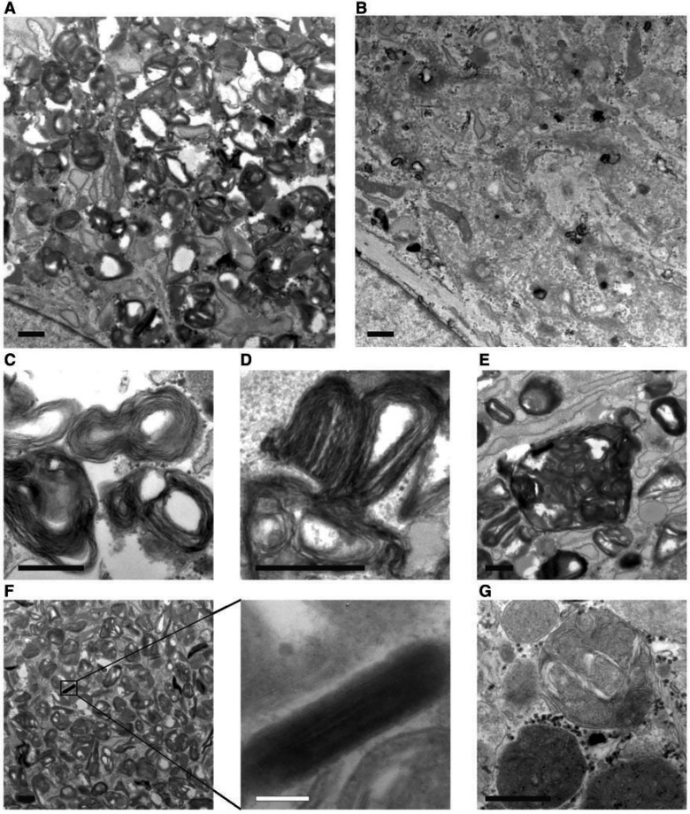
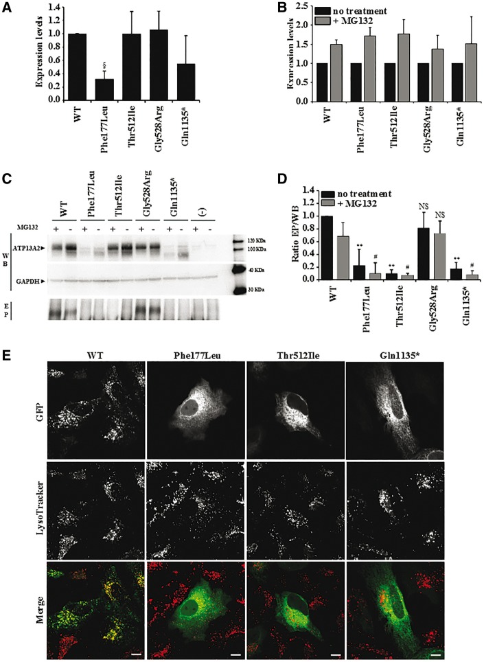
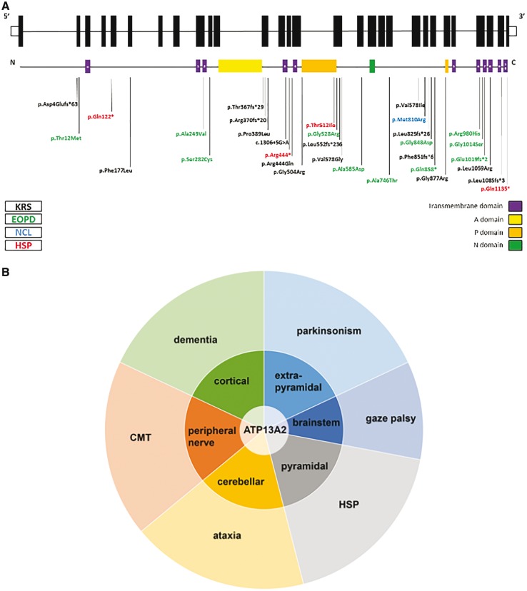
Hereditary spastic paraplegias are heterogeneous neurodegenerative disorders characterized by progressive spasticity of the lower limbs due to degeneration of the corticospinal motor neurons. In a Bulgarian family with three siblings affected by complicated hereditary spastic paraplegia, we performed whole exome sequencing and homozygosity mapping and identified a homozygous p.Thr512Ile (c.1535C > T) mutation in ATP13A2. Molecular defects in this gene have been causally associated with Kufor-Rakeb syndrome (#606693), an autosomal recessive form of juvenile-onset parkinsonism, and neuronal ceroid lipofuscinosis (#606693), a neurodegenerative disorder characterized by the intracellular accumulation of autofluorescent lipopigments. Further analysis of 795 index cases with hereditary spastic paraplegia and related disorders revealed two additional families carrying truncating biallelic mutations in ATP13A2. ATP13A2 is a lysosomal P5-type transport ATPase, the activity of which critically depends on catalytic autophosphorylation. Our biochemical and immunocytochemical experiments in COS-1 and HeLa cells and patient-derived fibroblasts demonstrated that the hereditary spastic paraplegia-associated mutations, similarly to the ones causing Kufor-Rakeb syndrome and neuronal ceroid lipofuscinosis, cause loss of ATP13A2 function due to transcript or protein instability and abnormal intracellular localization of the mutant proteins, ultimately impairing the lysosomal and mitochondrial function. Moreover, we provide the first biochemical evidence that disease-causing mutations can affect the catalytic autophosphorylation activity of ATP13A2. Our study adds complicated hereditary spastic paraplegia (SPG78) to the clinical continuum of ATP13A2-associated neurological disorders, which are commonly hallmarked by lysosomal and mitochondrial dysfunction. The disease presentation in our patients with hereditary spastic paraplegia was dominated by an adult-onset lower-limb predominant spastic paraparesis. Cognitive impairment was present in most of the cases and ranged from very mild deficits to advanced dementia with fronto-temporal characteristics. Nerve conduction studies revealed involvement of the peripheral motor and sensory nerves. Only one of five patients with hereditary spastic paraplegia showed clinical indication of extrapyramidal involvement in the form of subtle bradykinesia and slight resting tremor. Neuroimaging cranial investigations revealed pronounced vermian and hemispheric cerebellar atrophy. Notably, reduced striatal dopamine was apparent in the brain of one of the patients, who had no clinical signs or symptoms of extrapyramidal involvement.

Keywords: Kufor-Rakeb syndrome; hereditary spastic paraplegia (HSP); lysosomes; neuronal ceroid lipofuscinosis; parkinsonism.


Figures

Figure 1
Figure 1
Novel mutations in ATP13A2. (A) Graphic representation of three autosomal recessive families with complicated HSP in which ATP13A2 mutations were identified. Sanger sequencing traces confirming the presence of the mutations are presented and their segregation in available family members is demonstrated. The symbol (+) represents wild-type. Blackened symbols indicate affected individuals and symbols with slashes depict deceased individuals. (B) Amino acid conservation of ATP13A2 polypeptide in different species, the black frame delimits the autophosphorylation motif (508DKTGT), catalytic conserved residues Asp508 and Thr512 in human ATP13A2 are indicated by black arrows. Below the alignment, asterisk indicates fully conserved, colon indicates strongly similar, and full stop indicates weakly similar amino acids.
Figure 2
Figure 2
Cranial imaging in ATP13A2 HSP cases. Routine MRI in Patients HIH22132.II.1 (A) (disease duration 7 years), HSP84.II.3 (B) (disease duration 7 years), and HIH21480.II.3 (C) (disease duration 11 years). Images demonstrate vermian (white arrow) as well as hemispheric cerebellar atrophy (white arrowheads) that was progressive over time in Patient HIH22132.II.1 (not shown). Generalized cortical atrophy was present in all cases. In Patients HIH22132.II.1 (A) and HSP84.II.3 (B) an ‘ear of the lynx’ sign (T2 hyperintensity of the anterior fornix of the corpus callosum, marked by an asterisk) was present, while periventricular white matter changes were more pronounced in Patient HIH21480.II.3 (C). The latter case also demonstrated thinning of the corpus callosum (C, marked by black arrow). (D) Transversal fused 123I-FP-CIT SPECT and T2-weighted MRI images of three representative sections of the striatum in Patient HIH21480.II.3 (red box) and a representative section of the striatum from a healthy adult (green box, far right). Patient HIH21480.II.3 demonstrates severely reduced striatal dopamine.
Figure 3
Figure 3
Functional characterization of ATP13A2 mutant proteins. (A) Lysosomes of control and patient-derived (Patient HSP84.II.2) fibroblasts were stained with LysoTracker® (left), along with quantification of the lysosomal area ratio (lysosomal area/total cell area) (right). (B) Lysosome function was analysed using Lysotracker® Red and (C) DQ-BSA. DQ-BSA requires enzymatic cleavage in acidic lysosomal compartments to generate a highly fluorescent product. (D) Mitochondria of control and Patient HSP84.II.2-derived fibroblasts were stained with MitoTracker®, and mitochondrial circularity is shown on the right. (E) We used the mitochondrial-specific dye MitoTracker® Deep RED FM, which binds mitochondrial membrane independently of the membrane potential, and thus staining intensity has been considered an index of mitochondrial mass (MFI, mean fluorescence intensity). (F) Tetra-methylrhodamine (TMRM, 1 µM) treatment for quantifying changes in mitochondrial membrane potential. For A and D data are represented by average ± standard error of the mean. For B, C, E and F data are represented by average ± SD. §t-test P = 0.009, **P < 0.05; ***P < 0.001; NS = not significant. Scale bar = 10 µm.
Figure 4
Figure 4
Ultrastructure of cultured Thr512Ile-HSP fibroblasts. Lysosomes are excessively present in Thr512Ile-HSP fibroblasts (A) compared to controls (B). The majority of lysosomes contain aberrant storage material consisting of whirls and stacks of membranes (C), in some cases forming fingerprint bodies (D) or large clusters of membrane containing vesicles (E). In addition, cytoplasmic inclusions of tightly packed membranes were frequently observed (F). In contrast, the content of lysosomes in control fibroblasts consisted of a normal mix of material (G). Scale bars = 500 nm, except in F = 100 nm.
Figure 5
Figure 5
Expression levels and subcellular localization of wild-type and mutant ATP13A2. (A) Relative expression levels of ATP13A2 WT, Phe177Ile, Thr512Ile, Gly528Arg and Gln1135*, which were normalized to wild-type (WT) in COS-1 cells. (B) Expression levels of ATP13A2 WT, Phe177Ile, Thr512Ile, Gly528Arg and Gln1135* in COS-1 cells following MG132 treatment were normalized to the levels without MG132 treatment. (C) Semi-quantitative western blotting of ATP13A2 WT, Phe177Ile, Thr512Ile, Gly528Arg and Gln1135* overexpressed in COS-1 cells, which were either treated (+) or not treated (−) with proteasome inhibitor MG132. EP (phospho-enzyme) represents the autoradiogram displaying the relative autophosphorylation levels of wild-type and mutant ATP13A2. (D) Graph bar representing the ratio of the autophosphorylation and expression levels (EP/WB). For A, B and D data are represented by average ± SD. ANOVA test with post hoc Bonferroni correction, §P = 0.01, **P < 0.001 with respect to wild-type with no treatment; #P < 0.001 with respect to wild-type treated with MG132. NS = not significant. (E) Subcellular localization of GFP‐N‐ATP13A2 WT, Phe177Ile, Thr512Ile and Gln1135* mutant proteins, transiently expressed in HeLa cells and stained with LysoTracker®. Scale bar = 10 µm.
Figure 6
Figure 6
Clinical spectrum of ATP13A2 mutations. (A) Schematic representation of the genomic and protein lay out of ATP13A2 and location of currently known disease-associated mutations. Topology of ATP13A2 represents 10 transmembrane domains (1–10) and an additional membrane-associated segment in the N-terminus (a). In black, mutations associated with KRS; in green, mutations associated with early-onset parkinsonism (EOPD); in blue, mutation associated with NCL; in red, mutations identified in this study, which are associated with complicated HSP. (B) Schematic representation of the neuronal systems affected by ATP13A2 dysfunction and resulting signs and symptoms apparent on a clinical level. Loss of ATP13A2 function in the cortex, extrapyramidal system, brainstem, pyramidal system, cerebellum, and the peripheral nerves leads to a variety of signs and symptoms including dementia, parkinsonism, vertical gaze palsy, spasticity, ataxia, and peripheral neuropathy. Predominance of extrapyramidal involvement or spasticity has led to the classification of ATP13A2-related disease as parkinsonism or HSP, respectively. We hypothesize that other carriers of ATP13A2 mutations might display predominance of other ATP13A2-dependent systems and therefore manifest as cerebellar ataxia, dementia or Charcot–Marie–Tooth disease.

Comment in

References

    1. Adzhubei IA, Schmidt S, Peshkin L, Ramensky VE, Gerasimova A, Bork P, et al.A method and server for predicting damaging missense mutations. Nat Methods 2010; 7: 248–9. - PMC - PubMed
    1. Anheim M, Lagier-Tourenne C, Stevanin G, Fleury M, Durr A, Namer IJ, et al.SPG11 spastic paraplegia: a new cause of juvenile parkinsonism. J Neurol 2009; 256: 104–8. - PubMed
    1. Arif B, Kumar KR, Seibler P, Vulinovic F, Fatima A, Winkler S, et al.A novel OPA3 mutation revealed by exome sequencing: an example of reverse phenotyping. JAMA Neurol 2013; 70: 783–7. - PubMed
    1. Behrens MI, Bruggemann N, Chana P, Venegas P, Kagi M, Parrao T, et al.Clinical spectrum of Kufor-Rakeb syndrome in the Chilean kindred with ATP13A2 mutations. Mov Disord 2010; 25: 1929–37. - PubMed
    1. Bras J, Verloes A, Schneider SA, Mole SE, Guerreiro RJ. Mutation of the parkinsonism gene ATP13A2 causes neuronal ceroid-lipofuscinosis. Hum Mol Genet 2012; 21: 2646–50. - PMC - PubMed

MeSH terms

Substances