. 2017 Jan 31;135(5):476-489.
doi: 10.1161/CIRCULATIONAHA.116.025684.

The Role of Efferocytosis in Atherosclerosis

Affiliations

The Role of Efferocytosis in Atherosclerosis

Yoko Kojima et al. Circulation. .

Abstract

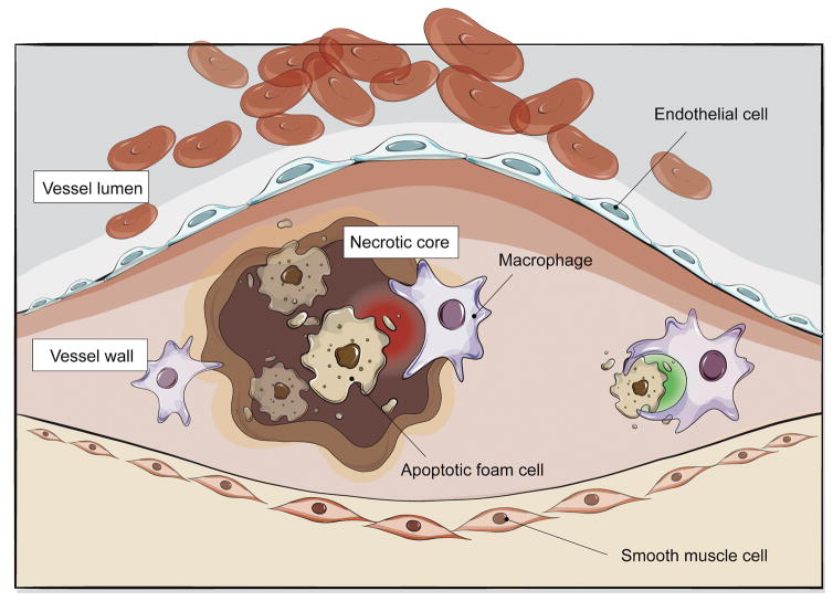
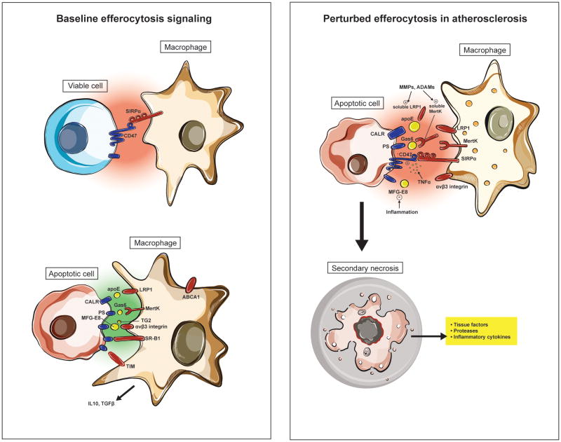
The necrotic core has long been a hallmark of the vulnerable atherosclerotic plaque. Although apoptotic cells are cleared quickly in almost all other tissue beds, their removal appears to be significantly impaired in the diseased blood vessel. Emerging evidence indicates that this phenomenon is caused by a defect in efferocytosis, the process by which apoptotic tissue is recognized for engulfment by phagocytic cells such as macrophages. Genetic and experimental data suggest that efferocytosis is impaired during atherogenesis caused by dysregulation of so-called eat me ligands, which govern the edibility of cells undergoing programmed cell death. The following is a summary of recent data indicating that efferocytosis is a major unappreciated driver of lesion expansion but also a reversible defect that can potentially be targeted as a means to prevent plaque progression.

Keywords: atherosclerosis; efferocytosis; macrophage; necrotic core; vascular biology.


Figures

Figure 1
Figure 1. Impaired efferocytosis contributes to atherosclerosis
Diseased and apoptotic cells in the growing atherosclerotic plaque are not recognized for efficient phagocytic clearance by lesional macrophages. While the mechanisms which drive this pathology are still an area of active investigation, emerging data suggests that this defect may be due to impaired ‘eat me’ (green) and ‘don’t eat me’ (red) signaling that renders these cells ‘inedible’. As a result, foam cells accumulate to promote lesion expansion and apoptotic tissue undergoes secondary necrosis to accelerate vascular inflammation and lesion instability.
Figure 2
Figure 2. Impaired efferocytosis signaling in vascular disease
Experimental data suggests that pro-phagocytic signals (including calreticulin, Mfg-e8 and Mertk) are reduced in atherosclerosis due to inflammation, post-translational modifications and/or genetic variability. Exacerbating this loss of ‘eat me’ signaling is a concomitant upregulation of the CD47-Sirp-α ‘don’t eat me’ pathway, which furthers decreases the ‘edibility’ of cells within the necrotic core. The end result is that apoptotic cells in the growing plaque becomes poor substrates for phagocytic cells such as macrophages and dendritic cells. Such uncleared cells become secondarily necrotic and release additional proinflammatory stimuli, thus promoting a positive feedback loop.

References

    1. Kinchen JM, Ravichandran KS. Phagocytic signaling: You can touch, but you can’t eat. Curr Biol. 2008;18:R521–524. - PMC - PubMed
    1. Thorp EB. Mechanisms of failed apoptotic cell clearance by phagocyte subsets in cardiovascular disease. Apoptosis : an international journal on programmed cell death. 2010;15:1124–1136. - PMC - PubMed
    1. Fadok VA, Voelker DR, Campbell PA, Cohen JJ, Bratton DL, Henson PM. Exposure of phosphatidylserine on the surface of apoptotic lymphocytes triggers specific recognition and removal by macrophages. J Immunol. 1992;148:2207–2216. - PubMed
    1. Ravichandran KS, Lorenz U. Engulfment of apoptotic cells: Signals for a good meal. Nat Rev Immunol. 2007;7:964–974. - PubMed
    1. Hoffmann PR, deCathelineau AM, Ogden CA, Leverrier Y, Bratton DL, Daleke DL, Ridley AJ, Fadok VA, Henson PM. Phosphatidylserine (ps) induces ps receptor-mediated macropinocytosis and promotes clearance of apoptotic cells. The Journal of cell biology. 2001;155:649–659. - PMC - PubMed

Publication types