. 2017 Mar 1;37(9):2435-2448.
doi: 10.1523/JNEUROSCI.2557-16.2017. Epub 2017 Jan 30.

An Ultrastructural Study of the Thalamic Input to Layer 4 of Primary Motor and Primary Somatosensory Cortex in the Mouse

Affiliations

An Ultrastructural Study of the Thalamic Input to Layer 4 of Primary Motor and Primary Somatosensory Cortex in the Mouse

Rita Bopp et al. J Neurosci. .

Abstract

The traditional classification of primary motor cortex (M1) as an agranular area has been challenged recently when a functional layer 4 (L4) was reported in M1. L4 is the principal target for thalamic input in sensory areas, which raises the question of how thalamocortical synapses formed in M1 in the mouse compare with those in neighboring sensory cortex (S1). We identified thalamic boutons by their immunoreactivity for the vesicular glutamate transporter 2 (VGluT2) and performed unbiased disector counts from electron micrographs. We discovered that the thalamus contributed proportionately only half as many synapses to the local circuitry of L4 in M1 compared with S1. Furthermore, thalamic boutons in M1 targeted spiny dendrites exclusively, whereas ∼9% of synapses were formed with dendrites of smooth neurons in S1. VGluT2+ boutons in M1 were smaller and formed fewer synapses per bouton on average (1.3 vs 2.1) than those in S1, but VGluT2+ synapses in M1 were larger than in S1 (median postsynaptic density areas of 0.064 μm2 vs 0.042 μm2). In M1 and S1, thalamic synapses formed only a small fraction (12.1% and 17.2%, respectively) of all of the asymmetric synapses in L4. The functional role of the thalamic input to L4 in M1 has largely been neglected, but our data suggest that, as in S1, the thalamic input is amplified by the recurrent excitatory connections of the L4 circuits. The lack of direct thalamic input to inhibitory neurons in M1 may indicate temporal differences in the inhibitory gating in L4 of M1 versus S1.SIGNIFICANCE STATEMENT Classical interpretations of the function of primary motor cortex (M1) emphasize its lack of the granular layer 4 (L4) typical of sensory cortices. However, we show here that, like sensory cortex (S1), mouse M1 also has the canonical circuit motif of a core thalamic input to the middle cortical layer and that thalamocortical synapses form a small fraction (M1: 12%; S1: 17%) of all asymmetric synapses in L4 of both areas. Amplification of thalamic input by recurrent local circuits is thus likely to be a significant mechanism in both areas. Unlike M1, where thalamocortical boutons typically form a single synapse, thalamocortical boutons in S1 usually formed multiple synapses, which means they can be identified with high probability in the electron microscope without specific labeling.

Keywords: VGluT2; barrel cortex; electron microscopy; layer 4; motor cortex; thalamocortical.


Figures

Figure 1.
Figure 1.
Thalamorecipient layers in sensory and motor areas revealed by dual labeling of VGluT2 and NeuN. AC, Coronal section (0.48 mm anterior of the bregma) containing M2, M1, and S1. Scale bar, 1 mm. A, Merge of micrographs of VGluT2- and NeuN-stained sections with cortical layers 1–6 indicated. B, VGluT2 label occurs in three distinct bands. The L4 band is most salient in S1 and can be followed medially into M1 and M2, where it becomes fainter and thinner. C, NeuN staining for neuronal somata revealing cytoarchitecture, L4 in S1 is composed of small and densely packed cells; in M1, it is present but less easily identified. DF, Coronal section containing vibrissal barrel cortex (0.48 mm posterior of bregma). Scale bar, 500 μm. D, Merge of micrographs of VGluT2- and NeuN-stained sections with cortical layers 1–6 indicated. E, VGluT2 fluorescence is strongest in L4 barrels and less intense in the septa. VGluT2 label is also present in L1, L2/3, upper L5 (brackets), and at the L5/6 border. F, NeuN staining revealing the cytoarchitecture of barrel cortex.
Figure 2.
Figure 2.
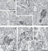
Electron micrographs of VGluT2-labeled boutons in L4 of motor cortex (AC) and vibrissal barrel cortex (DF). Scale bars in all panels, 1 μm. A, VGluT2+ bouton forming an asymmetric synapse (arrowhead) with a spine (sp) in L4 of M1. VGluT2 label appears as electron-dense material on synaptic vesicles. Unlabeled boutons in the surrounding neuropil also form asymmetric synapses with spines (arrowheads). B, Single VGluT2+ boutons often appeared large in cross-section, contained mitochondria (m), and formed synapses with multiple target spines (sp; arrowheads). A smooth (putative GABAergic) dendrite (smooth d) traverses and forms three asymmetric synapses (arrowheads) and one symmetric synapse (arrow) with unlabeled boutons. C, A VGluT2+ bouton forms a perforated synapse (two arrowheads) with a spine head containing a spine apparatus (asterisk). The spine originates from a dendrite traversing through (spiny d). D, A large VGluT2+ bouton in L4 of vibrissal barrel cortex forms two perforated synapses (arrowheads) on spines (sp), one of which contains a spine apparatus (asterisk). A second elongated VGluT2-labeled bouton (left) formed no synapses in this section. E, A VGluT2+ boutons forms a synapse (arrowhead) with a smooth dendrite (smooth d). The dendrite forms another asymmetric synapse with an unlabeled bouton (arrowhead) and a symmetric synapse (arrow) with an unlabeled (putative GABAergic) bouton, which contains pleomorphic vesicles. F, Some target spine heads are enveloped completely by the VGluT2+ bouton. In the same section, a VGluT2+ bouton forms an asymmetric synapse (arrowhead) with a smooth dendrite (smooth d).
Figure 3.
Figure 3.
Summary of the unbiased disector counts. A, Thalamocortical pathway to M1 L4 provides approximately half as many synapses compared with S1. The density of thalamocortical synapses in the neuropil of a layer (NVVGluT2 ± SEM) and the absolute thickness of the target layer (L4 ± SEM) determine how many synapses (syn) that thalamic projection contributes to the respective area. To compare the absolute number of VGluT2 synapses in M1 and S1 L4, we constructed hypothetical cuboid tissue blocks of M1 and S1 with a base area of 1 μm2. By multiplying NVVGluT2 by the volume of the respective L4 (VL4), we derived an absolute number of VGluT2+ synapses for M1 and S1 L4. In M1, the absolute number of VGluT2+ synapses was only half that of S1 (the statistical range is determined by the accumulated SEM of NVVGluT2 and L4 thickness). Scale bars indicate scales for cortical surface and individual layers. B, Postsynaptic targets of VGluT2+ boutons in unbiased physical disector counts for M1 and S1. The respective percentage is indicated above each bar (spine synapses: n = 173 in M1 and 231 in S1, smooth shaft synapses: n = 1 in M1 and 24 in S1, spiny shaft synapses: n = 0 in M1 and 1 in S1).
Figure 4.
Figure 4.
Representative 3D reconstructions of VGluT2+ boutons and their postsynaptic densities and targets in L4 of M1 and S1. AD, VGluT2+ boutons in M1 L4. E, F, VGluT2+ bouton in S1 L4. A, Electron micrograph of a VGluT2+ bouton forming a single perforated synapse (arrowheads) on a dendritic spine (sp). B Left, Reconstruction of the VGluT2+ bouton from A (blue), mitochondrion inside the bouton (green), PSD (red), and dendritic spine with a segment of its parent dendrite (black). Right, en face representation of the PSD. C, Electron micrograph of a VGluT2+ bouton in L4 of M1, forming two synapses, which are numbered. D Left, Reconstruction of the VGluT2+ bouton and its targets shown in C. Spines are indicated with numbers corresponding to the synapses that they receive, as seen in C. Right, en face representations of the two PSDs. E, Electron micrographs of a VGluT2+ bouton in L4 of S1, forming four synapses (numbered). Synapse 3 was formed with the shaft of a smooth dendrite. F Left, Reconstruction of the VGluT2+ bouton and its postsynaptic targets shown in E (smooth dendrite in gray). Right, En face representations of the four PSDs made by this bouton. Scale bars, 1 μm.
Figure 5.
Figure 5.
Comparison of the number of synapses made by VGluT2+ boutons in M1 and S1 L4. A, VGluT2+ boutons in M1 L4 (n = 61) predominantly form a single synapse, as did unlabeled boutons forming asymmetric synapses (VGluT2, n = 82). B, VGluT2+ boutons in S1 L4 (n = 72) predominantly form multiple synapses (VGluT2, n = 64). gives the arithmetic mean. The nonparametric M–W test was used for statistical analysis. **p ≤ 0.01; ****p ≤ 0.0001.
Figure 6.
Figure 6.
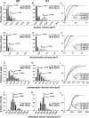
Morphological features of reconstructed VGluT2+ and VGluT2 boutons in M1 and S1 L4. AC, Distributions of the total volume of boutons in M1 (A; VGluT2+, n = 61; VGluT2, n = 82) and in S1 (B; VGluT2+, n = 72; VGluT2, n = 64); is the median. C, Cumulative histogram of the data in A and B. DF, Distributions of the mitochondria volume inside reconstructed boutons in M1 (D; VGluT2+, n = 61; VGluT2, n = 82) and in S1 (E; VGluT2+, n = 72; VGluT2, n = 64); is the median. F, Cumulative histogram of the data in D and E. GI, Distributions of the area of the PSDs in M1 (G; VGluT2+, n = 79; VGluT2, n = 83) and in S1 (H; VGluT2+, n = 138, VGluT2, n = 67); is the median. Synapses on dendritic shafts were excluded for comparability. I, Cumulative histogram of the data in G and H. JL, Vesicle density estimates for VGluT2+ and VGluT2 boutons in M1 (J; VGluT2+, n = 44; VGluT2, n = 59) and in S1 (K; VGluT2+, n = 68; VGluT2, n = 54); is the median. L, Cumulative histogram of the data in J and K. Statistical analysis was performed using the nonparametric M–W test, the results are shown in the box insets of the rightmost panels. ns: p > 0.05; *p ≤ 0.05; **p ≤ 0.01; ***p ≤ 0.001; ****p ≤ 0.0001.
Figure 7.
Figure 7.
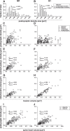
Relationships of various morphological features of VGluT2+ and VGluT2 boutons in M1 and S1 L4. A, B, Comparison of PSD sizes made by VGluT2+ boutons that either formed single synapses (unisynaptic) or multiple synapses (multisynaptic) in M1 (A; unisynaptic, n = 44; multisynaptic, n = 37) and in S1 (B; unisynaptic, n = 24; multisynaptic, n = 125); is the median (M–W test). C, D, Scatter plots showing the relationship between the bouton volume and all individual PSDs made by a bouton in M1 (C; VGluT2+, n = 81; VGluT2, n = 90) and in S1 (D; VGluT2+, n = 149; VGluT2, n = 79). In CH, boutons are marked by different symbols indicating whether they were unisynaptic or multisynaptic (see box inset). E, F, Scatter plots showing the relationship between the bouton volume and the sum of all PSDs made by a bouton in M1 (E; VGluT2+, n = 61; VGluT2, n = 82), and in S1 (F; VGluT2+, n = 72; VGluT2, n = 64). G, H, Scatter plots showing the relationship between bouton volume and volume of mitochondria they contain in M1 (G; VGluT2+, n = 61; VGluT2, n = 82) and in S1 (H; VGluT2+, n = 72; VGluT2, n = 64). I, J, Scatter plots of the relationship between spine head volume and the PSD area made on the spine head in M1 (I; VGluT2+, n = 79; VGluT2, n = 83) and in S1 (J; VGluT2+, n = 138; VGluT2, n = 67). Synapses formed with dendritic shafts were excluded for comparability. In EJ, r is the nonparametric Spearman correlation coefficient (p ≤ 0.0001 for all, t test).
Figure 8.
Figure 8.
Proximal portion of a smooth, putatively GABAergic dendrite reconstructed from a tissue block in L4 of S1. The dendrite emerged directly from its parent soma (thickening to the right). We reconstructed a total length of 31 μm, including two branch points and a local varicosity (to the left). A, Dendrite fragment formed a total of 90 synapses: 19 with VGluT2+ boutons (blue), 51 with VGluT2 boutons (purple), and 20 symmetric synapses with putative GABAergic boutons (green). B, To visualize individual postsynaptic densities, the dendrite was made transparent. Asymmetric synapses made by VGluT2+ boutons are shown in blue, asymmetric synapses made by VGluT2 boutons in purple, and symmetric synapses in green. Scale bar, 2 μm.
Figure 9.
Figure 9.
Schematic comparison of thalamic input to L4 of S1 and M1. A, In L4 of M1, ∼12.1% of synapses originate from the thalamus, most of them presumably from VL and Po; other termination zones include L1 (from VA and VM) and the L5/6 border (from VL and Po). B, Thalamic boutons (blue) in M1 L4 usually form one synapse, innervate pyramidal cells (Pyr) (Yamawaki et al., 2014) due to the absence of spiny stellate cells, make on average larger PSDs than in S1, and almost completely avoid dendrites of smooth (putative GABAergic) cells. C, In S1, VPM projects into the L4 barrels, where it makes ∼17.2% of all asymmetric synapses, and to the L5/6 border. Po projects to all laminae, in particular to L1 and into the septa and upper L5. D, Thalamic boutons in S1 L4 are larger compared with M1 and usually form >1 synapse. They innervate spiny stellate cells (SSCs) and the dendrites of inhibitory cells, yet their PSDs are smaller compared with M1.

References

    1. Abercrombie M. (1946) Estimation of nuclear population from microtome sections. Anat Rec 94:239–247. 10.1002/ar.1090940210 - DOI - PubMed
    1. Agmon A, Connors BW (1992) Correlation between intrinsic firing patterns and thalamocortical synaptic responses of neurons in mouse barrel cortex. J Neurosci 12:319–329. - PMC - PubMed
    1. Ahmed B, Anderson JC, Douglas RJ, Martin KA, Nelson JC (1994) Polyneuronal innervation of spiny stellate neurons in cat visual cortex. J Comp Neurol 341:39–49. 10.1002/cne.903410105 - DOI - PubMed
    1. Ahmed B, Anderson JC, Martin KA, Nelson JC (1997) Map of the synapses onto layer 4 basket cells of the primary visual cortex of the cat. J Comp Neurol 380:230–242. - PubMed
    1. Arellano JI, Benavides-Piccione R, Defelipe J, Yuste R (2007) Ultrastructure of dendritic spines: correlation between synaptic and spine morphologies. Front Neurosci 1:131–143. 10.3389/neuro.01.1.1.010.2007 - DOI - PMC - PubMed

Publication types

MeSH terms

Substances

LinkOut - more resources