. 2017 Apr;16(4):591-600.
doi: 10.1158/1535-7163.MCT-16-0352. Epub 2017 Jan 30.

Combining Chk1/2 Inhibition with Cetuximab and Radiation Enhances In Vitro and In Vivo Cytotoxicity in Head and Neck Squamous Cell Carcinoma

Affiliations

Combining Chk1/2 Inhibition with Cetuximab and Radiation Enhances In Vitro and In Vivo Cytotoxicity in Head and Neck Squamous Cell Carcinoma

Ling Zeng et al. Mol Cancer Ther. 2017 Apr.

Abstract

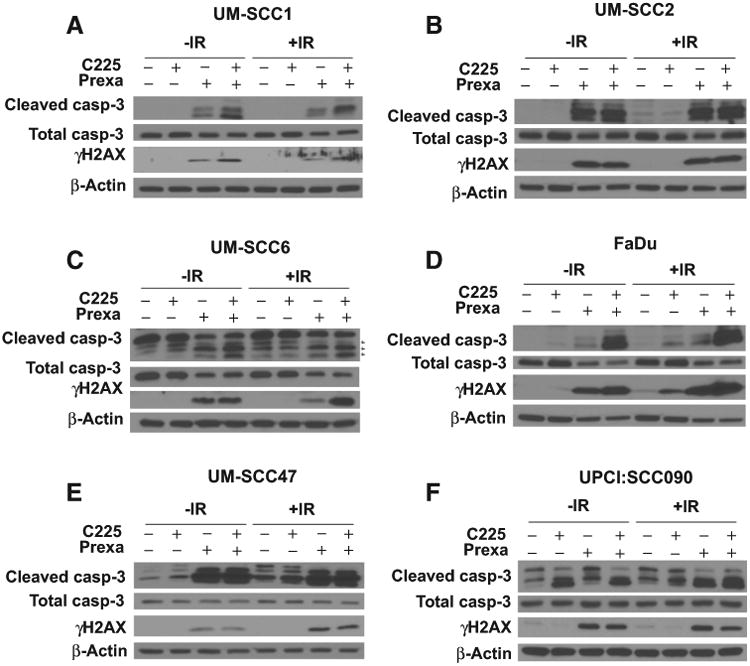
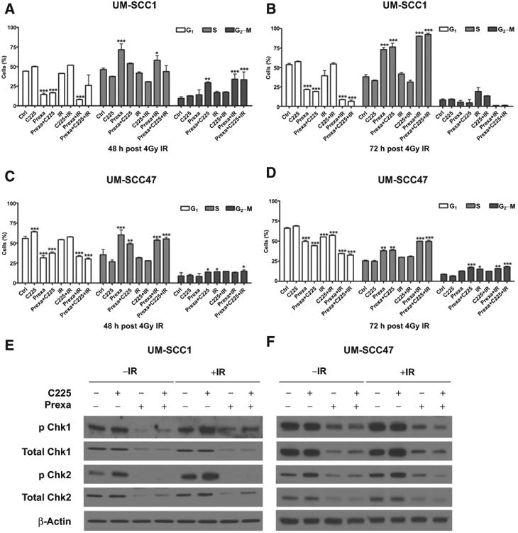
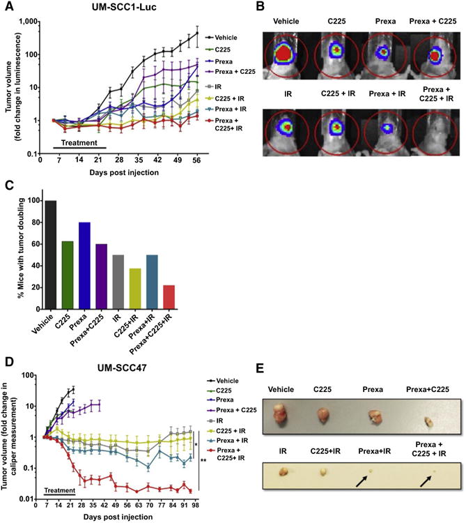
EGFR inhibition and radiotherapy are potent inducers of DNA damage. Checkpoint kinases 1 and 2 (Chk1/2) are critical regulators of the DNA-damage response, controlling cell-cycle checkpoints that may permit recovery from therapy-associated genomic stress. We hypothesized that Chk1/2 inhibition (CHKi) with prexasertib may enhance cytotoxicity from EGFR inhibition plus radiotherapy in head and neck squamous cell carcinoma (HNSCC). In this study, we found that the addition of CHKi to the EGFR inhibitor cetuximab with and without radiotherapy significantly decreased cell proliferation and survival fraction in human papillomavirus virus (HPV)-positive and HPV-negative HNSCC cell lines. Reduced proliferation was accompanied by decreased checkpoint activation, induced S-phase accumulation, persistent DNA damage, and increased caspase cleavage and apoptosis. Importantly, a significant tumor growth delay was observed in vivo in both HPV-positive and HPV-negative cell line xenografts receiving triple combination therapy with CHKi, cetuximab, and radiotherapy without a concomitant increase in toxicity as assessed by mouse body weight. Taken together, the combination of CHKi with cetuximab plus irradiation displayed significant antitumor effects in HNSCCs both in vitro and in vivo, suggesting that this combination therapy may increase clinical benefit. A clinical trial to test this treatment for patients with head and neck cancer is currently ongoing (NCT02555644). Mol Cancer Ther; 16(4); 591-600. ©2017 AACR.


Conflict of interest statement

Disclosure of Potential Conflicts of Interest: E.S. Yang reports receiving a commercial research grant from Eli Lilly and Company, has received speakers bureau honoraria, and is a consultant/advisory board member for Nanostring Technologies. No potential conflicts of interest were disclosed by the other authors.

Figures

Figure 1
Figure 1
Combination treatment with prexasertib, cetuximab, and IR decreases cell proliferation. UM-SCC1 (A), UM-SCC2 (B), UM-SCC6 (C), FaDu (D), UM-SCC47 (E), and UPCI:SCC090 cells (F) were treated with either vehicle or 0.25 μg/mL C225 for 16 hours, then 1 or 10 nmol/L prexasertib (prexa) for 2 hours, followed by sham or 2 Gy IR. Cell numbers were counted at 48, 72, and 96 hours after IR using the Beckman Z1 Coulter particle counter. Shown is the mean ± SEM from one of two independent experiments performed in triplicate;*, P < 0.05; **, P < 0.01; ***, P < 0.001.
Figure 2
Figure 2
Prexasertib with cetuximab and IR induces early apoptosis. Annexin V apoptotic assay in UM-SCC1 (A), UM-SCC2 (B), UM-SCC6 (C), FaDu (D), UM-SCC47 (E), and UPCI:SCC090 cells (F). Cells were treated with either vehicle or 0.25 mg/mL C225 for 16 hours, then 1 or 10 nmol/L prexasertib (prexa) for 2 hours, followed by sham or 2 Gy IR. Cells were harvested at 48 hours after IR and evaluated for Annexin V positivity by flow cytometry. Shown is the mean ± SEM from one of two independent experiments performed in triplicate; *, P < 0.05; **, P < 0.01; ***, P < 0.001.
Figure 3
Figure 3
Prexasertib with cetuximab and IR increases caspase-3 cleavage and induces persistent DNA damage. UM-SCC1 (A), UM-SCC2 (B), UM-SCC6 (C), FaDu (D), UM-SCC47 (E), and UPCI:SCC090 cells (F) were treated with either vehicle or 0.25 μg/mL C225 for 16 hours, then 1 or 10 nmol/L prexasertib (prexa) for 2 hours, followed by sham or 2 Gy IR. Cell lysates were harvested at 48 hours after IR and analyzed by Western blot analysis for total and cleaved caspase-3 and γ-H2AX protein induction. Shown are representative blots from at least two independent experiments, using β-actin as a loading control.
Figure 4
Figure 4
Prexasertib induces S-phase accumulation in cells and abrogates cetuximab and IR induced checkpoint activation. A-D, UM-SCC1 and UM-SCC47 cells were treated with either vehicle or 0.25 μg/mL C225 for 16 hours, then 1 nmol/L prexasertib (prexa) for 2 hours, followed by sham or 4 Gy IR. Cells were stained with propidium iodide (Pi) at (A and C) 48 or (B and D) 72 hours after IR and analyzed for cell-cycle distribution by flow cytometry. Shown is the mean ± SEM from one of three independent experiments performed in triplicate; *, P < 0.05; **, P < 0.01; ***, P < 0.001. UM-SCC1 (E) and UM-SCC47 (F) cells were treated, and cell lysates were harvested at 48 hours after IR and assessed for expression of Chk1 and Chk2, including both total and phosphorylated protein levels, by Western blot analysis. β-Actin was used as a loading control. Shown are representative blots from at least two independent experiments.
Figure 5
Figure 5
Prexasertib plus cetuximab-IR delays tumor growth of HPV-negative UM-SCC1-Luc orthotopic xenografts and HPV-positive UM-SCC47 heterotopic xenografts. A-D, The tongues of athymic nude mice were injected with UM-SCC1 luciferase-expressing cells (UM-SCC1-Luc), and tumor volume was measured by bioluminescence imaging twice weekly. A, UM-SCC1 tumor growth over time, normalized to luminescence measurement at the start of treatment on day 5. Shown is the mean fold change in tumor volume ± SEM. N = 8-10 mice for all treatment groups. B, Representative optical images of UM-SCC1 tumor luminescence at day 56. C, The percentage of mice with a two fold increase or greater in UM-SCC1 tumor volume in each treatment group at day 56. D-E, The flanks of athymic nude mice were injected subcutaneously with UM-SCC47 cells, and tumor volumes were measured by digital caliper twice a week and calculated using the equation: (width×length×height)/2. D, UM-SCC47 tumor growth over time, normalized to caliper measurement at the start of treatment on day 5. Shown is the mean fold change in tumor volume ± SEM. N = 8-10 mice for all treatment groups; *, P < 0.05, IR versus prexa + IR; **, P <0.01, IR versus prexa + C225 + IR. E, Representative images of harvested UM-SCC47 tumors for each treatment group. Vehicle, C225, prexasertib, and C225 + prexasertib were harvested at day 20. IR, IR + C225, IR + prexasertib, IR + C225 + prexasertib were harvested at day 49.

References

    1. Chaturvedi AK, Engels EA, Pfeiffer RM, Hernandez BY, Xiao W, Kim E, et al. Human papillomavirus and rising oropharyngeal cancer incidence in the United States. J Clin Oncol. 2011;29:4294–301. - PMC - PubMed
    1. Ang KK, Harris J, Wheeler R, Weber R, Rosenthal DI, Nguyen-Tan PF, et al. Human papillomavirus and survival of patients with oropharyngeal cancer. N Engl J Med. 2010;363:24–35. - PMC - PubMed
    1. Fakhry C, Westra WH, Li S, Cmelak A, Ridge JA, Pinto H, et al. Improved survival of patients with human papillomavirus-positive head and neck squamous cell carcinoma in a prospective clinical trial. J Natl Cancer Inst. 2008;100:261–9. - PubMed
    1. Bonner JA, Harari PM, Giralt J, Azarnia N, Shin DM, Cohen RB, et al. Radiotherapy plus cetuximab for squamous-cell carcinoma of the head and neck. N Engl J Med. 2006;354:567–78. - PubMed
    1. Chung CH, Ely K, McGavran L, Varella-Garcia M, Parker J, Parker N, et al. Increased epidermal growth factor receptor gene copy numberis associated with poor prognosis in head and neck squamous cell carcinomas. J Clin Oncol. 2006;24:4170–6. - PubMed

MeSH terms

Associated data