. 2017 May;23(5):655-672.
doi: 10.1261/rna.060368.116. Epub 2017 Jan 30.

RNA-Puzzles Round III: 3D RNA structure prediction of five riboswitches and one ribozyme

Affiliations

RNA-Puzzles Round III: 3D RNA structure prediction of five riboswitches and one ribozyme

Zhichao Miao et al. RNA. 2017 May.

Abstract

RNA-Puzzles is a collective experiment in blind 3D RNA structure prediction. We report here a third round of RNA-Puzzles. Five puzzles, 4, 8, 12, 13, 14, all structures of riboswitch aptamers and puzzle 7, a ribozyme structure, are included in this round of the experiment. The riboswitch structures include biological binding sites for small molecules (S-adenosyl methionine, cyclic diadenosine monophosphate, 5-amino 4-imidazole carboxamide riboside 5'-triphosphate, glutamine) and proteins (YbxF), and one set describes large conformational changes between ligand-free and ligand-bound states. The Varkud satellite ribozyme is the most recently solved structure of a known large ribozyme. All puzzles have established biological functions and require structural understanding to appreciate their molecular mechanisms. Through the use of fast-track experimental data, including multidimensional chemical mapping, and accurate prediction of RNA secondary structure, a large portion of the contacts in 3D have been predicted correctly leading to similar topologies for the top ranking predictions. Template-based and homology-derived predictions could predict structures to particularly high accuracies. However, achieving biological insights from de novo prediction of RNA 3D structures still depends on the size and complexity of the RNA. Blind computational predictions of RNA structures already appear to provide useful structural information in many cases. Similar to the previous RNA-Puzzles Round II experiment, the prediction of non-Watson-Crick interactions and the observed high atomic clash scores reveal a notable need for an algorithm of improvement. All prediction models and assessment results are available at http://ahsoka.u-strasbg.fr/rnapuzzles/.

Keywords: 3D prediction; X-ray structures; bioinformatics; force fields; models; structure quality.


Figures

FIGURE 1.
FIGURE 1.
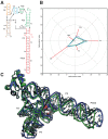
Puzzle 4: the SAM-I riboswitch aptamer. (A) Secondary structure and (B) module-based deformation profile values for the three predicted models from different groups with lowest RMSD: Chen model 2 (green), Bujnicki model 1 (blue), and Santalucia model 1 (cyan). (Radial red lines) The minimum, maximum, and mean DP values for each domain. (C) Structure superimposition between native structure (green) and best predicted model (blue, Chen model 2).
FIGURE 2.
FIGURE 2.
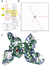
Puzzle 8: SAM-I/IV riboswitch. (A) Secondary structure and (B) module-based deformation profile values for the three predicted models from different groups with lowest RMSD: Das model 3 (green), Bujnicki model 7 (blue), and Chen model 3 (cyan). (Radial red lines) The minimum, maximum, and mean DP values for each domain. (C) Structure superimposition between native structure (green) and best predicted model (blue, Das model 3).
FIGURE 3.
FIGURE 3.
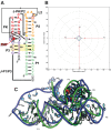
Puzzle 12: ydaO riboswitch. (A) Secondary structure and (B) module-based deformation profile values for the three predicted models from different groups with lowest RMSD: Ding model 12 (green), Bujnicki model 8 (blue), and Das model 7 (cyan). (Radial red lines) The minimum, maximum, and mean DP values for each domain. (C) Structure superimposition between native structure (green) and best predicted model (blue, Ding model 12).
FIGURE 4.
FIGURE 4.
Puzzle 13: ZTP riboswitch. (A) Secondary structure and (B) module-based deformation profile values for the three predicted models from different groups with lowest RMSD: Das model 7 (green), Chen model 5 (blue), and Bujnicki model 3 (cyan). (Radial red lines) The minimum, maximum, and mean DP values for each domain. (C) Structure superimposition between native structure (green) and best predicted model (blue, Das model 7). The bound ZMP is shown in red.
FIGURE 5.
FIGURE 5.
Puzzle 14: L-glutamine riboswitch. (A) Secondary structure and (B) module-based deformation profile values, measured from free state structure, for the three predicted models from different groups with lowest RMSD: Das post-experiment model 2 (green), Ding post-experiment model 8 (blue), and Chen post-experiment model 2 (cyan). (Radial red lines) The minimum, maximum, and mean DP values for each domain. (C) Module-based deformation profile values, measured from bound state structure, for the three predicted models from different groups with lowest RMSD: Bujnicki pre-experiment model 2 (green), Ding post-experiment model 8 (blue), and Chen post-experiment model 2 (cyan). (D) Free state structure superimposition between native structure (green) and best predicted model (blue, Das post-experiment model 2). (E) Bound state structure superimposition between native structure (green) and best predicted model (blue, Bujnicki pre-experiment model 2).
FIGURE 6.
FIGURE 6.
Puzzle 7: Varkud satellite ribozyme. (A) Secondary structure and (B) module-based deformation profile values for the three predicted models from different groups with lowest RMSD: Das model 1 (green), Chen model 5 (blue), and Bujnicki model 5 (cyan). (Radial red lines) The minimum, maximum, and mean DP values for each domain. (C) Structure superimposition between native structure (green) and best predicted model (blue, Das model 1).

References

    1. Ames TD, Breaker RR. 2011. Bacterial aptamers that selectively bind glutamine. RNA Biol 8: 82–89. - PMC - PubMed
    1. Antczak M, Zok T, Popenda M, Lukasiak P, Adamiak RW, Blazewicz J, Szachniuk M. 2014. RNApdbee—a webserver to derive secondary structures from pdb files of knotted and unknotted RNAs. Nucleic Acids Res 42: W368–W372. - PMC - PubMed
    1. Antczak M, Popenda M, Zok T, Sarzynska J, Ratajczak T, Adamiak RW, Szachniuk M. 2016. New functionality of RNAComposer: application to shape the axis of miR160 precursor structure. Acta Biochim Pol 63: 737–744. - PubMed
    1. Baird NJ, Zhang J, Hamma T, Ferré-D'Amaré AR. 2012. YbxF and YlxQ are bacterial homologs of L7Ae and bind K-turns but not K-loops. RNA 18: 759–770. - PMC - PubMed
    1. Boniecki MJ, Lach G, Dawson WK, Tomala K, Lukasz P, Soltysinski T, Rother KM, Bujnicki JM. 2016. SimRNA: a coarse-grained method for RNA folding simulations and 3D structure prediction. Nucleic Acids Res 44: e63. - PMC - PubMed

Publication types

MeSH terms