

Retinoic Acid Enhances the Differentiation of Adipose-Derived Stem Cells to Keratocytes In Vitro
- PMID: 28138416
- PMCID: PMC5270625
- DOI: 10.1167/tvst.6.1.6
Retinoic Acid Enhances the Differentiation of Adipose-Derived Stem Cells to Keratocytes In Vitro
Abstract
Purpose: All-trans retinoic acid (RA) supplementation was investigated as a method of enhancing the differentiation of human adipose-derived stem cells (ASCs) to corneal keratocytes in vitro, in combination with a chemically defined serum-free medium.
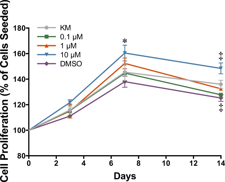
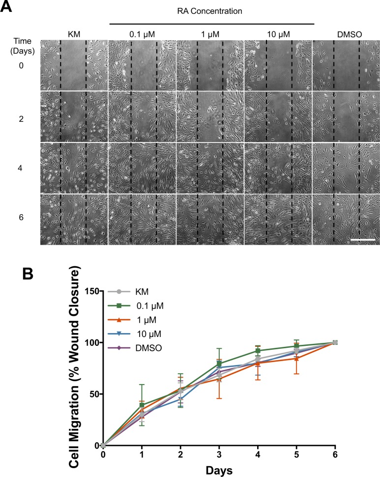
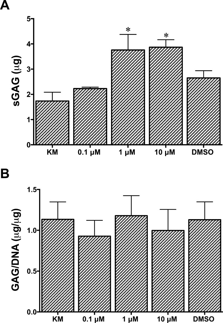
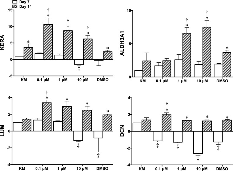
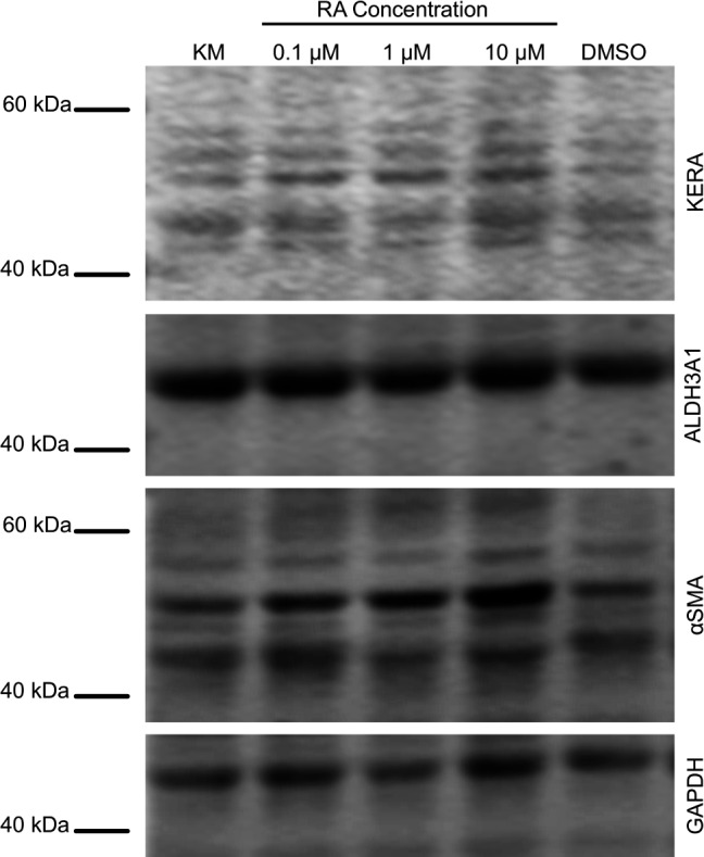
Methods: Adipose-derived stem cells were cultured in monolayer and supplemented with 0.1, 1, or 10 μM RA for 14 days. The effects of RA on cell proliferation, migration, and extracellular matrix (ECM) accumulation were evaluated. In addition, the expression of phenotypic keratocyte markers was examined by reverse transcription polymerase chain reaction (PCR), immunocytochemistry, and Western blotting.
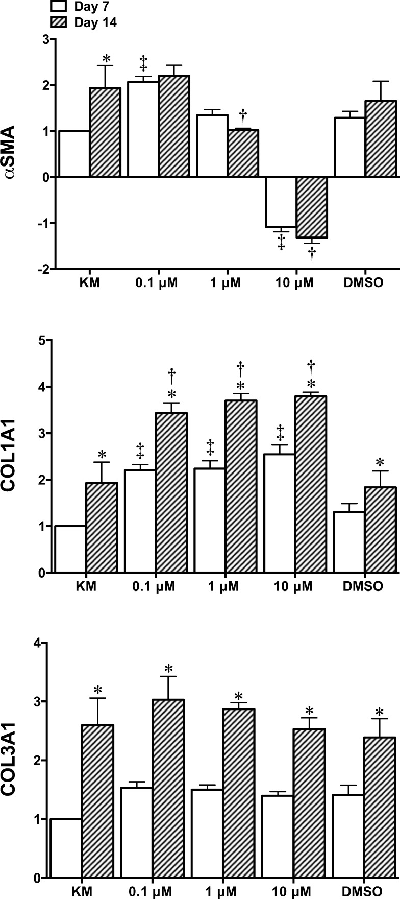
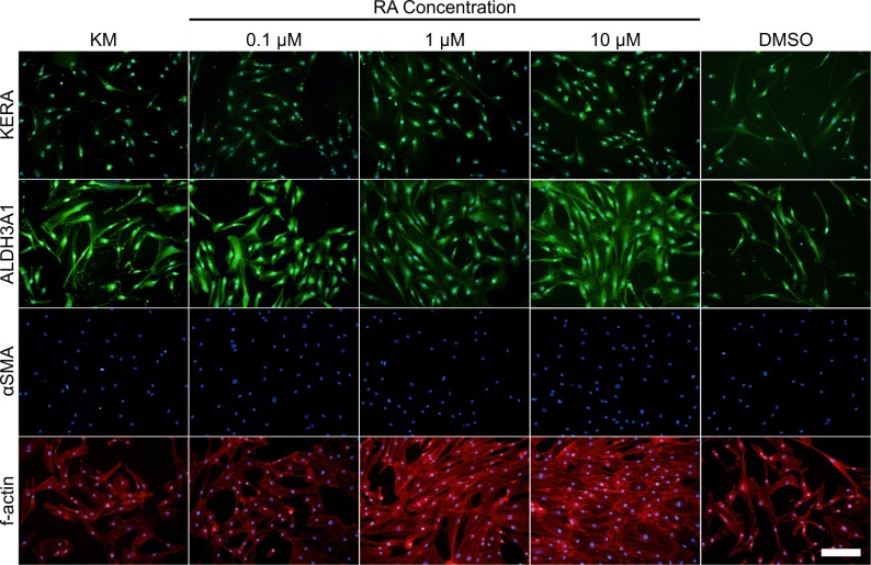
Results: Adipose-derived stem cells cultured with RA showed improved cell proliferation and ECM production. In addition, RA enhanced the expression of keratocyte-specific markers, keratocan, aldehyde dehydrogenase 3A1, lumican, and decorin, when compared to serum-free media alone. Furthermore, the presence of RA increased the amount of collagen type I while reducing the expression of fibrotic marker, α-smooth muscle actin.
Conclusions: These findings indicate that RA is a useful supplement for promoting a keratocyte phenotype in ASC.
Translational relevance: This study is particularly important for the generation of biological corneal substitutes and next generation cell based therapies for corneal conditions.
Keywords: differentiation; keratocytes; retinoic acid; stem cells.
Figures
Similar articles
-
The effect of growth factor supplementation on corneal stromal cell phenotype in vitro using a serum-free media.Exp Eye Res. 2016 Oct;151:26-37. doi: 10.1016/j.exer.2016.07.015. Epub 2016 Jul 22. Exp Eye Res. 2016. PMID: 27456135
-
The effects of retinoic acid on human corneal stromal keratocytes cultured in vitro under serum-free conditions.Invest Ophthalmol Vis Sci. 2013 Nov 13;54(12):7483-91. doi: 10.1167/iovs.13-13092. Invest Ophthalmol Vis Sci. 2013. PMID: 24150763
-
Corneal keratocyte transition to mesenchymal stem cell phenotype and reversal using serum-free medium supplemented with fibroblast growth factor-2, transforming growth factor-β3 and retinoic acid.J Tissue Eng Regen Med. 2018 Jan;12(1):e203-e215. doi: 10.1002/term.2316. Epub 2017 Mar 30. J Tissue Eng Regen Med. 2018. PMID: 27685949
-
Application of retinoic acid improves form and function of tissue engineered corneal construct.Organogenesis. 2015;11(3):122-36. doi: 10.1080/15476278.2015.1093267. Organogenesis. 2015. PMID: 26496651 Free PMC article.
-
Human mesenchymal stem cells differentiate into keratocyte-like cells in keratocyte-conditioned medium.Exp Eye Res. 2012 Aug;101:16-26. doi: 10.1016/j.exer.2012.05.009. Epub 2012 Jun 7. Exp Eye Res. 2012. PMID: 22683947
Cited by
-
The Effect of Calcium and Glucose Concentration on Corneal Epithelial Cell Lines Differentiation, Proliferation, and Focal Adhesion Expression.Biores Open Access. 2019 Jun 5;8(1):74-83. doi: 10.1089/biores.2018.0036. eCollection 2019. Biores Open Access. 2019. PMID: 31179162 Free PMC article.
-
Therapeutic efficacy of BSA formulated hydrogels in corneal wound healing and epithelial cell regeneration: an ex vivo study.Sci Rep. 2025 Jun 6;15(1):19956. doi: 10.1038/s41598-025-04408-3. Sci Rep. 2025. PMID: 40481091 Free PMC article.
-
Advances in Regulatory Strategies of Differentiating Stem Cells towards Keratocytes.Stem Cells Int. 2022 Jan 31;2022:5403995. doi: 10.1155/2022/5403995. eCollection 2022. Stem Cells Int. 2022. PMID: 35140792 Free PMC article. Review.
-
Differentiation Capacity of Human Mesenchymal Stem Cells into Keratocyte Lineage.Invest Ophthalmol Vis Sci. 2019 Jul 1;60(8):3013-3023. doi: 10.1167/iovs.19-27008. Invest Ophthalmol Vis Sci. 2019. PMID: 31310658 Free PMC article.
-
Cornea-Specific Human Adipose Stem Cell-Derived Extracellular Matrix for Corneal Stroma Tissue Engineering.ACS Appl Mater Interfaces. 2024 Apr 3;16(13):15761-15772. doi: 10.1021/acsami.3c17803. Epub 2024 Mar 21. ACS Appl Mater Interfaces. 2024. PMID: 38513048 Free PMC article.
References
-
- Pascolini D,, Mariotti SP. Global estimates of visual impairment: 2010. Br J Ophthalmol. 2012. ; 96: 614–618. - PubMed
-
- Liu Y,, Gan L,, Carlsson DJ,, et al. A simple, cross-linked collagen tissue substitute for corneal implantation. Invest Ophthalmol Vis Sci. 2006. ; 47: 1869–1875. - PubMed
-
- Berryhill BL,, Kader R,, Kane B,, Birk DE,, Feng J,, Hassell JR. Partial restoration of the keratocyte phenotype to bovine keratocytes made fibroblastic by serum. Invest Ophthalmol Vis Sci. 2002. ; 43: 3416–3421. - PubMed
LinkOut - more resources
Full Text Sources
Other Literature Sources
Miscellaneous
