

Evaluation of early-phase [18F]-florbetaben PET acquisition in clinical routine cases
- PMID: 28138429
- PMCID: PMC5257027
- DOI: 10.1016/j.nicl.2016.10.005
Evaluation of early-phase [18F]-florbetaben PET acquisition in clinical routine cases
Abstract
Objectives: In recent years several [18F]-labelled amyloid PET tracers have been developed and have obtained clinical approval. There is accumulating evidence that early (post injection) acquisitions with these tracers are equally informative as conventional blood flow and metabolism studies for diagnosis of Alzheimer's disease, but there have been few side-by-side studies. Therefore, we investigated the performance of early acquisitions of [18F]-florbetaben (FBB) PET compared to [18F]-fluorodeoxyglucose (FDG) PET in a clinical setting.
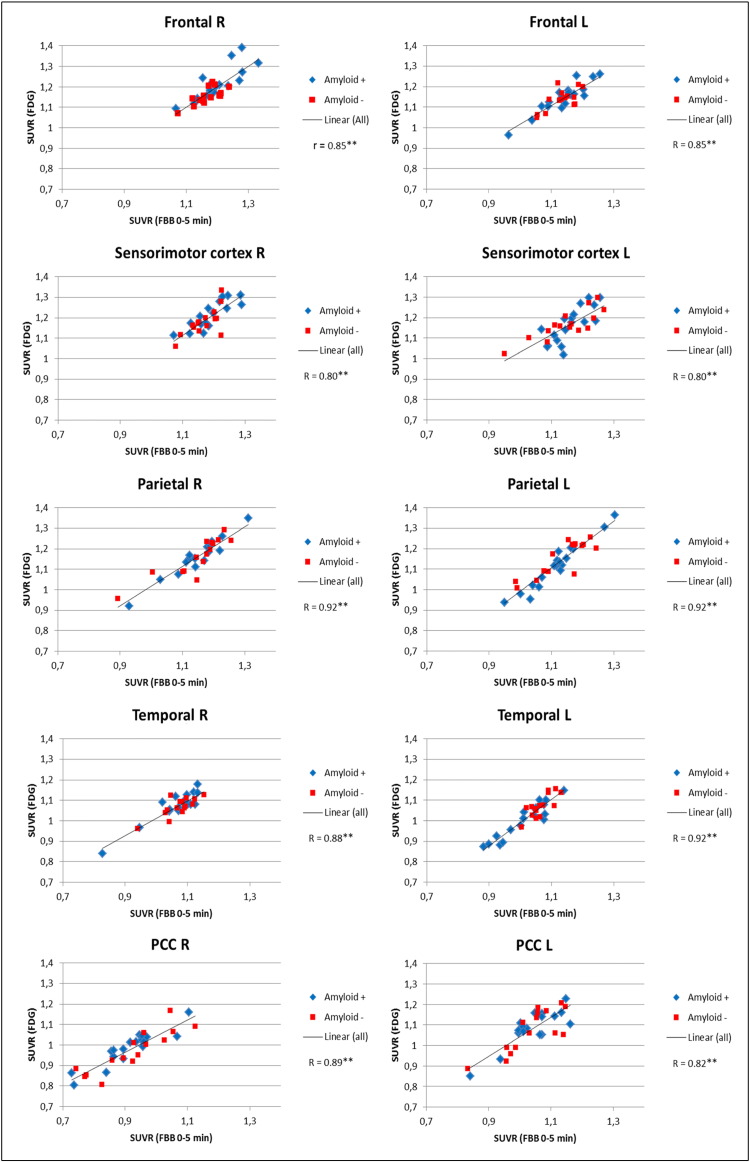
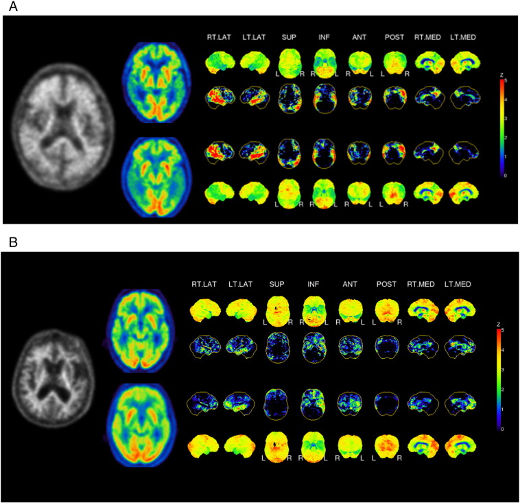
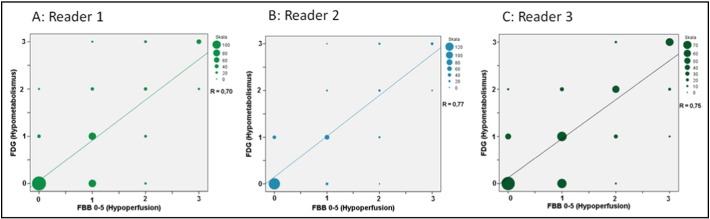
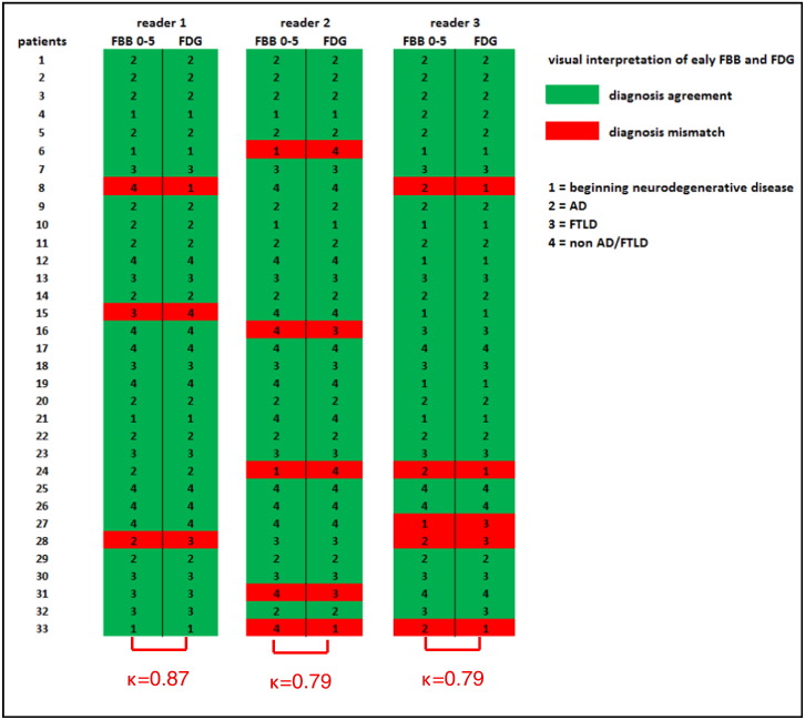
Methods: All subjects were recruited with clinical suspicion of dementia due to neurodegenerative disease. FDG PET was undertaken by conventional methods, and amyloid PET was performed with FBB, with early recordings for the initial 10 min (early-phase FBB), and late recordings at 90-110 min p.i. (late-phase FBB). Regional SUVR with cerebellar and global mean normalization were calculated for early-phase FBB and FDG PET. Pearson correlation coefficients between FDG and early-phase FBB were calculated for predefined cortical brain regions. Furthermore, a visual interpretation of disease pattern using 3-dimensional stereotactic surface projections (3D-SSP) was performed, with assessment of intra-reader agreement.
Results: Among a total of 33 patients (mean age 67.5 ± 11.0 years) included in the study, 18 were visually rated amyloid-positive, and 15 amyloid-negative based on late-phase FBB scans. Correlation coefficients for early-phase FBB vs. FDG scans displayed excellent agreement in all target brain regions for global mean normalization. Cerebellar normalization gave strong, but significantly lower correlations. 3D representations of early-phase FBB visually resembled the corresponding FDG PET images, irrespective of the amyloid-status of the late FBB scans.
Conclusions: Early-phase FBB acquisitions correlate on a relative quantitative and visual level with FDG PET scans, irrespective of the amyloid plaque density assessed in late FBB imaging. Thus, early-phase FBB uptake depicts a metabolism-like image, suggesting it as a valid surrogate marker for synaptic dysfunction, which could ultimately circumvent the need for additional FDG PET investigation in diagnosis of dementia.
Keywords: 3D-SSP, 3-dimensional stereotactic surface projections; AD, Alzheimer's disease; Alzheimer's disease; CBF, cerebral blood flow; CBL, cerebellum; CN, cognitively normal; FBB, [18F]florbetaben; FDG Pet; FDG, [18F]-fluorodeoxyglucose; FTLD, frontotemporal lobar degeneration; GLM, global mean; L, left; MCI, mild cognitive impairment; MNI, Montreal Neurological Institute; Metabolism; PCC, posterior cingulate cortex; PET, Positron emission tomography; Perfusion; R, right; SPECT, single photon emission computed tomography; SUVR, standardized uptake value ratio; VOI, volume of interest; [18F]-florbetaben PET; p.i., post injection; ß-amyloid.
Figures
Similar articles
-
Early [(18)F]florbetaben and [(11)C]PiB PET images are a surrogate biomarker of neuronal injury in Alzheimer's disease.Eur J Nucl Med Mol Imaging. 2016 Aug;43(9):1700-9. doi: 10.1007/s00259-016-3353-1. Epub 2016 Mar 30. Eur J Nucl Med Mol Imaging. 2016. PMID: 27026271
-
Early-Phase 18F-Florbetaben PET as an Alternative Modality for 18F-FDG PET.Clin Nucl Med. 2020 Jan;45(1):e8-e14. doi: 10.1097/RLU.0000000000002768. Clin Nucl Med. 2020. PMID: 31524679 Clinical Trial.
-
Usefulness of Dual-Point Amyloid PET Scans in Appropriate Use Criteria: A Multicenter Study.J Alzheimers Dis. 2018;65(3):765-779. doi: 10.3233/JAD-180232. J Alzheimers Dis. 2018. PMID: 30103321
-
[(18)F]Florbetaben: a review in β-amyloid PET imaging in cognitive impairment.CNS Drugs. 2015 Jul;29(7):605-13. doi: 10.1007/s40263-015-0258-7. CNS Drugs. 2015. PMID: 26175116 Review.
-
Brain fluorodeoxyglucose (FDG) PET in dementia.Ageing Res Rev. 2016 Sep;30:73-84. doi: 10.1016/j.arr.2016.02.003. Epub 2016 Feb 11. Ageing Res Rev. 2016. PMID: 26876244 Review.
Cited by
-
Spatial distributions of cholinergic impairment and neuronal hypometabolism differ in MCI due to AD.Neuroimage Clin. 2019;24:101978. doi: 10.1016/j.nicl.2019.101978. Epub 2019 Aug 9. Neuroimage Clin. 2019. PMID: 31422337 Free PMC article.
-
Ageing stereotypes and prodromal Alzheimer's disease (AGING): study protocol for an ongoing randomised clinical study.BMJ Open. 2019 Oct 7;9(10):e032265. doi: 10.1136/bmjopen-2019-032265. BMJ Open. 2019. PMID: 31594904 Free PMC article.
-
A comparison of advanced semi-quantitative amyloid PET analysis methods.Eur J Nucl Med Mol Imaging. 2022 Oct;49(12):4097-4108. doi: 10.1007/s00259-022-05846-1. Epub 2022 Jun 2. Eur J Nucl Med Mol Imaging. 2022. PMID: 35652962 Free PMC article.
-
3D conditional generative adversarial networks for high-quality PET image estimation at low dose.Neuroimage. 2018 Jul 1;174:550-562. doi: 10.1016/j.neuroimage.2018.03.045. Epub 2018 Mar 20. Neuroimage. 2018. PMID: 29571715 Free PMC article.
-
Longitudinal evaluation of surrogates of regional cerebral blood flow computed from dynamic amyloid PET imaging.J Cereb Blood Flow Metab. 2020 Feb;40(2):288-297. doi: 10.1177/0271678X19830537. Epub 2019 Feb 12. J Cereb Blood Flow Metab. 2020. PMID: 30755135 Free PMC article. Clinical Trial.
References
-
- Barthel H., Sabri O. Florbetaben to trace amyloid-beta in the Alzheimer brain by means of PET. J. Alzheimers Dis. 2011;26(Suppl 3):117–121. - PubMed
-
- Barthel H., Gertz H.J., Dresel S., Peters O., Bartenstein P., Buerger K., Hiemeyer F., Wittemer-Rump S.M., Seibyl J., Reininger C., Sabri O. Cerebral amyloid-beta PET with florbetaben ((18)F) in patients with Alzheimer's disease and healthy controls: a multicentre phase 2 diagnostic study. Lancet Neurol. 2011;10(5):424–435. - PubMed
-
- Braak H., Braak E. Neuropathological stageing of Alzheimer-related changes. Acta Neuropathol. 1991;82(4):239–259. - PubMed
-
- Dishino D.D., Welch M.J., Kilbourn M.R., Raichle M.E. Relationship between lipophilicity and brain extraction of C-11-labeled radiopharmaceuticals. J. Nucl. Med. 1983;24(11):1030–1038. - PubMed
MeSH terms
Substances
LinkOut - more resources
Full Text Sources
Other Literature Sources
Medical
Miscellaneous
