. 2016 Nov 16;2(11):e1601185.
doi: 10.1126/sciadv.1601185. eCollection 2016 Nov.

Epidermal mechano-acoustic sensing electronics for cardiovascular diagnostics and human-machine interfaces

Affiliations

Epidermal mechano-acoustic sensing electronics for cardiovascular diagnostics and human-machine interfaces

Yuhao Liu et al. Sci Adv. .

Abstract

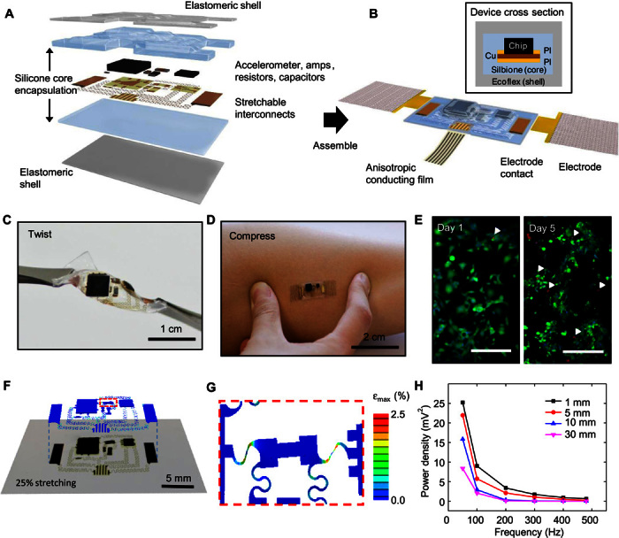
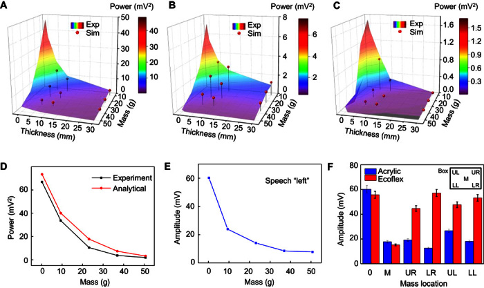
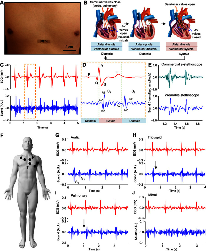
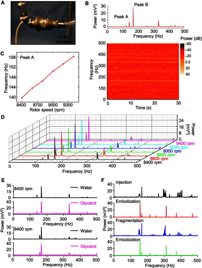
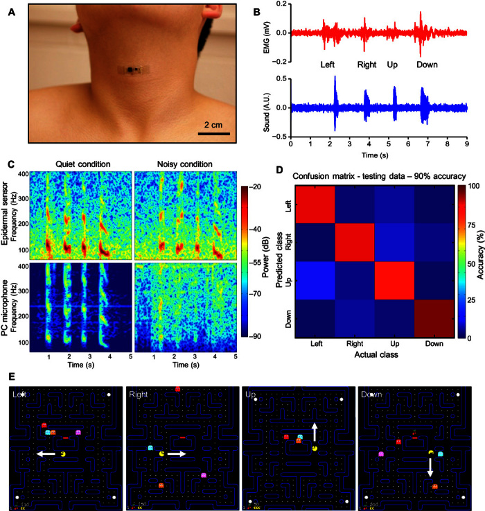
Physiological mechano-acoustic signals, often with frequencies and intensities that are beyond those associated with the audible range, provide information of great clinical utility. Stethoscopes and digital accelerometers in conventional packages can capture some relevant data, but neither is suitable for use in a continuous, wearable mode, and both have shortcomings associated with mechanical transduction of signals through the skin. We report a soft, conformal class of device configured specifically for mechano-acoustic recording from the skin, capable of being used on nearly any part of the body, in forms that maximize detectable signals and allow for multimodal operation, such as electrophysiological recording. Experimental and computational studies highlight the key roles of low effective modulus and low areal mass density for effective operation in this type of measurement mode on the skin. Demonstrations involving seismocardiography and heart murmur detection in a series of cardiac patients illustrate utility in advanced clinical diagnostics. Monitoring of pump thrombosis in ventricular assist devices provides an example in characterization of mechanical implants. Speech recognition and human-machine interfaces represent additional demonstrated applications. These and other possibilities suggest broad-ranging uses for soft, skin-integrated digital technologies that can capture human body acoustics.

Keywords: Epidermal; accelerometer; acoustic; cardiovascular; flexible; human-machine interface; seismocardiology; stretchable; ventricle assist device.


Figures

Fig. 1
Fig. 1. Schematic illustration of an epidermal mechano-acoustic–electrophysiological measurement device.
(A) Exploded view diagram of the overall design structure of the system. (B) Illustration of the assembled device and its interface with soft EP measurement electrodes and flexible cable for power supply and data acquisition. A cross-sectional view appears in the upper inset. (C) Device held by tweezers in a twisted configuration. (D) Device mounted on skin while compressed by pinching. (E) Fluorescence micrographs of cells cultured on the surface of a device to illustrate its biocompatibility. Green and red regions correspond to live and dead cells, respectively. The white arrowheads highlight the latter. Scale bars, 200 μm. (F) Overlay of optical image and finite element simulation results for a device under biaxial stretching to a strain of 25%. (G) Magnified view of modeling results for the part of the interconnect structures that experiences the highest strain. (H) Vibrational response summarized in a plot of spectral power measured while mounted on a layer of chicken breast, to simulate tissue, on a vibration source.
Fig. 2
Fig. 2. Summary of the experimental and computational studies of the effects of device mass, modulus, tissue thickness, and signal frequency on measured mechano-acoustic responses.
Experimentally measured spectral power and simulation results associated with the mechano-acoustic response of a device mounted in an acrylic box placed on a tissue sample on a vibrational source at frequencies of 50 Hz (A), 100 Hz (B), and 200 Hz (C). (D) Comparison of measured (experiment) and computed (analytical) dependence of spectral power on mass. (E) Measured maximum signal amplitude recorded with a device mounted on the neck as the subject said the word “left,” as a function of the mass of the device. (F) Amplitude measured using a device in a rigid box and on a thin substrate of Ecoflex, as a function of spatial location of the added mass.
Fig. 3
Fig. 3. Application of mechano-acoustic–electrophysiological sensing with an epidermal device in diagnosing cardiovascular health status.
(A) Image of an epidermal device mounted on the chest. (B) Schematic diagram of cardiac cycle: (left) artrial and ventricular diastole, (middle) artrial systole and ventricular diastole, and (right) ventricular systole and atrial diastole. (C) Plot of ECG (top) and heart sound (bottom) signals measured simultaneously. A.U., arbitrary units. (D) Magnified view of ECG (top) and heart sound (bottom) signals measured in (C). MC, mitral valve closure; AO, aortic valve opening; RE, rapid ventricular ejection; AC, aortic valve closure; MO, mitral valve opening; RF, rapid ventricular filling. (E) Comparison of heart sound signals measured using a commercial electronic stethoscope and the reported device. (F) Schematic illustration of the measurement site: A, aortic; P, pulmonary; T, tricuspid; M, mitral. Representative measurement from a 78-year-old female patient with diagnosed mild pulmonary and tricuspid regurgitation at the aortic (G), tricuspid (H), pulmonary (I), and mitral (J) sites.
Fig. 4
Fig. 4. Application of mechano-acoustic sensing with an epidermal device in diagnosing VAD operation.
(A) Image of the experimental circulation loop with the device mounted on the VAD (HeartMate II). (B) Fast Fourier transform (FFT) of the vibration response (top) and spectrogram (bottom) associated with the operation of the VAD at 8400 rpm in a water circulation loop. (C) FFT spectral power of the vibration response for operating frequencies between 8400 and 9400 rpm. Distinctive changes with VAD speed occur only on the peak around 150 Hz. (D) Comparison of vibrational responses in a circulation loop with water and with glycerol at 8400 rpm (top) and 9400 rpm (bottom). (E) Demonstration of changes in acoustic signature associated with circulation of a blood clot (500 μl) in the glycerol loop during stages of initial injection of the blood clot, first few circulation passes without decomposition, subsequent complete decomposition, and circulation of tiny blood clots.
Fig. 5
Fig. 5. Application of mechano-acoustic sensing with an epidermal device for speech recognition.
(A) Image of an epidermal device mounted on the vocal cords. (B) Plot of EMG (top) and vocal vibrational (bottom) signals measured simultaneously from the neck. (C) Comparison of speech recorded with the reported device (top) and with an external microphone (bottom). The left and right columns represent recordings made under quiet and noisy conditions, respectively. (D) Confusion matrix that describes the performance of the speech classification. (E) Demonstration of speech recognition and classification in a Pac-Man game with left, right, up, and down instruction.

References

    1. Fan J. A., Yeo W.-H., Su Y., Hattori Y., Lee W., Jung S.-Y., Zhang Y., Liu Z., Cheng H., Falgout L., Bajema M., Coleman T., Gregoire D., Larsen R. J., Huang Y., Rogers J. A., Fractal design concepts for stretchable electronics. Nat. Commun. 5, 3266 (2014). - PubMed
    1. Jang K.-I., Han S. Y., Xu S., Mathewson K. E., Zhang Y., Jeong J.-W., Kim G.-T., Webb R. C., Lee J. W., Dawidczyk T. J., Kim R. H., Song Y. M., Yeo W. H., Kim S., Cheng H., Rhee S. I., Chung J., Kim B., Chung H. U., Lee D., Yang Y., Cho M., Gaspar J. G., Carbonari R., Fabiani M., Gratton G., Huang Y., Rogers J. A., Rugged and breathable forms of stretchable electronics with adherent composite substrates for transcutaneous monitoring. Nat. Commun. 5, 4779 (2014). - PubMed
    1. Jeong J.-W., McCall J. G., Shin G., Zhang Y., Al-Hasani R., Kim M., Li S., Sim Joo Y., Jang K.-I., Shi Y., Hong D. Y., Liu Y., Schmitz G. P., Xia L., He Z., Gamble P., Ray W. Z., Huang Y., Bruchas M. R., Rogers J. A., Wireless optofluidic systems for programmable in vivo pharmacology and optogenetics. Cell 162, 662–674 (2015). - PMC - PubMed
    1. Kim D.-H., Lu N., Ma R., Kim Y.-S., Kim R.-H., Wang S., Wu J., Won S. M., Tao H., Islam A., Yu K. J., Kim T.-i., Chowdhury R., Ying M., Xu L., Li M., Chung H.-J., Keum H., McCormick M., Liu P., Zhang Y.-W., Omenetto F. G., Huang Y., Coleman T., Rogers J. A., Epidermal electronics. Science 333, 838–843 (2011). - PubMed
    1. Wang S., Li M., Wu J., Kim D.-H., Lu N., Su Y., Kang Z., Huang Y., Rogers J. A., Mechanics of epidermal electronics. J. Appl. Mech. 79, 031022 (2012).