. 2017 Jan 26;2(2):e90565.
doi: 10.1172/jci.insight.90565.

Ganglionic GFAP + glial Gq-GPCR signaling enhances heart functions in vivo

Ganglionic GFAP + glial Gq-GPCR signaling enhances heart functions in vivo

Alison Xiaoqiao Xie et al. JCI Insight. .

Abstract

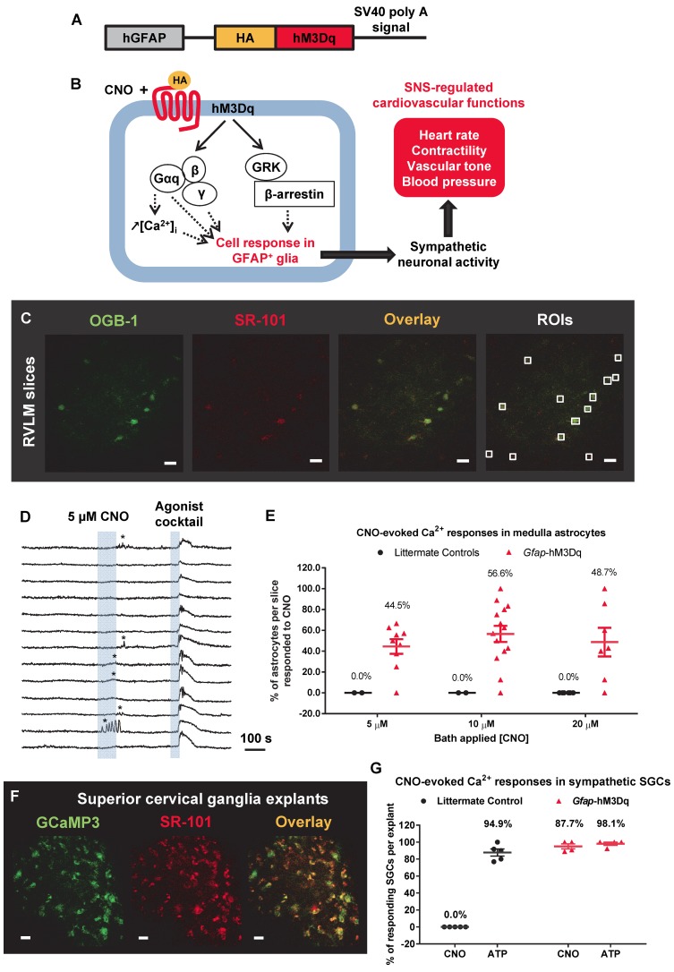
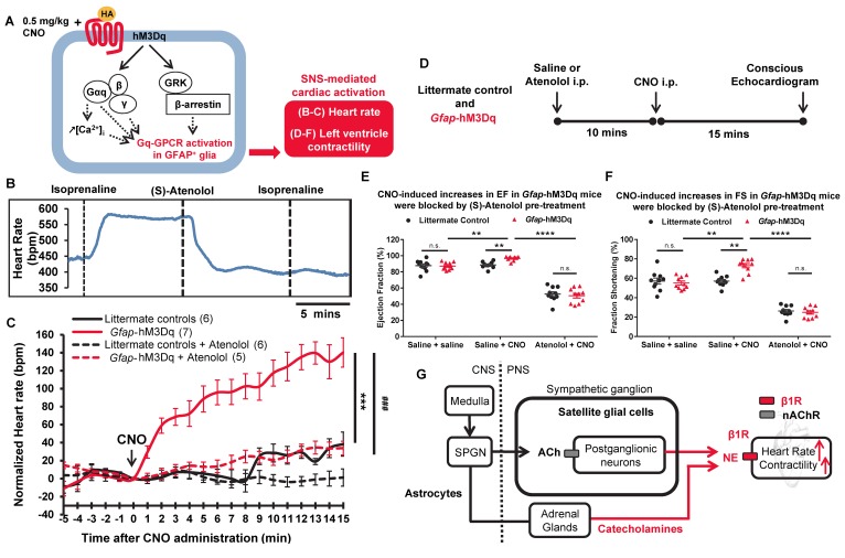
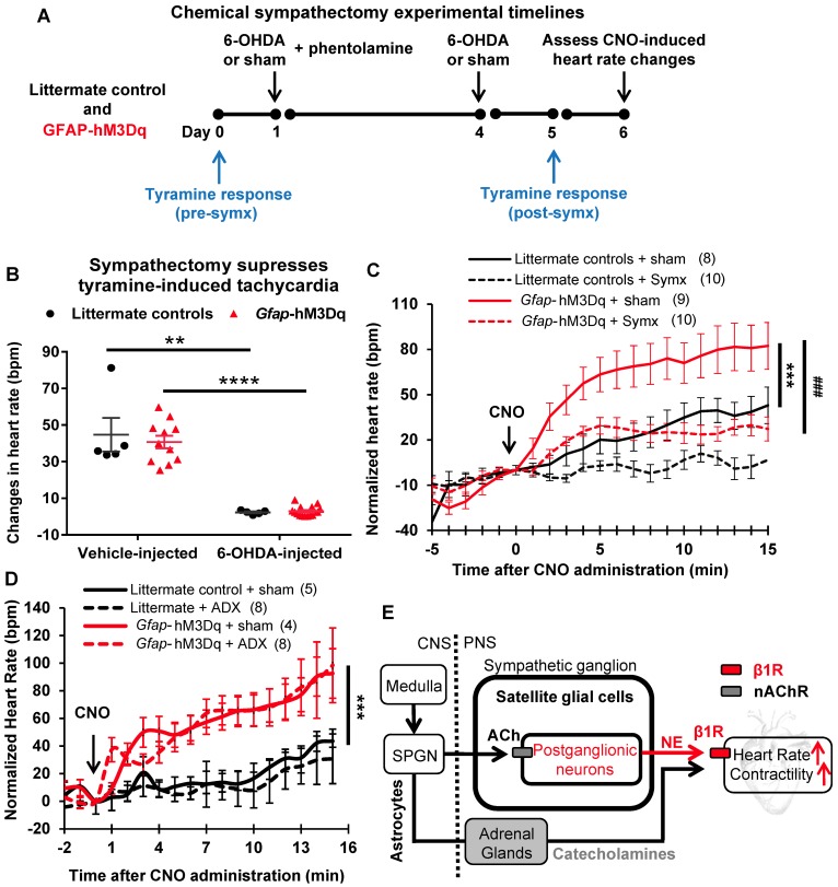
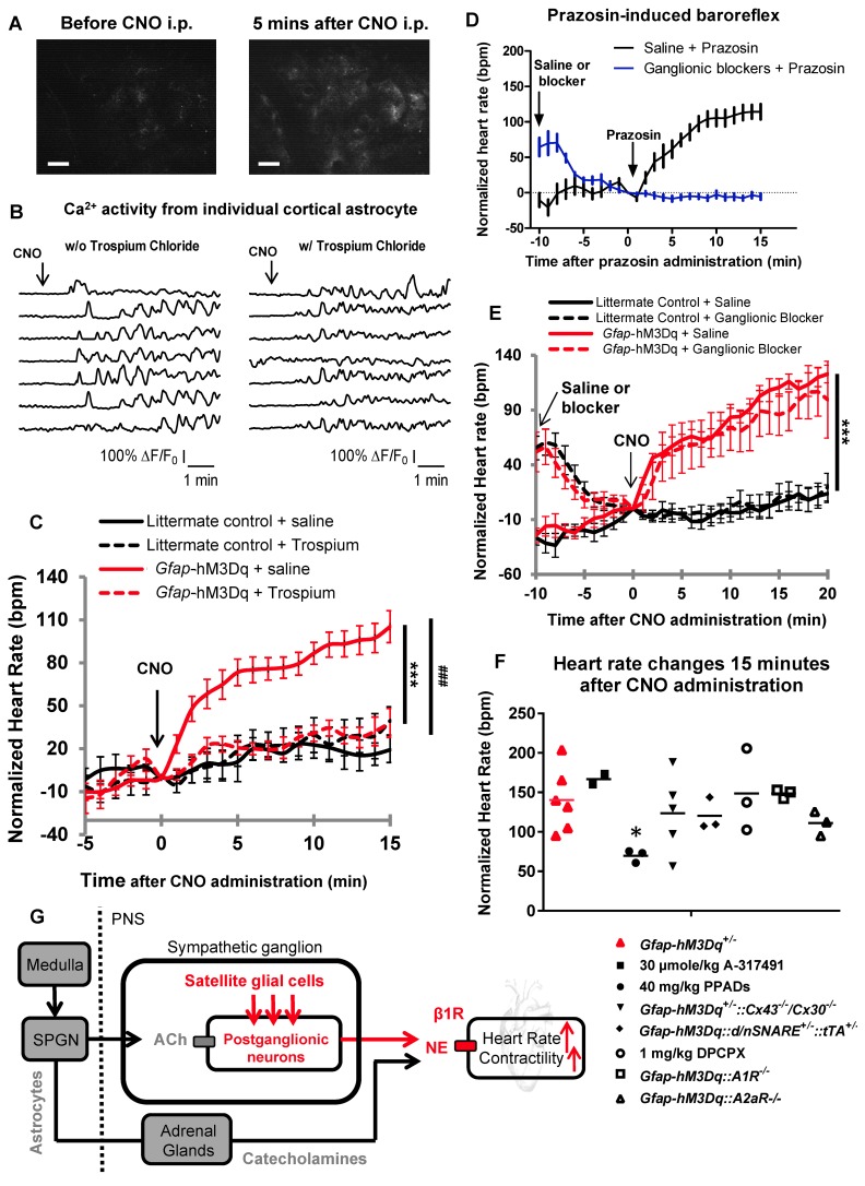
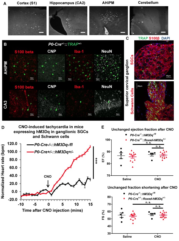
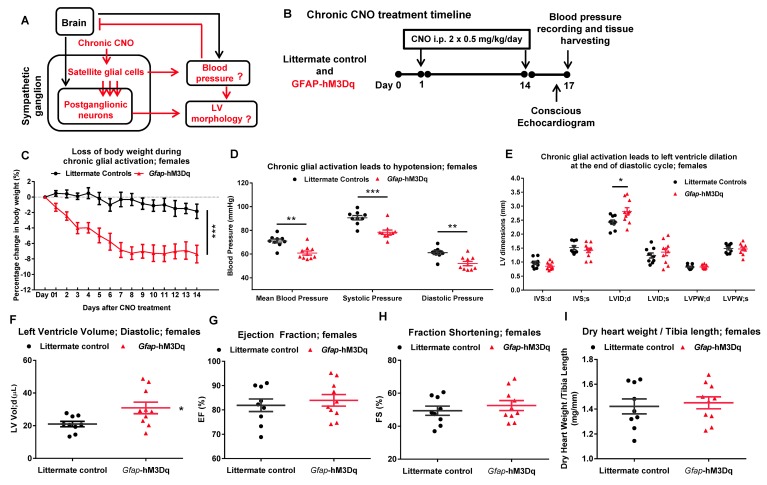
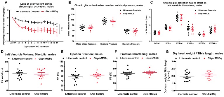
The sympathetic nervous system (SNS) accelerates heart rate, increases cardiac contractility, and constricts resistance vessels. The activity of SNS efferent nerves is generated by a complex neural network containing neurons and glia. Gq G protein-coupled receptor (Gq-GPCR) signaling in glial fibrillary acidic protein-expressing (GFAP+) glia in the central nervous system supports neuronal function and regulates neuronal activity. It is unclear how Gq-GPCR signaling in GFAP+ glia affects the activity of sympathetic neurons or contributes to SNS-regulated cardiovascular functions. In this study, we investigated whether Gq-GPCR activation in GFAP+ glia modulates the regulatory effect of the SNS on the heart; transgenic mice expressing Gq-coupled DREADD (designer receptors exclusively activated by designer drugs) (hM3Dq) selectively in GFAP+ glia were used to address this question in vivo. We found that acute Gq-GPCR activation in peripheral GFAP+ glia significantly accelerated heart rate and increased left ventricle contraction. Pharmacological experiments suggest that the glial-induced cardiac changes were due to Gq-GPCR activation in satellite glial cells within the sympathetic ganglion; this activation led to increased norepinephrine (NE) release and beta-1 adrenergic receptor activation within the heart. Chronic glial Gq-GPCR activation led to hypotension in female Gfap-hM3Dq mice. This study provides direct evidence that Gq-GPCR activation in peripheral GFAP+ glia regulates cardiovascular functions in vivo.


Conflict of interest statement

The authors have declared that no conflict of interest exists.

Figures

Figure 1
Figure 1. Selective and functional expression of hM3Dq by GFAP+ glia in Gfap-hM3Dq mice.
(A) Schematic of transgene containing hemagglutinin-tagged (HA-tagged) hM3Dq driven by the fragment of human glial fibrillary acidic protein promoter (hGFAP). (B) Schematic of the hypothesis that pharmacogenetic activation of Gq-GPCR signaling pathways in GFAP+ glia leads to changes in sympathetic neuronal activity and changes in cardiovascular functions in vivo. GRK, GPCR kinase. (C) OGB-1 loading in SR-101–labeled medulla astrocytes in acute medulla slices (original magnification, 60×; scale bars: 20 μm). RVLM, rostral ventrolateral medulla. (D) Representative data of bath-applied CNO–induced intracellular Ca2+ elevations in a subset of medulla astrocytes in acute brainstem slices from Gfap-hM3Dq mice (9 experiments conducted). (E) Percentage of medulla astrocytes that responded to CNO with intracellular Ca2+ elevations in Gfap-hM3Dq mice and littermate control mice. (F) Confocal image showing GCaMP3 signal from SR-101–labeled GFAP+ SGCs in superior cervical ganglia explant culture isolated from Gfap-GCaMP3+/–::Gfap-hM3Dq+/– mice (original magnification, 60×; scale bars: 20 μm). (G) Quantification of percentage of superior cervical ganglionic SGCs that exhibited intracellular Ca2+ elevations in response to CNO and ATP (10 μM each) from both Gfap-GCaMP3+/–::Gfap-hM3Dq+/– mice and Gfap-GCaMP3+/– littermate control mice (Gfap-GCaMP3+/–::Gfap-hM3Dq+/–: 48 cells/4 explants/2 mice; Gfap-GCaMP3+/– littermate controls: 57 cells/5 explants/2 mice).
Figure 2
Figure 2. Acute activation of hM3Dq in GFAP+ glia led to robust, beta adrenergic receptor–mediated increases in heart rate and left ventricular contractility in Gfap-hM3Dq mice.
(A) Schematic of in vivo model: CNO-induced hM3Dq activation in GFAP+ glia leads to changes in SNS-driven cardiovascular functions in Gfap-hM3Dq mice. (B) Heart rate recordings showed that 10 mg/kg (S)-atenolol blocked isoprenaline-induced increases in heart rate in C56BL/6J mice in vivo (6 recordings/2 mice). (C) CNO-induced significant increases in heart rate in Gfap-hM3Dq mice over 15 minutes compared with littermate controls (n = 5–7 mice in each group; 2-way ANOVA, ***P < 0.0001, time and genotype interaction between Gfap-hM3Dq and littermate controls), which were blocked by (S)-atenolol in vivo (2-way ANOVA, ###P < 0.0001, time and treatment interaction between two Gfap-hM3Dq groups). Saline or atenolol were injected 10 minutes before CNO. (D) CNO-induced increases in left ventricle functions 15 minutes after CNO injections. Saline or atenolol was injected 10 minutes before CNO. (E) Significant increases in ejection fraction (EF) and (F) fraction shortening (FS) after CNO administration in Gfap-hM3Dq mice (unpaired t test; **P < 0.01 between littermate control and Gfap-hM3Dq mice, also between two Gfap-hM3Dq groups; n = 10 for each genotype), which were blocked by (S)-atenolol pretreatment (unpaired t test; ****P < 0.00001 between the two Gfap-hM3Dq groups; n = 10 for each genotype). (G) Schematic model for potential mechanisms (red lines) underlying CNO-induced cardiac changes in vivo in Gfap-hM3Dq mice. nAChR, nicotinic acetylcholine receptors; SPGN, spinal preganglionic neurons.
Figure 3
Figure 3. CNO-induced tachycardia is due to neural released NE, not adrenally released catecholamines.
(A) Experimental timeline of 6-OHDA–induced chemical sympathectomy (symx). (B) Peripheral chemical sympathectomy largely suppressed tyramine-induced tachycardia in both littermate control mice (**P = 0.0017) and Gfap-hM3Dq mice (****P < 0.0001), indicating successful sympathectomy (unpaired t test; n = 5–16 for each group). (C) Peripheral chemical sympathectomy blocked CNO-induced tachycardia (n = 8–10 for each group; 2-way ANOVA, ***P = 0.0003, time and genotype interaction between vehicle-injected Gfap-hM3Dq mice and littermate controls; ###P < 0.0001, time and treatment interaction between vehicle- and 6-OHDA–injected Gfap-hM3Dq mice). (D) Bilateral adrenalectomy did not block CNO-induced changes in heart rate (n = 7–10 for each group; 2-way ANOVA, ***P < 0.0001, time and genotype interaction between vehicle-injected Gfap-hM3Dq mice and littermate controls). (E) Schematic model for CNO-induced, SNS-regulated cardiac changes in vivo in Gfap-hM3Dq mice. Red highlights the mechanism underlying CNO-induced changes in heart rate and left ventricle functions.
Figure 4
Figure 4. CNO-induced changes in cardiac functions are due to hM3Dq activation in peripheral glial cells, likely ganglionic SGCs.
(A) AAV8-GFAP-GCaMP6–expressing astrocytes in visual cortex visualized through chronic polished window using 2-photon imaging before (left) and after (right) CNO i.p. injection in Gfap-hM3Dq mice (original magnification, 60×; scale bars: 40 μm). (B) Intracellular Ca2+ activity in cortical astrocytes in response to CNO i.p. administration, with or without i.p. trospium chloride injection (7 cells per condition, 2 repeats). (C) Trospium chloride, when administrated i.p., abolished CNO-induced increases in tachycardia (2-way ANOVA, n = 8–11 for each group; ***P < 0.0001, interaction between saline-treated Gfap-hM3Dq mice and littermate controls; ###P < 0.0001, interaction between saline- and trospium-treated Gfap-hM3Dq mice). (D) A cocktail of ganglionic blockers decreased baseline heart rates and blocked prazosin-induced tachycardia in both Gfap-hM3Dq and littermate control mice (n = 8–9 for each group). (E) The cocktail of ganglionic blockers did not block CNO-induced tachycardia in Gfap-hM3Dq mice (2-way ANOVA, ***P < 0.0001, interaction between blocker-treated Gfap-hM3Dq mice and littermate controls, n = 8–10 for each group). (F) CNO administration (0.5 mg/kg, s.c.) led to increases in heart rate in Gfap-hM3Dq+/–::Cx43–/–/Cx30–/– mice (n = 6), Gfap-hM3Dq+/–::d/nSNARE+/– mice (n = 3), Gfap-hM3Dq+/–::A1R–/– mice (n = 3), and Gfap-hM3Dq+/–::A2aR–/– mice (n = 3). Perturbing purinergic signaling with selective antagonists failed to block CNO-induced tachycardia in Gfap-hM3Dq mice; antagonists included P2X3 and P2X2/3 receptor antagonist (A-317491; n = 3) and adenosine A1 receptor antagonist (DPCPX; n = 3). Pre-block using nonselective P2 purinergic antagonist reduced but did not abolish CNO-induced tachycardia (PPADs; Mann-Whitney U test, *P < 0.01, n = 3). (G) Schematic model for CNO-induced, SNS-regulated cardiac changes in vivo in Gfap-hM3Dq mice.
Figure 5
Figure 5. CNO administration increases heart rate in P0-Cre+/–::hM3Dq+/– mice.
(A) Cre recombinase activity can be detected in a very small subset of neuronal-like cells in the adult brain of P0-Cre+/–::TRAP+/– mice (original magnification, 20×; scale bars: 100 μm). (B) Cre recombinase activity detected in AHiPM and hippocampus is restricted to neurons in the brain, and not detected in astrocytes, oligodendrocytes, or microglia. AHiPM, posteromedial hippocampal amygdala. (C) A small subset of SGCs in the sympathetic ganglia also exhibit Cre recombinase activity in P0-Cre mice. Scale bars: 40 μm. (D) CNO administration (0.5 mg/kg, i.p.) led to increases in heart rate in P0-Cre+/–::hM3Dq+/– mice but not in littermate control floxed-hM3Dq mice (2-way ANOVA, ***P < 0.0001, P0-Cre–/–::hM3Dq–/fl, n = 11; P0-Cre+/–::hM3Dq+/–, n = 15). (E) CNO administration (0.5 mg/kg, i.p.) did not result in changes in left ventricular contractility in P0-Cre+/–::hM3Dq+/– mice (unpaired t test, P0-Cre–/–::hM3Dq–/fl, n = 5; P0-Cre+/-::hM3Dq+/–, n = 11).
Figure 6
Figure 6. Chronic glial activation leads to hypotension and left ventricle dilation in female Gfap-hM3Dq mice.
Littermate controls, n = 9; Gfap-hM3Dq, n = 10 throughout the figure. (A) Schematic model for chronic activation of ganglionic SGCs and potential cardiovascular outcomes. (B) Chronic CNO treatment experiment timeline. (C) Chronic CNO-induced glial activation led to a significant decrease in body weight in female Gfap-hM3Dq mice but not female littermate controls at the end of the treatment period (2-way ANOVA, ***P < 0.0001, time and genotype interaction). (D) Chronic CNO-induced glial activation led to hypotension in female Gfap-h3Dq animals relative to littermate controls (Mann-Whitney U test, **P < 0.01, ***P < 0.001). (E) Echocardiogram recordings showed that chronic CNO treatment led to increased left ventricle diameter at the end of the diastolic cycle (left ventricular internal diameter end diastole [LVID;d]) in female Gfap-hM3Dq mice compared with female littermate controls (Mann-Whitney U test, P < 0.05). IVS;d and IVS;s, interventricular septal end diastole and end systole. LVPW;d and LVPW;s, left ventricular posterior wall end diastole and end systole. (F) The volume of the left ventricle at the end of diastolic cycle was significantly increased in female Gfap-hM3Dq mice after 2 weeks of CNO treatment (Mann-Whitney U test, *P < 0.05). (G and H) Chronic CNO treatment did not result in changes in ejection fraction (G) or fraction shortening (H) in female Gfap-hM3Dq animals compared with littermate controls (Mann-Whitney U test). (I) The weight of the hearts of female Gfap-hM3Dq animals did not change after chronic CNO treatment (Mann-Whitney U test).
Figure 7
Figure 7. Chronic CNO treatment in male Gfap-hM3Dq mice did not lead to changes in cardiovascular phenotypes as observed in CNO-treated female Gfap-hM3Dq mice.
Littermate controls, n = 14; Gfap-hM3Dq, n = 11 throughout the figure. (A) Chronic CNO treatment led to a decrease in the body weight of male Gfap-hM3Dq mice (2-way ANOVA, ***P < 0.0001). (B) In contrast to what was observed in female Gfap-hM3Dq animals, chronic CNO treatment did not induce hypotension in male Gfap-hM3Dq mice (Mann-Whitney U test). (C and D) In contrast to what was observed in female Gfap-hM3Dq mice, chronic CNO treatment did not induce changes in left ventricle diameter (C) or volume (D) at the end of the diastolic cycle (Mann-Whitney U test). (E and F) Left ventricle functions did not change after chronic CNO treatment in male Gfap-hM3Dq mice compared with their littermate controls (Mann-Whitney U test). (G) Chronic CNO treatment in male Gfap-hM3Dq mice did not lead to changes in heart weight when normalized to tibia length (Mann-Whitney U test).

References

    1. Thomas GD. Neural control of the circulation. Adv Physiol Educ. 2011;35(1):28–32. doi: 10.1152/advan.00114.2010. - DOI - PubMed
    1. Guyenet PG. The sympathetic control of blood pressure. Nat Rev Neurosci. 2006;7(5):335–346. - PubMed
    1. Kaye DM, Esler MD. Autonomic control of the aging heart. Neuromolecular Med. 2008;10(3):179–186. doi: 10.1007/s12017-008-8034-1. - DOI - PubMed
    1. Malpas SC. Sympathetic nervous system overactivity and its role in the development of cardiovascular disease. Physiol Rev. 2010;90(2):513–557. doi: 10.1152/physrev.00007.2009. - DOI - PubMed
    1. Schlaich MP, et al. Sympathetic augmentation in hypertension: role of nerve firing, norepinephrine reuptake, and angiotensin neuromodulation. Hypertension. 2004;43(2):169–175. doi: 10.1161/01.HYP.0000103160.35395.9E. - DOI - PubMed

Publication types

MeSH terms

Substances