Case Reports
. 2017 May;139(5):1629-1640.e2.
doi: 10.1016/j.jaci.2016.11.022. Epub 2017 Jan 27.

Ruxolitinib reverses dysregulated T helper cell responses and controls autoimmunity caused by a novel signal transducer and activator of transcription 1 (STAT1) gain-of-function mutation

Affiliations
Case Reports

Ruxolitinib reverses dysregulated T helper cell responses and controls autoimmunity caused by a novel signal transducer and activator of transcription 1 (STAT1) gain-of-function mutation

Katja G Weinacht et al. J Allergy Clin Immunol. 2017 May.

Abstract

Background: Gain-of-function (GOF) mutations in the human signal transducer and activator of transcription 1 (STAT1) manifest in immunodeficiency and autoimmunity with impaired TH17 cell differentiation and exaggerated responsiveness to type I and II interferons. Allogeneic bone marrow transplantation has been attempted in severely affected patients, but outcomes have been poor.

Objective: We sought to define the effect of increased STAT1 activity on T helper cell polarization and to investigate the therapeutic potential of ruxolitinib in treating autoimmunity secondary to STAT1 GOF mutations.

Methods: We used in vitro polarization assays, as well as phenotypic and functional analysis of STAT1-mutated patient cells.

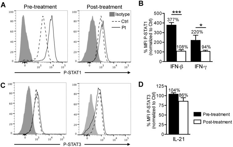
Results: We report a child with a novel mutation in the linker domain of STAT1 who had life-threatening autoimmune cytopenias and chronic mucocutaneous candidiasis. Naive lymphocytes from the affected patient displayed increased TH1 and follicular T helper cell and suppressed TH17 cell responses. The mutation augmented cytokine-induced STAT1 phosphorylation without affecting dephosphorylation kinetics. Treatment with the Janus kinase 1/2 inhibitor ruxolitinib reduced hyperresponsiveness to type I and II interferons, normalized TH1 and follicular T helper cell responses, improved TH17 differentiation, cured mucocutaneous candidiasis, and maintained remission of immune-mediated cytopenias.

Conclusions: Autoimmunity and infection caused by STAT1 GOF mutations are the result of dysregulated T helper cell responses. Janus kinase inhibitor therapy could represent an effective targeted treatment for long-term disease control in severely affected patients for whom hematopoietic stem cell transplantation is not available.

Keywords: IFN-γ; STAT1 gain of function; T helper cell polarization; T(H)1 cell; T(H)17 cell; autoimmunity; follicular T helper cell; ruxolitinib.


Figures

Figure 1
Figure 1. Hemoglobin and platelet count in response to pharmacotherapy
Graph depicts serum hemoglobin concentration in g/dL (gray curve) and platelet count in 103 cells/μL (black curve) in response to changes in steroid dose, as well as pharmacotherapy with anti-thymocyte globulin (ATG), the IL-1 receptor antagonist anakinra, cyclosporine A and ruxolitinib. Timing of packed red blood cell (PRBC) transfusions is indicated in dark gray. The prednisone dose is expressed in mg/kg body weight.
Figure 2
Figure 2. STAT1 E545K mutation leads to hyperphosphorylation gain of function
(A) Sanger sequencing revealed a monoallelic 1633G>A substitution in STAT1. (B) Structural alignment between STAT1 structure and modeled E545K mutant structure (white). WT domains are colored (SH2 in green, dimerization domain in cyan, and IFN-γ peptide in orange) to highlight their position relative to residue 545 (WT structure in yellow and mutant in magenta). (C) Total STAT1 expression in CD4+ T cells by flow cytometry in pt 1 and control. (D) Phospho-STAT1 expression in CD4+ T cells stimulated with IFN-β (20 ng/mL) and IFN-γ (20 ng/mL) in patient and control (top) and dose response curve with increasing interferon concentrations (bottom). (E) Dephosphorylation kinetics of phospho-STAT1 in response to deprivation of IFN-β and IFN-γ in CD4+ T cells represented as absolute MFI (top) and normalized to maximum expression prior to deprivation (bottom). *** p<0.001 by two-way ANOVA.
Figure 3
Figure 3
STAT1E545K GOF mutation results in exacerbated TH1/TC1 and TFH responses and impaired TH17/Tc17 immunity. (A) Representative flow cytometric analyses of IL-17 and IFN-γ secretion by peripheral CD4+ and CD8+ T cells from STAT1E545K patient and controls. (B) Dot plots represent frequencies of IFN-γ and IL-17 producing CD4+ and CD8+ T cells in each group. N=8-9 individual controls and the STAT1E545K patient at three different time points. (C) CXCR5 and PD1 expression in CD4+ T cells from patient 1 (STAT1E545K) and patient 2 (STAT1T385M) compared to healthy controls. (D) CXCR3 and CCR6 expression in CD4+CXCR5+PD1+ T cells from patients 1, 2 and healthy controls (gates shown above).
Figure 4
Figure 4. Different Janus kinase inhibitors variably inhibit STAT1 and STAT3 phosphorylation in vitro
(A) Phospho-STAT1 expression upon IFN-β stimulation in CD4+ T cells from control and STAT1E545K patient treated in vitro with 10nM and 100nM concentrations of ruxolitinib (red curve) and tofacitinib (blue curve) or vehicle (DMSO, black curve). Plain grays correspond to unstimulated cells. (B) Phospho-STAT1 mean fluorescence intensity (MFI) expressed as percent of maximum vehicle-treated control CD4+ T cells shown in (A). (C) Phospho-STAT3 expression upon IL-21 stimulation in CD4+ T cells from control and STAT1E545K patient treated in vitro with 10nM and 100nM concentrations of ruxolitinib (red curve) and tofacitinib (blue curve) or vehicle. Plain grays correspond to unstimulated cells. (D) Phospho-STAT3 mean fluorescence intensity (MFI) expressed as percent of maximum vehicle-treated control CD4+ T cells shown in (C). ** p<0.001 and *** p<0.0001 two-way ANOVA with post test analysis.
Figure 5
Figure 5. Janus kinase inhibitor treatment controls STAT1 phosphorylation
(A) Expression of phospho-STAT1 in CD4+ T cells of STAT1E545K patient and control pre-and post in vivo therapy with ruxolitinib after IFN-β stimulation. (B) Histogram represents phospho-STAT1 MFI normalized to expression in control CD4+ T cells after IFN-β or IFN-γ stimulation pre- and post in vivo therapy with ruxolitinib. (C) Expression of phospho-STAT3 in CD4+ T cells of STAT1E545K patient and control pre- and post in vivo therapy with ruxolitinib after IL-21 stimulation. (D) Histogram represents phospho-STAT3 MFI normalized to expression in control CD4+ T cells after IL-21 stimulation pre-and post in vivo therapy with ruxolitinib.
Figure 6
Figure 6. Ruxolitinib treatment normalizes amplified TH1 and strengthens impaired TH17 responses
(A) Expression of IFN-γ and IL-17 in CD4+ T cells of patient pre- and post in vivo therapy with ruxolitinib at target dose after 3-4 months and >12 months, respectively. (B) Histograms represent frequencies of IFN-γ and IL-17 producing CD4+ T cells shown in panel (A). ** p<0.01 and *** p<0.001 by unpaired two-tailed Student's t-test. (C) Expression of IFN-γ and IL-17 in control and STAT1E545K patient naïve CD4+ T cells cultured under TH0, TH1 and TH17 conditions before and after initiation of ruxolitinib treatment. (D) Histograms represent frequency of IFN-γ and IL-17 producing CD4+ T cells under TH0, TH1 and TH17 conditions respectively. The pre-treatment samples were obtained at least 6 months post ATG and rituximab treatment. The post-treatment samples were obtained after at least 12 months of ruxolitinib treatment at target dose. One representative experiment out of 2 is shown. * p<0.05, ** p<0.01 and *** p<0.001 by unpaired two-tailed Student's t-test (B) or two-way ANOVA (D) with post-test analysis.
Figure 7
Figure 7. Ruxolitinib treatment corrects exacerbated TFH response in patient with STAT1E545K mutation
(A) CXCR5 and PD1 expression in CD4+ T cells in STAT1E545K patient pre- and post-treatment with ruxolitinib at target dose for 3 months compared to control. (B) Histograms represent frequencies of TFH cells in STAT1E545K patient pre-and post-treatment compared to control. (C) CXCR5 and ICOS expression in in vitro-differentiated TFH-like cells from patient and control stimulated with TGF-β1, IL-12 and IL-23 in the presence or absence of ruxolitinib. (D) Histograms represent frequencies of in vitro-differentiated TFH cells with and without ruxolitinib in patient and control. (E) ICOS MFI in in vitro-differentiated TFH cells with and without ruxolitinib in patient and control. * p<0.05, ** p<0.01 and *** p<0.001 by one-way (B, D, E) and two-way (D, E) ANOVA with post-test analysis.

References

    1. Boisson-Dupuis S, Kong XF, Okada S, Cypowyj S, Puel A, Abel L, et al. Inborn errors of human STAT1: allelic heterogeneity governs the diversity of immunological and infectious phenotypes. Curr Opin Immunol. 2012;24:364–78. - PMC - PubMed
    1. Subramaniam PS, Torres BA, Johnson HM. So many ligands, so few transcription factors: a new paradigm for signaling through the STAT transcription factors. Cytokine. 2001;15:175–87. - PubMed
    1. O'Shea JJ, Holland SM, Staudt LM. JAKs and STATs in immunity, immunodeficiency, and cancer. N Engl J Med. 2013;368:161–70. - PMC - PubMed
    1. vdV FL. STAT1 Mutations in Autosomal Dominant Chronic Mucocutaneous Candidiasis. N Engl J Med. 2011:54–61. - PubMed
    1. Hori T, Ohnishi H, Teramoto T, Tsubouchi K, Naiki T, Hirose Y, et al. Autosomal-dominant chronic mucocutaneous candidiasis with STAT1-mutation can be complicated with chronic active hepatitis and hypothyroidism. J Clin Immunol. 2012;32:1213–20. - PubMed

Publication types

MeSH terms