. 2017 Jan 31:7:41862.
doi: 10.1038/srep41862.

Lysosomal dysfunction and autophagy blockade contribute to IMB-6G-induced apoptosis in pancreatic cancer cells

Affiliations

Lysosomal dysfunction and autophagy blockade contribute to IMB-6G-induced apoptosis in pancreatic cancer cells

Lu Liu et al. Sci Rep. .

Abstract

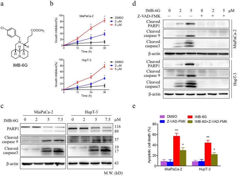
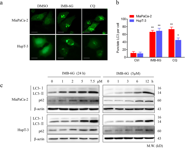
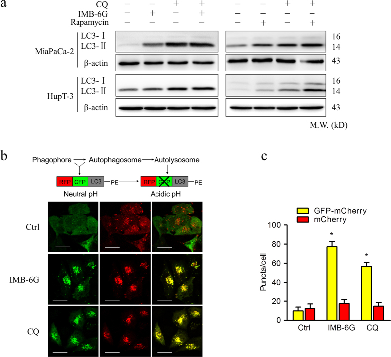
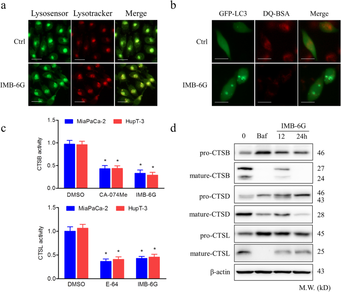
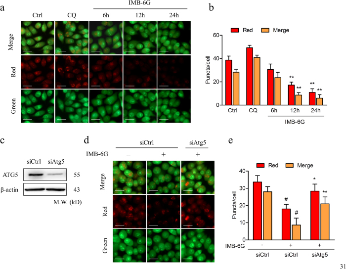
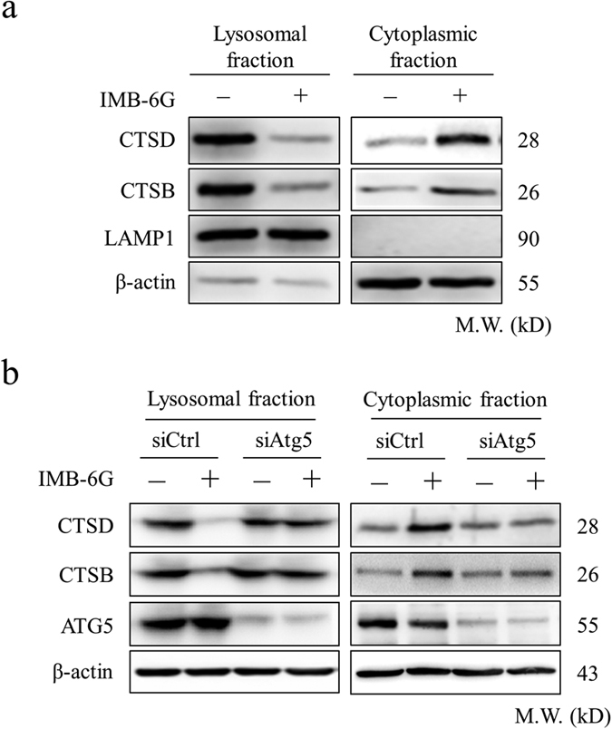
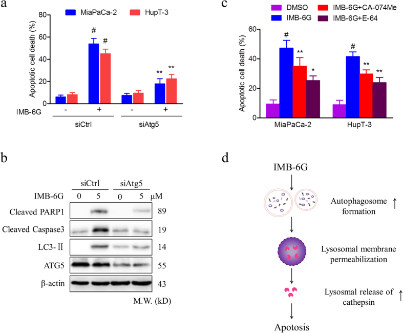
Targeting the autophagic pathway is currently regarded as an attractive strategy for cancer drug discovery. Our previous work showed that IMB-6G is a novel N-substituted sophoridinic acid derivative with potent cytotoxicity against tumor cells, yet the effect of IMB-6G on autophagy and pancreatic cancer cell death remains unknown. Here, we show that IMB-6G inhibits the growth of MiaPaCa-2 and HupT-3 pancreatic cancer cells and induces caspase-mediated apoptosis, which is correlated with an accumulation of autophagic vacuoles. IMB-6G promotes autophagosome accumulation from the early stage of treatment but blocks autophagic flux in the degradation stage, mainly through attenuation of lysosomal cathepsin activity in pancreatic cancer cells. Moreover, IMB-6G triggers lysosomal membrane permeabilization (LMP), followed by cathepsin B/CTSB and cathepsin D/CTSD release from lysosomes into the cytoplasm. Inhibition of autophagosome formation with siRNA against autophagy protein 5 (Atg5) attenuates IMB-6G-induced LMP and apoptosis. Furthermore, cathepsin inhibitors relieve IMB-6G-induced apoptosis as well. Altogether, our findings demonstrate that IMB-6G is a novel autophagy inhibitor, which induces autophagy-dependent apoptosis through autophagosomal-cathepsin axis in pancreatic cancer cells and indicate the potential value of IMB-6G as a novel antitumor drug candidate.


Conflict of interest statement

The authors declare no competing financial interests.

Figures

Figure 1
Figure 1. IMB-6G induces cytotoxicity and apoptosis in pancreatic cancer cells.
(a) Structure of the IMB-6G molecule. (b) MiaPaCa-2 and HupT-3 cells were treated with various concentrations of IMB-6G for the indicated time. Cell viability was measured by MTT assay. (c) MiaPaCa-2 and HupT-3 cells were treated with indicated concentrations of IMB-6G for 24 h. The protein expression levels of cleaved caspase 9, cleaved caspase 3, and poly (ADP-ribose) polymerases (PARP) were detected by immunoblotting. (d) MiaPaCa-2 and HupT-3 cells were treated with IMB-6G in the presence or absence of Z-VAD-FMK (20 μM). Cleaved PARP1, caspase 9 and caspase 3 were detected by immunoblotting. (e) MiaPaCa-2 and HupT-3 cells were stained with Annexin V-FITC and PI after incubated with indicated concentrations of IMB-6G or DMSO in the presence or absence of Z-VAD-FMK for 24 h, the numbers of apoptotic cells were analyzed by flow cytometry. Annexin V-positive cells were accepted as apoptotic cells. The results are presented as mean ± SD and represent three individual experiments. *p < 0.05, **p < 0.01 compared with the untreated control group.
Figure 2
Figure 2. IMB-6G induces autophagosome accumulation in pancreatic cancer cells.
(a) MiaPaCa-2 and HupT-3 cells were transfected with the EGFP-LC3 plasmid. After 24 h, the cells were incubated with IMB-6G (5 μM), CQ (50 μM) or DMSO (Ctrl) for 6 h and visualized with confocal microscopy (upper panel; scale bars, 20 μm). (b) The number of punctate EGFP-LC3 in each cell was counted, and at least 100 cells were included for each group (lower panel). Data were the mean value of three independent experiments with each count of no less than 100 cells.*p < 0.05, **p < 0.01 compared with the untreated control group. (c) MiaPaCa-2 and HupT-3 cells were treated with IMB-6G at the indicated concentrations for 24 h, or treated with IMB-6G (5 μM) for indicated time points, the lipidation of LC3 and the levels of p62/SQSTM1 were detected by immunoblotting using corresponding antibodies.
Figure 3
Figure 3. IMB-6G blocks autophagic flux in the degradation stage.
(a) MiaPaCa-2 and HupT-3 cells were pretreated with CQ (50 μM) or rapamycin (200 nM) for 2 hours, followed by IMB-6G (5 μM) treatment for 12 h. The LC3 turnovers in both cells were detected by immunoblotting. (b) MiaPaCa-2 cells were transfected with mCherry-EGFP-LC3 plasmid, followed by treatment with IMB-6G (5 μM) or CQ (50 μM) for 6 h. Representative fluorescent images are visualized with confocal microscopy (scale bars, 20 μm). (c) Quantification of GFP/mCherry double-positive and mCherry single-positive puncta per cell in control or cells treated with CQ or IMB-6G. Data were the mean value of three independent experiments with each count of no less than 100 cells. Values are expressed as the mean ± SD, *p < 0.05, **p < 0.01 vs. untreated control.
Figure 4
Figure 4. IMB-6G blocks autophagic flux through attenuation of cathepsin activity in pancreatic cancer cells.
(a) MiaPaCa-2 cells were incubated with IMB-6G (5 μM) for 12 h, followed by staining with LysoTracker Red (100 nM) for 30 min, cells were incubated with a LysoSensor Green dye (2 μM) for 10 min. Merged (yellow) images are indicative of an acidic pH (scale bars, 20 μm). (b) MiaPaCa-2 cells were transfected with the EGFP-LC3 plasmid. After 24 h, the cells were treated with IMB-6G (5 μM) for 12 h before being incubated with DQ-Red BSA (10 μg/ml) for 30 min. The cells were fixed and analyzed for fluorescence microscopy (scale bars, 20 μm). (c) Enzymatic activity of CTSB and CTSL in IMB-6G treated MiaPaCa-2 and HupT-3 cells. Cells were treated with DMSO, IMB-6G (5 μM), CA-074Me (2 μM, CTSB inhibitor) or E-64 (10 μM, CTSB and CTSL inhibitor) for 24 h. Enzymatic activity was analyzed using fluorogenic kits. Data are presented as the mean ± SD from 3 independent experiments. (d) MiaPaCa-2 cells were treated with 5 μM IMB-6G for 12 or 24 h, the precursor and the mature form of CTSB, CTSD and CTSL were determined by immunoblotting. Bafilomycin (Baf, 200 nM) was used as a positive control.
Figure 5
Figure 5. IMB-6G induced autophagy-dependent LMP.
(a) MiaPaCa-2 cells were treated with 2 μM IMB-6G for 6 h, 12 h, or 24 h. Lysosomal membrane stability was measured by AO staining under a fluorescence microscopy. CQ (50 μM) was used as a positive control (scale bars, 20 μm). (b) Quantification of red and yellow fluorescence intensity of AO in MiaPaCa-2 cells. Data were the mean value of three independent experiments with each count of no less than 100 cells. Values are expressed as the mean ± SD, **p < 0.01 vs. untreated control. (c–e) MiaPaCa-2 cells were transfected with either 50 nM siRNA against human Atg5 (siAtg5) or control siRNA (siCtrl), and then treated with 5 μM IMB-6G for 12 h. (c) siRNA transfection efficiency was assessed by immunoblotting after 48 h of transfection. β-actin was used as an internal control. (d) Lysosomal membrane stability was measured by AO staining under a fluorescence microscopy (scale bars, 20 μm). (e) The quantification of red and yellow fluorescence intensity of AO was shown. Values are expressed as the mean ± SD, #p < 0.01 compared with IMB-6G-untreated siCtrl group, *p < 0.05, **p < 0.01 compared with IMB-6G-treated siCtrl group.
Figure 6
Figure 6. Inhibition of autophagosome formation alleviated IMB-6G-induced lysosomal release of cathepsin.
(a) MiaPaCa-2 cells were treated with 5 μM IMB-6G for 12 h, cell fragments were performed to separate lysosomal and cytosolic fraction from DMSO- or IMB-6G-treated cells. CTSB and CTSD levels were detected by immunoblotting of the different fractions. Lysosomal-associated membrane protein 1(LAMP1) was as a lysosomal marker, β-actin was a cytosolic marker. (b) MiaPaCa-2 cells were transfected with control siRNA or siAtg5, followed by IMB-6G treatment for 12 h, lysosomal and cytosolic fraction were separated and CTSB, CTSD levels were determined by immunoblotting.
Figure 7
Figure 7. Blunting autophagosome formation and cathepsin activity protects cells from IMB-6G-induced cell death.
MiaPaCa-2 and HupT-3 cells were transfected with control siRNA or siAtg5, followed by IMB-6G (5 μM) treatment for 24 h. (a) Apoptotic cell death was measured by flow cytometry. Values are expressed as the mean ± SD, #p < 0.01 compared with IMB-6G-untreated siCtrl group, **p < 0.01 compared with IMB-6G-treated siCtrl group. (b) Cleaved PARP1/caspase3, LC3-II, and ATG5 were detected by immunoblotting. (c) MiaPaCa-2 and HupT-3 cells were treated with CA-074Me (CTSB specific inhibitor) or E-64 (CTSB and CTSL inhibitor), followed by IMB-6G (5 μM) treatment for 24 h, the apoptotic cell death was measured by flow cytometry. Data are mean ± SD of 3 independent experiments, #p < 0.01 compared with DMSO-treated group, *p < 0.05, **p < 0.01 compared with IMB-6G-treated group. (d) The proposed pathway of IMB-6G-induced autophagy-dependent apoptosis in pancreatic cancer cells. IMB-6G-activated autophagosome formation is an upstream event that may trigger LMP. IMB-6G-induced LMP causes lysosomal release of cathepsin and eventually triggers apoptosis.

Similar articles

Cited by

References

    1. Hidalgo M. Pancreatic cancer. N. Engl. J. Med. 362, 1605–1617, doi: 10.1056/NEJMra0901557 (2010). - DOI - PubMed
    1. Jemal A., Siegel R., Xu J. & Ward E. Cancer statistics, 2010. CA Cancer J. Clin. 60, 277–300, doi: 10.3322/caac.20073 (2010). - DOI - PubMed
    1. Neuzillet C. et al.. State of the art and future directions of pancreatic ductal adenocarcinoma therapy. Pharmacol. Ther. 155, 80–104, doi: 10.1016/j.pharmthera.2015.08.006 (2015). - DOI - PubMed
    1. Gomez V. E., Giovannetti E. & Peters G. J. Unraveling the complexity of autophagy: Potential therapeutic applications in Pancreatic Ductal Adenocarcinoma. Semin. Cancer Biol. 35, 11–19, doi: 10.1016/j.semcancer.2015.09.011 (2015). - DOI - PubMed
    1. Anding A. L. & Baehrecke E. H. Autophagy in Cell Life and Cell Death. Curr. Top. Dev. Biol. 114, 67–91, doi: 10.1016/bs.ctdb.2015.07.012 (2015). - DOI - PubMed

Publication types