. 2017 Jan 31:7:41715.
doi: 10.1038/srep41715.

Histone H4 expression is cooperatively maintained by IKKβ and Akt1 which attenuates cisplatin-induced apoptosis through the DNA-PK/RIP1/IAPs signaling cascade

Affiliations

Histone H4 expression is cooperatively maintained by IKKβ and Akt1 which attenuates cisplatin-induced apoptosis through the DNA-PK/RIP1/IAPs signaling cascade

Ruixue Wang et al. Sci Rep. .

Abstract

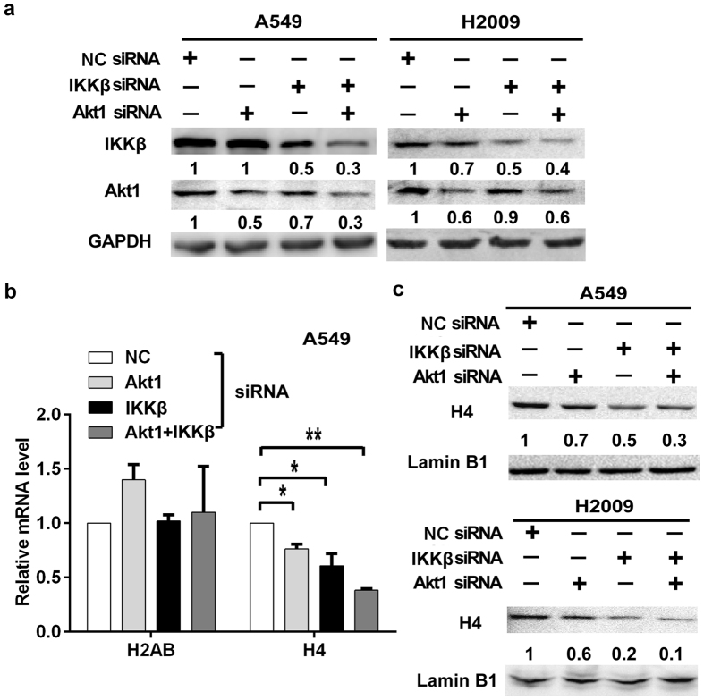
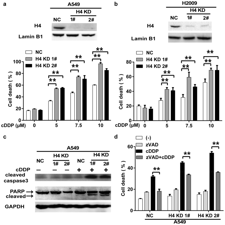
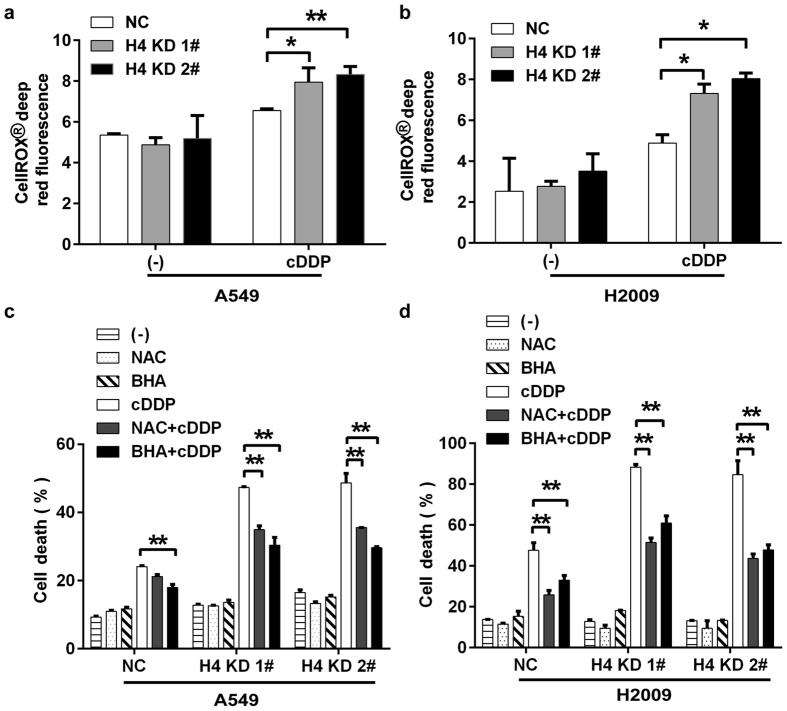
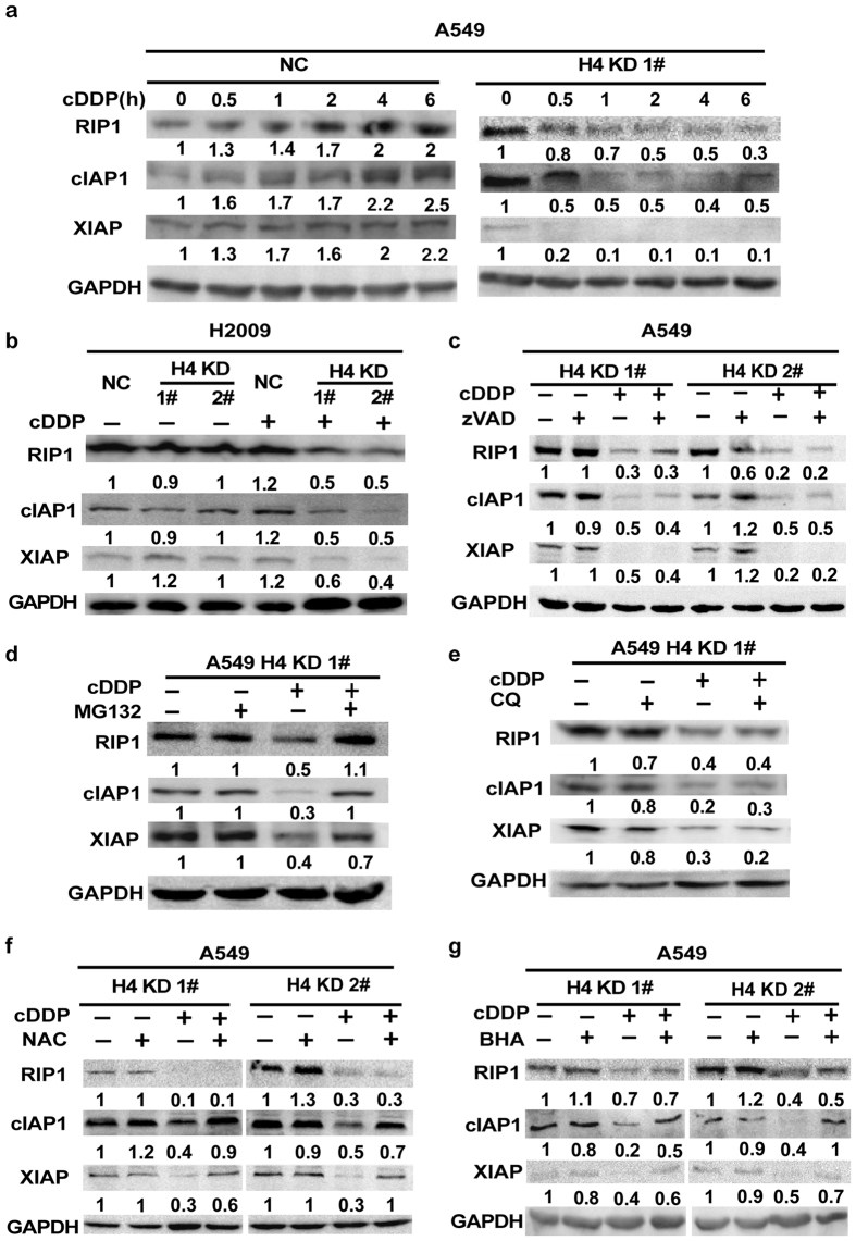
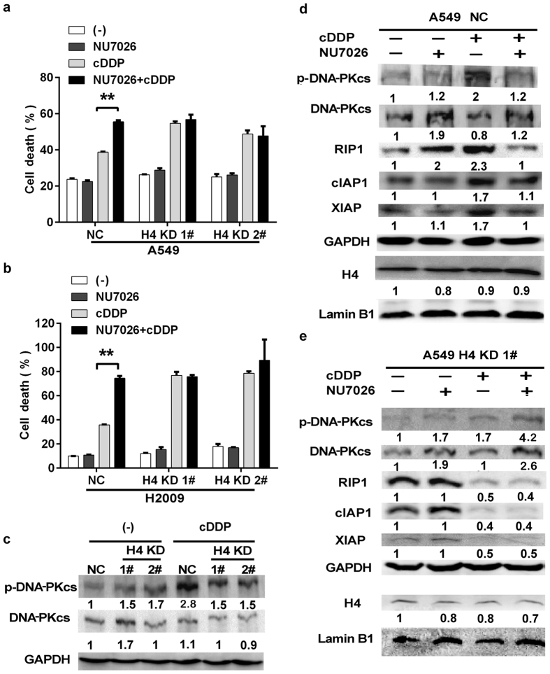
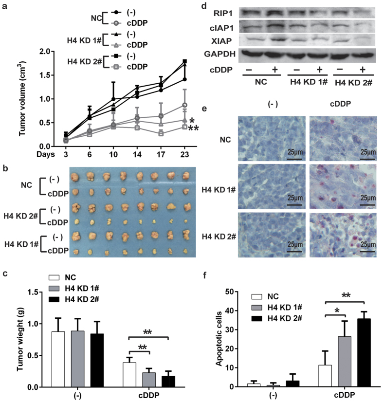
While chromatin remodeling mediated by post-translational modification of histone is extensively studied in carcinogenesis and cancer cell's response to chemotherapy and radiotherapy, little is known about the role of histone expression in chemoresistance. Here we report a novel chemoresistance mechanism involving histone H4 expression. Extended from our previous studies showing that concurrent blockage of the NF-κB and Akt signaling pathways sensitizes lung cancer cells to cisplatin-induced apoptosis, we for the first time found that knockdown of Akt1 and the NF-κB-activating kinase IKKβ cooperatively downregulated histone H4 expression, which increased cisplatin-induced apoptosis in lung cancer cells. The enhanced cisplatin cytotoxicity in histone H4 knockdown cells was associated with proteasomal degradation of RIP1, accumulation of cellular ROS and degradation of IAPs (cIAP1 and XIAP). The cisplatin-induced DNA-PK activation was suppressed in histone H4 knockdown cells, and inhibiting DNA-PK reduced expression of RIP1 and IAPs in cisplatin-treated cells. These results establish a novel mechanism by which NF-κB and Akt contribute to chemoresistance involving a signaling pathway consisting of histone H4, DNA-PK, RIP1 and IAPs that attenuates ROS-mediated apoptosis, and targeting this pathway may improve the anticancer efficacy of platinum-based chemotherapy.


Conflict of interest statement

The authors declare no competing financial interests.

Figures

Figure 1
Figure 1. Concurrent knockdown of Akt1 and IKKβ down-regulates histone H4 expression.
(a) A549 and H2009 cells were transfected with Akt1-, IKKβ-siRNA or both. Negative control (NC) siRNA transfected cells were used as a negative control. The knockdown efficacy of IKKβ and Akt1 was confirmed by Western blotting. GAPDH was used as a loading control. (b) Reduced histone H4 mRNA expression was detected by qRT-PCR. H2AB was detected as a negative control (*p < 0.05; **p < 0.01). (c) The expression of H4 protein was detected by Western blot. Lamin B1 was detected as a loading control.
Figure 2
Figure 2. H4 knockdown sensitizes cancer cells to cisplatin-induced apoptosis.
(a,b) The H4 knockdown cell clones (A549-H4 KD 1# and 2#, and H2009-H4 KD 1# and 2#) were established. H4 protein expression was detected by Western blot. The cells were treated with indicated concentrations of cisplatin. Cell death was measured by LDH releasing assay 72 h after cisplatin treatment. (c) Negative control (NC) A549 cells and its H4 knockdown clones were treated with cisplatin (7.5 μM) for 30 h. Active caspase-3 and PARP were detected by Western blot. GAPDH was detected as a loading control. (d) The cells were pretreated with zVAD-fmk (20 μM) for 1 h or remained untreated, and then treated with cisplatin (7.5 μM) for 72 h. Cell death was measured by LDH releasing assay. Columns, mean of three experiments; bars, SD. **p < 0.01.
Figure 3
Figure 3. Cisplatin-induced cellular ROS accumulation contributes to the increased cytotoxicity in H4 KD cells.
(a,b) The cells were treated with cisplatin (7.5 μM) for 7 h followed by incubating with CellRox dye for 30 min. Cellular fluorescence was measured with the Varioskan Flash Multimode Reader. (c,d) The cells were pre-treated with NAC (1 mM) or BHA (100 μM) for 1 h, followed by cisplatin (7.5 μM) treatment for another 72 h. Cell death was measured by LDH releasing assay. Columns, mean of three experiments; bars, SD. *p < 0.05; **p < 0.01.
Figure 4
Figure 4. Histone H4 suppresses proteasomal degradation of RIP1 and IAPs induced by cisplatin.
(a) The cells were treated with cisplatin (7.5 μM) for indicate times, and the expression of the indicated proteins was detected by Western blot. GAPDH was detected as a loading control. (b) The cells were treated with cisplatin (7.5 μM) for 2 h. The expression of the indicated proteins was detected by Western blot. GAPDH was detected as a loading control. (c–g) The cells were pretreated with zVAD (20 μM), MG132 (10 μM), chloroquine (CQ, 20 μM), NAC (1 mM) or BHA (100 μM) for 1 h. Then the cells were treated with cisplatin (7.5 μM) for another 2 h. The expression of the indicated proteins was detected by Western blot. GAPDH was detected as a loading control.
Figure 5
Figure 5. Suppression of cisplatin-induced DNA-PK activation contributes to RIP1 and IAPs downregulation in H4 knockdown cells.
(a,b) The cells were pre-treated with DNA-PK inhibitor NU7026 (10 μM) for 1 h or left untreated and treated with cisplatin (7.5 μM) treatment for another 72 h. Cell death was measured by LDH releasing assay. Columns, mean of three experiments; bars, SD. **p < 0.01. (c) The cells were treated with cisplatin (7.5 μM) for 30 min. The expression of the indicated proteins was detected by Western blot. GAPDH was detected as a loading control. (d,e) The cells were pre-treated with NU7026 (10 μM) for 1 h or left untreated followed by cisplatin (7.5 μM) treatment for another 2 h. The expression of the indicated proteins was detected by Western blot. GAPDH and Lamin B1 were detected as a loading control.
Figure 6
Figure 6. H4 knockdown enhances cisplatin’s antitumor activity in vivo.
(a) A549-NC, A549-KD 1# or -KD 2#) cells (1 × 106) were inoculated subcutaneously into the flanks of athymic nude mice. When the xenograft tumors were palpable, the mice were randomly divided into two groups receiving injection of cisplatin (i.p.) or PBS. The growth curve with the mean of tumor volume was shown. (b) Excised tumors. (c) Tumor weight. Columns, mean, bars, SD. (d) Expression of RIP1, cIAP1, and XIAP in tumor tissues was detected by Western blot. GAPDH was detected as a loading control. (e) Detection of apoptosis by TUNEL assay. Representative images were shown. (f) Apoptotic cells (TUNEL-positive cells) were counted in five fields (40×) from each group and the average of apoptotic cell numbers per field were shown as mean ± SD. *p < 0.05; **p < 0.01.

Similar articles

Cited by

References

    1. Basu A. & Krishnamurthy S. Cellular responses to Cisplatin-induced DNA damage. J Nucleic Acids 2010 (2010). - PMC - PubMed
    1. Fojo T. Cancer, DNA repair mechanisms, and resistance to chemotherapy. J. Natl. Cancer Inst. 93, 1434–1436 (2001). - PubMed
    1. Lin Y., Bai L., Chen W. & Xu S. The NF-kappaB activation pathways, emerging molecular targets for cancer prevention and therapy. Expert Opin. Ther. Targets 14, 45–55 (2010). - PMC - PubMed
    1. Aggarwal B. B. Nuclear factor-kappaB: the enemy within. Cancer Cell 6, 203–208 (2004). - PubMed
    1. Caporali S. et al.. AKT is activated in an ataxia-telangiectasia and Rad3-related-dependent manner in response to temozolomide and confers protection against drug-induced cell growth inhibition. Mol. Pharmacol. 74, 173–183 (2008). - PubMed

Publication types

MeSH terms