. 2017 Jan 31:7:41646.
doi: 10.1038/srep41646.

Independent movement of the voltage sensors in KV2.1/KV6.4 heterotetramers

Affiliations

Independent movement of the voltage sensors in KV2.1/KV6.4 heterotetramers

Elke Bocksteins et al. Sci Rep. .

Abstract

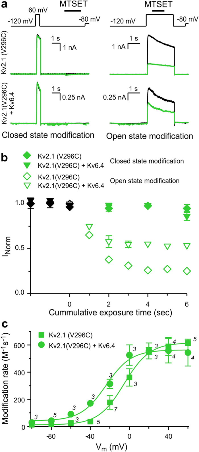
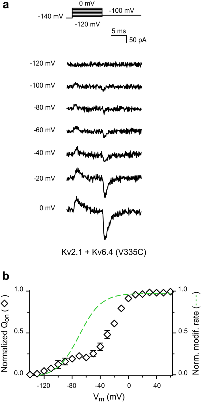
Heterotetramer voltage-gated K+ (KV) channels KV2.1/KV6.4 display a gating charge-voltage (QV) distribution composed by two separate components. We use state dependent chemical accessibility to cysteines substituted in either KV2.1 or KV6.4 to assess the voltage sensor movements of each subunit. By comparing the voltage dependences of chemical modification and gating charge displacement, here we show that each gating charge component corresponds to a specific subunit forming the heterotetramer. The voltage sensors from KV6.4 subunits move at more negative potentials than the voltage sensors belonging to KV2.1 subunits. These results indicate that the voltage sensors from the tetrameric channels move independently. In addition, our data shows that 75% of the total charge is attributed to KV2.1, while 25% to KV6.4. Thus, the most parsimonious model for KV2.1/KV6.4 channels' stoichiometry is 3:1.


Conflict of interest statement

The authors declare no competing financial interests.

Figures

Figure 1
Figure 1. Sequence alignment of the Shaker, KV2.1 and KV6.4 S4 region.
The underlined arginine residues in Shaker represent those that contribute to the gating charge. The bold valine residues were substituted for cysteines in KV2.1 (KV2.1(V296C)) and KV6.4 (KV6.4(V335C)). In red are shown residues that are conserved among the three channel sequences, while blue residues represent those conserved in only two sequences. The remaining ones are shown in black.
Figure 2
Figure 2. MTSET modification of KV2.1 subunits.
(a) Representative current recordings to determine whether KV2.1(V296C) homotetramers and KV2.1(V296C)/KV6.4 heterotetramers are modified by MTSET in the closed state (left) or open state (right). The applied pulse protocols and modification are given on top. (b) Time course of modification. Symbols represent normalized current reductions by MTSET modifications of KV2.1(V296C) (diamond) and KV2.1(V296C)/KV6.4 (triangle down) channels at +60 mV (open symbols) and −120 mV (closed symbols). Black symbols denote normalized values before modification (c) Voltage dependence of the modification rate of KV2.1(V296C) (square) and KV2.1(V296C)/KV6.4 (circle). The solid lines represent sigmoidal fits. The best-fit parameter values for V1/2 for the KV2.1(V296C) and KV2.1(V296C)/KV6.4 data were −5.4 mV and −23.9 mV, respectively. Numbers above symbols represent the number of cells analyzed at each voltage. Data are represented as the mean ± SEM (shown when it is larger than the size of the symbol).
Figure 3
Figure 3. Gating charge displacement in KV2.1(V296C) homotetramers and KV2.1(V296C)/KV6.4 heterotetramers.
(a) Representative gating current recordings of KV2.1(V296C) homotetramers (left) and KV2.1(V296C)/KV6.4 heterotetramers (right) at −120 mV, −100 mV, −80 mV, −60 mV, −40 mV, −20 mV and 0 mV. The applied pulse protocol is given on top. (b) QV distribution of KV2.1(V296C) (square) and KV2.1(V296C)/KV6.4 (circle) channels. The QV distributions were obtained by plotting the area under the recorded ON-gating currents as a function of voltage. For comparison, the normalized MTSET modification curves of KV2.1(V296C) and KV2.1(V296C)/KV6.4 (dashed and solid green line, respectively) are shown. Data are represented as the mean ± SEM (shown when it is larger than the size of the symbol).
Figure 4
Figure 4. MTSET modification of KV6.4 subunits.
(a) Representative current recordings to determine whether KV2.1/KV6.4(V335C) channels are modified by MTSET in the closed state (left) or open state (right). The applied pulse and modification protocols are given on top. (b) Time course of modification. Symbols represent current normalized to the value at time 0. Open symbols depict MTSET modifications at + 60 mV while closed symbols represent modification at −120 mV. Black symbols symbolize normalized values before modification. (c) Voltage dependence of the modification rate of KV2.1/KV6.4(V335C). Solid line represents a sigmoidal fit. The best-fit parameter values for V1/2 and k were −71.3 mV and 16.5, respectively. Numbers above symbols represent the number of cells analyzed at each voltage. Data are represented as the mean ± SEM (shown when it is larger than the size of the symbol).
Figure 5
Figure 5. Gating charge displacement in KV2.1/KV6.4(V335C) channels.
(a) Representative gating current recordings of KV2.1/KV6.4(V335C) channels at −120 mV, −100 mV, −80 mV, −60 mV, −40 mV, −20 mV and 0 mV. The applied pulse protocol is given on top. (b) QV distribution of KV2.1/KV6.4(V335C) channels. The QV distribution was determined as described in Fig. 3b. For comparison, the normalized MTSET modification curve (dashed green line) is shown. Data are represented as the mean ± SEM (shown when it is larger than the size of the symbol).

Similar articles

Cited by

References

    1. Hille B. Ionic channels of excitable membranes. 2 edn (Sinauer Associates, 1991).
    1. Bezanilla F. The voltage sensor in voltage-dependent ion channels. Physiol. Rev. 80, 555–592 (2000). - PubMed
    1. Papazian D. M., Timpe L. C., Jan Y. N. & Jan L. Y. Alteration of voltage-dependence of Shaker potassium channel by mutations in the S4 sequence. Nature 349, 305–310 (1991). - PubMed
    1. Long S. B., Campbell E. B. & MacKinnon R.. Crystal structure of a mammalian voltage-dependent Shaker family K+ channel. Science 309, 897–903 (2005). - PubMed
    1. Seoh S. A., Sigg D., Papazian D. M. & Bezanilla F. Voltage-sensing residues in the S2 and S4 segments of the Shaker K+ channel. Neuron 16, 1159–1167 (1996). - PubMed

Publication types

LinkOut - more resources