. 2017 Jan 31:7:41721.
doi: 10.1038/srep41721.

Roxatidine attenuates mast cell-mediated allergic inflammation via inhibition of NF-κB and p38 MAPK activation

Affiliations

Roxatidine attenuates mast cell-mediated allergic inflammation via inhibition of NF-κB and p38 MAPK activation

Minho Lee et al. Sci Rep. .

Abstract

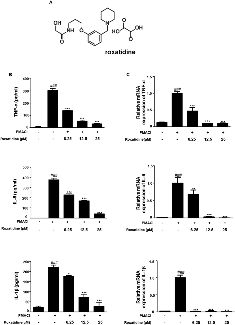
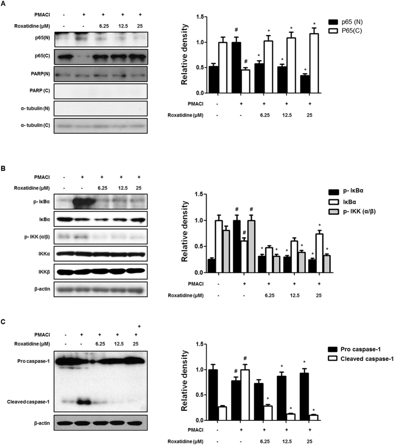
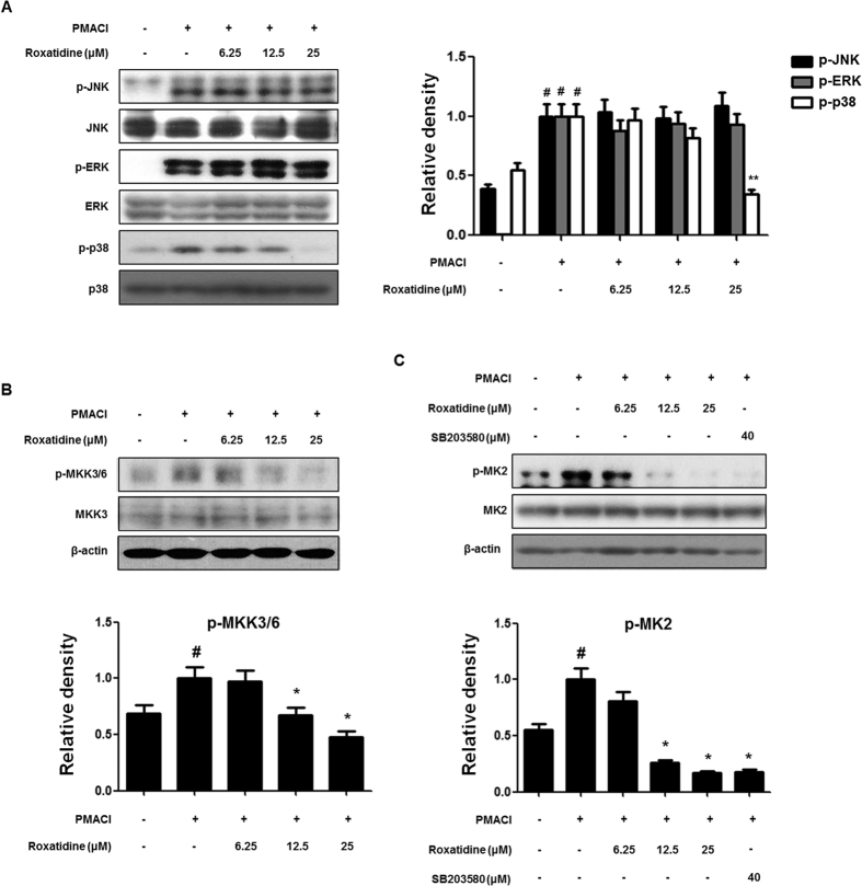
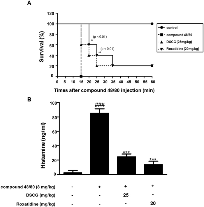
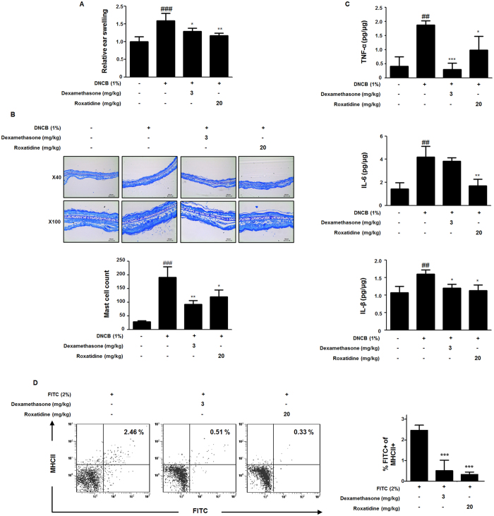
Roxatidine is an active metabolite of roxatidine acetate hydrochloride which is a histamine H2-receptor antagonist that is used to treat gastric and duodenal ulcers. In this study, we investigated the anti-allergic inflammatory effects and the underlying molecular mechanism of roxatidine in phorbol 12-myristate 13-acetate and calcium ionophore (PMACI)-stimulated human mast cells-1 (HMC-1), compound 48/80-induced anaphylactic animal model and chemical allergen-induced contact hypersensitivity (CHS) models. Roxatidine suppressed the mRNA and protein expression of inflammatory cytokines such as TNF-α, IL-6, and IL-1β in PMACI-stimulated HMC-1 and compound 48/80-induced anaphylactic mice. In addition, roxatidine attenuated PMACI-induced nuclear translocation of NF-κB and the phosphorylation of MKK3/6 and MK2, which are both involved in the p38 MAPK pathway. Furthermore, we observed that roxatidine suppressed the activation of caspase-1, an IL-1β converting enzyme, in PMACI-stimulated HMC-1 and compound 48/80-induced anaphylactic mice. In CHS model, roxatidine significantly reduced ear swelling, increased number of mast cells, production levels of cytokines and migration of dendritic cells. Our findings provide evidence that the anti-allergic inflammatory properties of roxatidine are mediated by the inhibition of NF-κB and caspase-1 activation, p38 MAPK pathway and mast cell-derived cytokine production. Taken together, the in vitro and in vivo anti-allergic inflammatory effects suggest a possible therapeutic application of roxatidine in allergic inflammatory diseases.


Conflict of interest statement

The authors declare no competing financial interests.

Figures

Figure 1
Figure 1. Effects of Roxatidine on PMACI-induced production of pro-inflammatory cytokines in HMC-1.
(A) Chemical structure of roxatidine. (B) Cells were treated with 6.25,12.5 and 25 μM of roxatidine for 30 min prior to the addition of PMACI, and the cells were further incubated for 24 h. Cytokine production was measured by ELISA. (C) Cells were pre-treated with roxatidine for 30 min prior to the addition of PMACI for 6 h. The mRNA level of TNF-α, IL 6 and IL-1β was determined by qRT-PCR. Values represent mean ± S.D. of three independent experiments. ###p < 0.001 vs. the control group; *p < 0.05, **p < 0.01, and ***p < 0.001 vs. PMACI-treated group.
Figure 2
Figure 2. Effects of roxatidine on PMACI-induced NF-κB and caspase-1 activation in HMC-1.
(A) Cells were pre-treated with roxatidine for 30 min prior to the addition of PMACI for 30 min. Nuclear (N) and cytosolic (C) extracts were isolated and the levels of p65 in each fraction were determined by western blot analysis. PARP and α- tubulin were used as internal controls. (B and C) Cells were pre-treated with roxatidine for 30 min prior to the addition of PMACI for 15 min. Nuclear and cytosolic extract and total proteins were prepared, and western blot analysis was performed by using specific antibodies. Densitometric analysis was performed using Bio-Rad Quantity One Software. The data shown represent mean ± SD of three independent experiments. #p < 0.05 vs. the control group; *p < 0.05 vs. PMACI-treated group.
Figure 3
Figure 3. Effects of roxatidine on PMACI-induced activation of MAPKs MKK3/6 and MK2 in HMC-1.
(A and B) Cells were pre-treated with roxatidine for 30 min prior to the addition of PMACI for 15 min. (C) Cells were pre-treated with roxatidine or SB203580 (40 μM) for 30 min prior to the addition of PMACI for 15 min. Total proteins were prepared, and western blot analysis was performed using specific antibodies. Densitometric analysis was performed using Bio-Rad Quantity One Software. The data shown represent mean ± SD of three independent experiments. #p < 0.05 vs. the control group; *p < 0.05 and **p < 0.01 vs. PMACI-treated group.
Figure 4
Figure 4. Effects of roxatidine on compound 48/80-induced mortality rate and histamine release in anaphylactic animal models.
Mice were administrated with roxatidine (20 mg/kg, p.o.), DSCG (25 mg/kg, p.o) and PBS as a vehicle (n = 6 per group) for 1 h before compound 48/80 injection (8 mg/kg i.p.). (A) Survival rates of these mice were monitored for 1 h. (B) Histamine concentrations were measured in culture medium by EIA. Densitometric analysis was performed using Bio-Rad Quantity One Software. The data shown represent mean ± SD of three independent experiments. ###p < 0.001 vs. the control group; **p < 0.01 and ***p < 0.001 vs. PMACI-treated group.
Figure 5
Figure 5. Effects of roxatidine on compound 48/80-induced cytokines and caspase-1 activation in anaphylactic animal models.
Mice were administrated with roxatidine (20 mg/kg, p.o.), DSCG (25 mg/kg, p.o) and PBS as a vehicle (n = 6 per group) for 1 h before compound 48/80 injection (8 mg/kg i.p.). (A) Serum level of TNF-α, IL-6 and IL-1β were determined by using EIA kits. (B) Total RNA prepared from the liver tissue and the level of TNF-α, IL-6 and IL-1β were determined by quantitative real-time PCR. (C) Expression of procaspae-1 was determined by western blot analysis using specific antibodies. Densitometric analysis was performed using Bio-Rad Quantity One Software. The data shown represent mean ± SD of three independent experiments. #p < 0.05 and ###p < 0.001 vs. the control group; *p < 0.05 and ***p < 0.001 vs. PMACI-treated group.
Figure 6
Figure 6. Effects of roxatidine on the sensitization phase in CHS.
(A) Mice were administrated with roxatidine (20 mg/kg, p.o.), dexamethasone (3 mg/kg, p.o.) and a vehicle (n = 6 per group) for 1 h before 1% DNCB-induced sensitization. Relative ear swelling was measured as the increases compared to prechallenge ear weight. (B) Sections were stained with toluidine blue to identify mast cells and mast cells were counted with a microscope at a magnification of 40× (scale bar: 250 μm) and 100× (scale bar: 100 μm), respectively. (C) The cytokine levels of TNF-α, IL-6 and IL-1β from the ear tissue were determined by EIA kits. (D) Mice were administrated with roxatidine, dexamethasone and a vehicle (n = 6 per group) for 1 h before 2% FITC-induced sensitization. The number of FITC+ MHC classII+ DCs in the draining LNs of mice 48 h after sensitization of FITC. The data shown represent mean ± SD of three independent experiments. ##p < 0.01 and ###p < 0.001 vs. the control group; *p < 0.05, **p < 0.01 and ***p < 0.001 vs. CHS-induced group.

Similar articles

Cited by

References

    1. Galli S. J., Tsai M. & Piliponsky A. M. The development of allergic inflammation. Nature 454, 445–454 (2008). - PMC - PubMed
    1. Kemp S. F. & Lockey R. F. Anaphylaxis: a review of causes and mechanisms. J Allergy Clin Immunol 110, 341–348 (2002). - PubMed
    1. Kim S. H. et al.. Gallic acid inhibits histamine release and pro-inflammatory cytokine production in mast cells. Toxicological sciences: an official journal of the Society of Toxicology 91, 123–131 (2006). - PubMed
    1. Baud V. & Karin M. Signal transduction by tumor necrosis factor and its relatives. Trends Cell Biol 11, 372–377 (2001). - PubMed
    1. Baeuerle P. A. & Baltimore D. I kappa B: a specific inhibitor of the NF-kappa B transcription factor. Science 242, 540–546 (1988). - PubMed

Publication types

MeSH terms