. 2017 Jan 31;7(1):e1020.
doi: 10.1038/tp.2016.296.

Glucose deficit triggers tau pathology and synaptic dysfunction in a tauopathy mouse model

Affiliations

Glucose deficit triggers tau pathology and synaptic dysfunction in a tauopathy mouse model

E Lauretti et al. Transl Psychiatry. .

Abstract

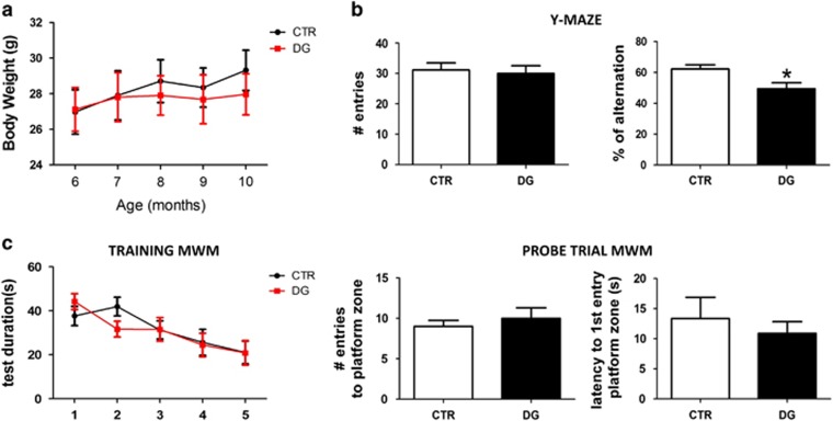
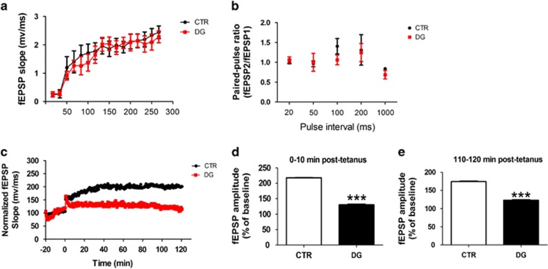
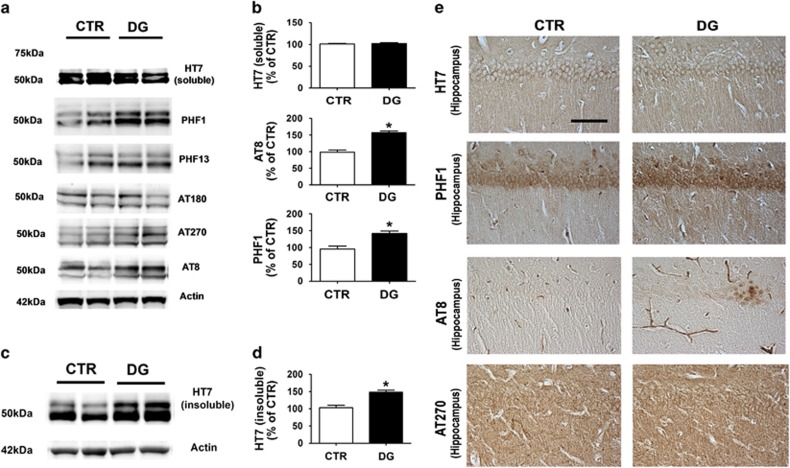
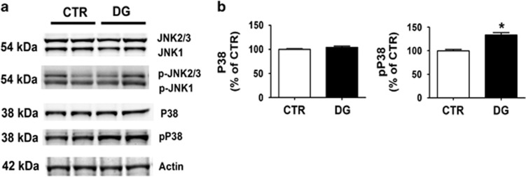
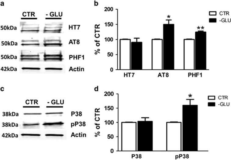
Clinical investigations have highlighted a biological link between reduced brain glucose metabolism and Alzheimer's disease (AD). Previous studies showed that glucose deprivation may influence amyloid beta formation in vivo but no data are available on the effect that this condition might have on tau protein metabolism. In the current paper, we investigated the effect of glucose deficit on tau phosphorylation, memory and learning, and synaptic function in a transgenic mouse model of tauopathy, the h-tau mice. Compared with controls, h-tau mice with brain glucose deficit showed significant memory impairments, reduction of synaptic long-term potentiation, increased tau phosphorylation, which was mediated by the activation of P38 MAPK Kinase pathway. We believe our studies demonstrate for the first time that reduced glucose availability in the central nervous system directly triggers behavioral deficits by promoting the development of tau neuropathology and synaptic dysfunction. Since restoring brain glucose levels and metabolism could afford the opportunity to positively influence the entire AD phenotype, this approach should be considered as a novel and viable therapy for preventing and/or halting the disease progression.


Figures

Figure 1
Figure 1
Glucose deprivation impairs cognition in the h-tau mice. (a) Body weights (g) of 2-deoxyglucose (DG)-treated and control (CTR) mice were recorded at the beginning of the study and each month until the end when the mice were 10 months old. (b) Total number of entries and percentage of alternation in the Y-maze for controls (CTR) and h-tau mice that underwent DG treatment (DG; *P< 0.05). (c) Morris water maze test results for controls (CTR) and h-tau mice undergoing DG treatment (DG). Mice were tested initially in the cue test and then trained for 5 days to reach a platform (training phase). After the last training session, for the probe trials phase mice were put back in the water maze and the number of entries to the target platform zone as well as the distance until the first entry in the target platform zone recorded. Results are mean±s.e.m. (n=9 mice per group).
Figure 2
Figure 2
Glucose deprivation impairs synaptic function in the h-tau mice. (a) Input/output (I/O) curves and representative field excitatory postsynaptic potentials (fEPSPs) at increasing stimulus strengths (0–300 μA) are shown for CTR and 2-deoxyglucose (DG) treated h-tau mice at 10 months of age. (b) Mean fEPSP slopes as a function of interpulse interval between the first and second fEPSPs evoked at CA3–CA1 synapses in slices from the same mice at 20, 50, 100, 200, and 1000 milliseconds in the same animals. (c) fEPSP slopes were recorded for 2 h and expressed as the percentage of pretetanus baseline in the same mice. (d) Long-term potentiation (LTP) magnitudes expressed as the percentages of baseline for 0 to 10 min post-tetanus (***P<0.001). (e) For the same groups of mice, LTP magnitudes expressed as the percentages of baseline for 110 to 120 min post-tetanus (***P<0001). Values represent mean±s.e.m.
Figure 3
Figure 3
Glucose deprivation increases tau phosphorylation. (a) Representative western blot analyses of soluble total tau (HT-7) and phosphorylated tau at residues S396/S404 (PHF-1), S396 (PHF-13), T231/S235 (AT180), T181 (AT270), and S202/T205 (AT8) in brain cortex homogenates of CTR and 2-deoxyglucose (DG)-treated h-tau mice. (b) Densitometric analyses of the immunoreactivities shown in the previous panel (*P<0.05). (c) Representative western blot analyses of sarkosyl-soluble tau (HT7) in brain cortex homogenates from CTR and DG-treated h-tau. (d) Densitometric analyses of the immunoreactivities shown in the previous panel (*P<0.05). (e) Representative images of immunohistochemistry analyses for soluble total tau (HT-7) and phosphorylated tau at residues S396/S404 (PHF-1), S202/T205 (AT8), and T181 (AT270) in brain sections of CTR and DG-treated h-tau mice (Scale bar: 100μm). Results are mean±s.e.m.
Figure 4
Figure 4
Glucose deprivation triggers P38 MAPK kinase activation. (a) Representative western blot analyses of JNK, pJNK, P38 and pP38 in brain cortex homogenates from CTR and 2-deoxyglucose (DG)-treated h-tau mice (*P<0.05). (b) Densitometric analyses of the immunoreactivities shown in the previous panel (*P<0.05). Results are mean±s.e.m.
Figure 5
Figure 5
Glucose deprivation induces synaptic pathology in h-tau mice. (a) Representative western blot analyses of synaptophysin (SYP), post-synaptic protein-95 (PSD-95), and MAP2 in brain cortex homogenates from CTR and DG-treated h-tau mice (*P<0.05). (b) Densitometric analyses of the immunoreactivities shown in the previous panel (*P<0.05; n=9 mice per group). Results are mean±s.e.m. (c) Representative images of immunohistochemistry analyses for SYP and MAP2 in brain sections of CTR and DG-treated h-tau mice (scale bar: 100μm).
Figure 6
Figure 6
Glucose deprivation-dependent neuronal apoptosis is mediated by caspases 12 and 3. (a) Representative western blot analysis for procaspase-12, procaspase-3, procaspase-7, caspase-12, caspase-3, and caspase-7 in brain cortex homogenates from CTR and DG-treated h-tau mice (*P<0.05). (b) Densitometric analyses of the immunoreactivities shown in the previous panel (*P<0.05). Results are mean±s.e.m.
Figure 7
Figure 7
Glucose deprivation modulates tau phosphorylation via P38 MAPK kinase. (a) Representative western blots of total tau (HT7) and phosphorylated tau at residues S202/T205 (AT8) and S396/S404 (PHF-1) in primary cortical neuronal cells from h-tau mice incubated with normal medium (CTR) or glucose-deprived (-GLU) medium. (b) Densitometric analyses of the immunoreactivities to the antibodies shown in the previous panel (*P<0.05, **P<0.01). (c) Representative western blot analyses for P38 and pP38 in primary cortical neurons from h-tau mice incubated with normal medium (CTR) or glucose-deprived (-GLU) medium. (d) Densitometric analyses of the immunoreactivities to the antibodies shown in the previous panel (*P<0.05). Values represent mean±s.e.m.

References

    1. Iqbal K, Liu F, Gong CX. Tau and neurodegenerative disease: the story so far. Nat Rev Neurol 2016; 12: 15–27. - PubMed
    1. Arriagada PV, Growdon JH, Hedley-Whyte ET, Hyman BT. Neurofibrillary tangles but not senile plaques parallel duration and severity of Alzheimer's disease. Neurology 1992; 42: 631–639. - PubMed
    1. Riley KP, Snowdon DA, Markesbery WR. Alzheimer's neurofibrillary pathology and the spectrum of cognitive function: findings from the Nun Study. Ann Neurol 2002; 51: 567–577. - PubMed
    1. Rissman RA. Stress-induced tau phosphorylation: functional neuroplasticity or neuronal vulnerability? J Alzheimer's Dis 2009; 18: 453–457. - PMC - PubMed
    1. De Felice FG, Lourenco MV. Brain metabolic stress and neuroinflammation at the basis of cognitive impairment in Alzheimer's disease. Front Aging Neurosci 2015; 7: 94. - PMC - PubMed

Publication types

MeSH terms

LinkOut - more resources