. 2017 Apr;163(4):488-501.
doi: 10.1099/mic.0.000443. Epub 2017 Apr 26.

Functional amyloids in Streptococcus mutans, their use as targets of biofilm inhibition and initial characterization of SMU_63c

Affiliations

Functional amyloids in Streptococcus mutans, their use as targets of biofilm inhibition and initial characterization of SMU_63c

Richard N Besingi et al. Microbiology (Reading). 2017 Apr.

Abstract

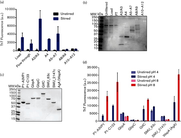
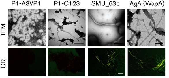
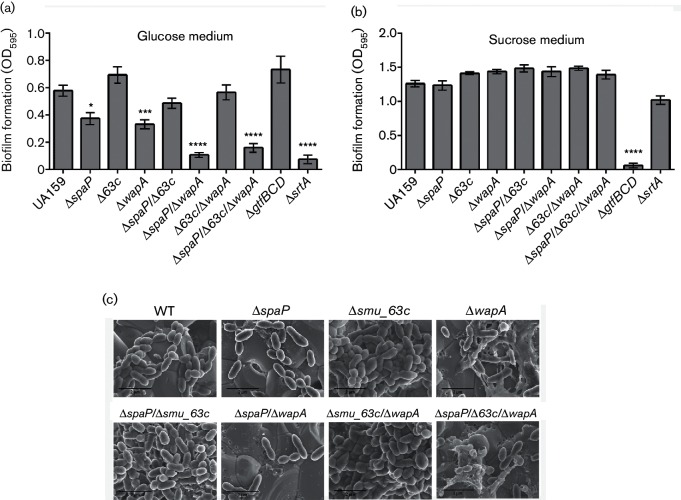
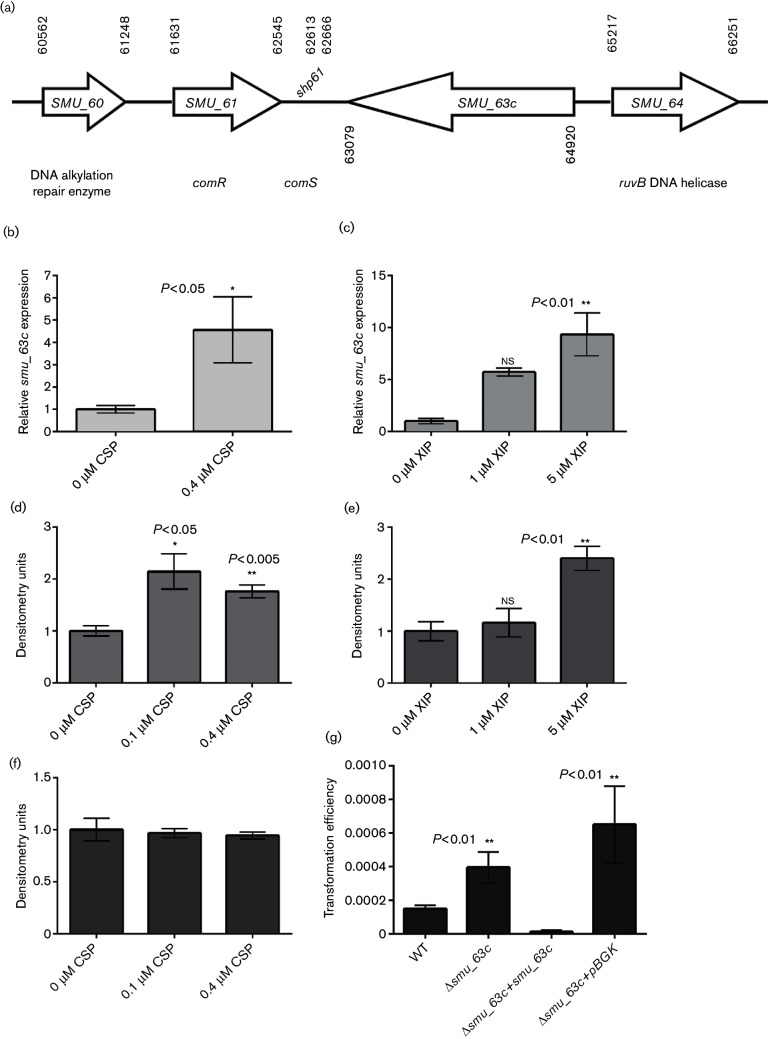
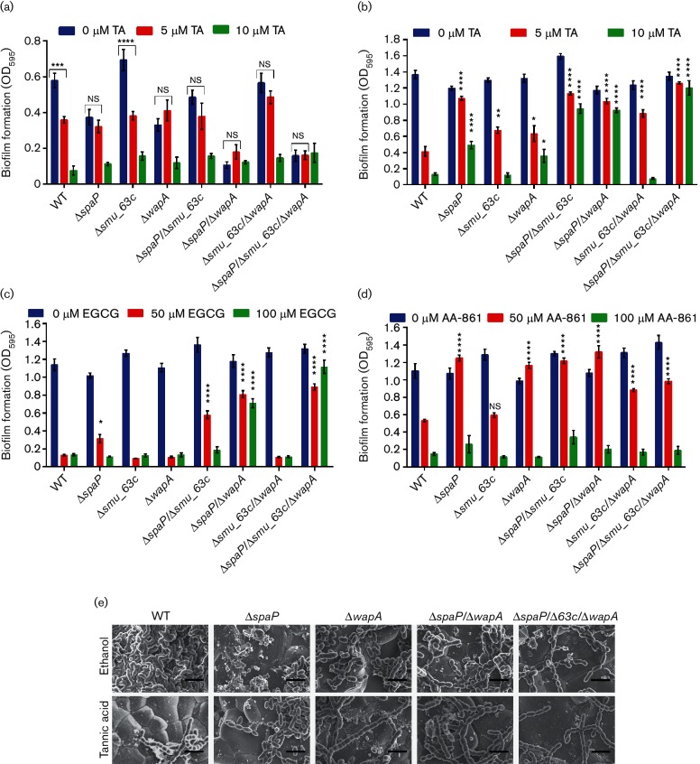
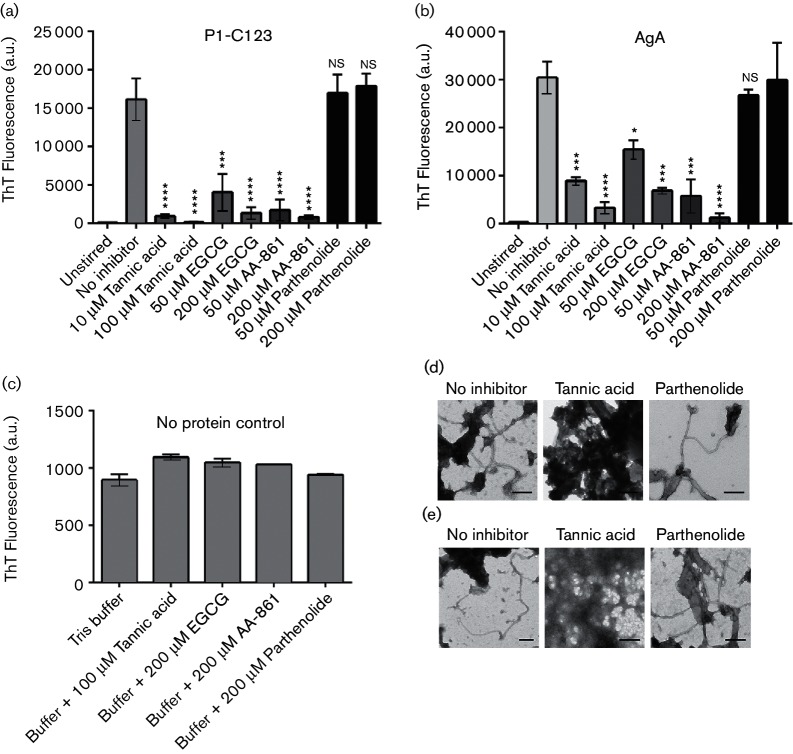
Amyloids have been identified as functional components of the extracellular matrix of bacterial biofilms. Streptococcus mutans is an established aetiologic agent of dental caries and a biofilm dweller. In addition to the previously identified amyloidogenic adhesin P1 (also known as AgI/II, PAc), we show that the naturally occurring antigen A derivative of S. mutans wall-associated protein A (WapA) and the secreted protein SMU_63c can also form amyloid fibrils. P1, WapA and SMU_63c were found to significantly influence biofilm development and architecture, and all three proteins were shown by immunogold electron microscopy to reside within the fibrillar extracellular matrix of the biofilms. We also showed that SMU_63c functions as a negative regulator of biofilm cell density and genetic competence. In addition, the naturally occurring C-terminal cleavage product of P1, C123 (also known as AgII), was shown to represent the amyloidogenic moiety of this protein. Thus, P1 and WapA both represent sortase substrates that are processed to amyloidogenic truncation derivatives. Our current results suggest a novel mechanism by which certain cell surface adhesins are processed and contribute to the amyloidogenic capability of S. mutans. We further demonstrate that the polyphenolic small molecules tannic acid and epigallocatechin-3-gallate, and the benzoquinone derivative AA-861, which all inhibit amyloid fibrillization of C123 and antigen A in vitro, also inhibit S. mutans biofilm formation via P1- and WapA-dependent mechanisms, indicating that these proteins serve as therapeutic targets of anti-amyloid compounds.


Conflict of interest statement

The authors declare that there are no conflicts of interest.

Figures

Fig. 1.
Fig. 1.
Identification and characterization of amyloid-forming proteins. Spent medium (TDM-glucose) from a P1-deficient S. mutans mutant was concentrated and fractionated by ion-exchange chromatography. (a) ThT fluorescence assay showing ThT uptake by unstirred and stirred (aggregated) samples of pooled fractions corresponding to ion exchange peaks, as well as load and flow-through (FT) fractions. (b) SDS-PAGE of unstirred samples shown in (a), as well as the starting material (load), FT fractions and starting material prior to filtration. (c) SDS-PAGE of purified recombinant protein candidates. (d) ThT fluorescence assay of unstirred and stirred recombinant proteins. ThT uptake was measured at pH 4 and pH 8. Error bars represent the mean±sem of four independent replicates.
Fig. 2.
Fig. 2.
CR-induced birefringence and visualization of amyloid fibrils by TEM. Aggregated recombinant proteins (stirred at pH 8 for A3VP1, C123 and AgA, and pH 4 for SMU_63c) were evaluated for the presence of amyloid by CR-induced birefringence (bottom panel) and TEM (top panel). Scale bars represent 50 µm (bottom panel), and 200 nm (top panel). Orange or yellow-green birefringence, and amyloid fibrils, were observed for the P1-C123, SMU_63c and WapA-AgA, but not for the P1-A3VP.
Fig. 3.
Fig. 3.
Evaluation of biofilms produced by S. mutans WT and single-, double- and triple-deletion mutants by CV staining and FE-SEM. (a and b) CV assays of cells grown in BM containing glucose (a) or sucrose (b) as the carbon source. (c) Visualization of biofilms grown on 16 mM glucose and 4 mM sucrose by FE-SEM. Scale bars represent 2 µm. *P<0.05; ***P<0.001; ****P<0.0001.
Fig. 4.
Fig. 4.
Characterization of SMU_63c. (a) Schematic representation of the genetic locus containing smu_63 c. (b and c) Quantitative real-time PCR analysis of smu_63 c expression with and without addition of CSP in THYE medium (b) or XIP in CDM-glucose (c). (d) Western blot densitometry analysis of SMU_63c present in cell surface SDS extracts of S. mutans in THYE, with and without addition of CSP. (e) Western blot densitometry analysis of SMU_63c in concentrated spent medium from CDM-glucose cultures grown with or without XIP. (f) Western blot analysis of WapA in the same samples as (d). (g) Evaluation of the genetic competence of S. mutans WT, ∆smu_63 c mutant, complemented mutant and pBGK integration vector control strain. Error bars represent the mean±sem of three or four independent replicates. Statistical significance was evaluated by Student's t-test or one-way ANOVA.
Fig. 5.
Fig. 5.
Inhibition of biofilm formation by TA, EGCG or AA-861. (a) CV assays of S. mutans WT and deletion-mutant biofilms grown in BM-glucose and in the presence of the indicated concentrations of TA. b–d: CV assays of the same biofilms grown in BM-sucrose in the presence of indicated concentrations of TA (b); EGCG (c); and AA-861 (d). Error bars represent the mean±sem of six independent replicates. Statistical significance was evaluated by two-way ANOVA using Tukey’s multiple comparisons. Asterisks indicate the statistical significance of the comparison of the mean±sem value of each deletion mutant to that of the WT under the same inhibitor concentration: *P<0.05; **P<0.01; ***P<0.001; ****P<0.0001. (e) FE-SEM of S. mutans WT and mutant biofilms grown in BM-glucose and sucrose with 10 µM TA (bottom panel) or the 0.1 % ethanol diluent control (top panel). Scale bars represent 2 µm.
Fig. 6.
Fig. 6.
Anti-biofilm compounds inhibit fibrillization of P1-C123 and WapA-AgA in vitro. (a and b) ThT uptake of C123 (a) or AgA (b) stirred in the absence or presence of the indicated inhibitors. (c) ThT fluorescence in the absence of added proteins. (d and e) Transmission electron micrographs of C123 (d) or AgA (e) stirred without inhibitor, or in the presence of 200 µM TA or 200 µM parthenolide. Scale bars represent 200 nm. Error bars represent mean±sem of three independent replicates. Statistical significance was evaluated by one-way ANOVA using Dunnett’s multiple comparisons. Asterisks indicate statistically significant differences between protein stirred in the presence of inhibitor compared to the absence of inhibitor: *P<0.05; **P<0.01; ***P<0.001; ****P<0.0001.

References

    1. Chiti F, Dobson CM. Protein misfolding, functional amyloid, and human disease. Annu Rev Biochem. 2006;75:333–366. doi: 10.1146/annurev.biochem.75.101304.123901. - DOI - PubMed
    1. Fowler DM, Kelly JW. Functional amyloidogenesis and cytotoxicity—insights into biology and pathology. PLoS Biol. 2012;10:e1001459. doi: 10.1371/journal.pbio.1001459. - DOI - PMC - PubMed
    1. Fowler DM, Koulov AV, Balch WE, Kelly JW. Functional amyloid—from bacteria to humans. Trends Biochem Sci. 2007;32:217–224. doi: 10.1016/j.tibs.2007.03.003. - DOI - PubMed
    1. Nilsson MR. Techniques to study amyloid fibril formation in vitro. Methods. 2004;34:151–160. doi: 10.1016/j.ymeth.2004.03.012. - DOI - PubMed
    1. Howie AJ, Brewer DB. Optical properties of amyloid stained by Congo red: history and mechanisms. Micron. 2009;40:285–301. doi: 10.1016/j.micron.2008.10.002. - DOI - PubMed

Publication types

MeSH terms

LinkOut - more resources