Review
. 2017 May;104(3):365-376.
doi: 10.1111/mmi.13634. Epub 2017 Mar 8.

Staphylococcus aureus biofilm: a complex developmental organism

Affiliations
Review

Staphylococcus aureus biofilm: a complex developmental organism

Derek E Moormeier et al. Mol Microbiol. 2017 May.

Abstract

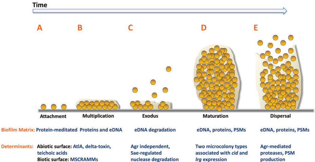
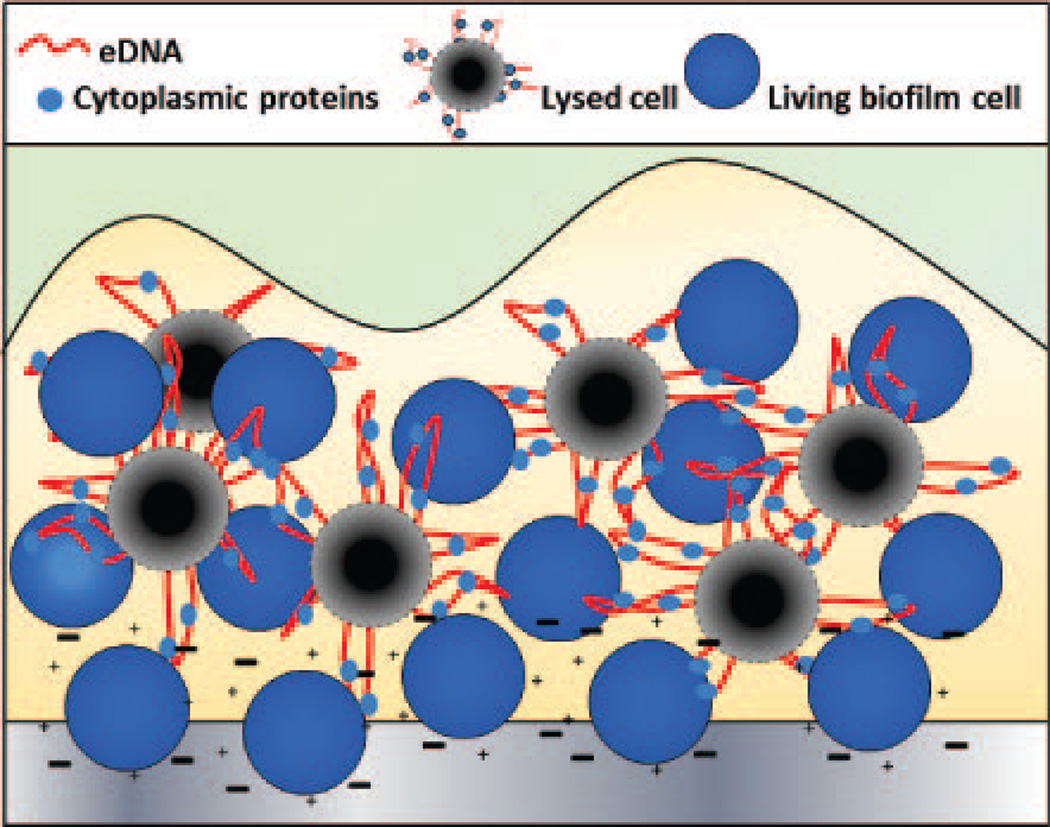
Chronic biofilm-associated infections caused by Staphylococcus aureus often lead to significant increases in morbidity and mortality, particularly when associated with indwelling medical devices. This has triggered a great deal of research attempting to understand the molecular mechanisms that control S. aureus biofilm formation and the basis for the recalcitrance of these multicellular structures to antibiotic therapy. The purpose of this review is to summarize our current understanding of S. aureus biofilm development, focusing on the description of a newly-defined, five-stage model of biofilm development and the mechanisms required for each stage. Importantly, this model includes an alternate view of the processes involved in microcolony formation in S. aureus and suggests that these structures originate as a result of stochastically regulated metabolic heterogeneity and proliferation within a maturing biofilm population, rather than a subtractive process involving the release of cell clusters from a thick, unstructured biofilm. Importantly, it is proposed that this new model of biofilm development involves the genetically programmed generation of metabolically distinct subpopulations of cells, resulting in an overall population that is better able to adapt to rapidly changing environmental conditions.


Figures

Figure 1
Figure 1. Model of Staphylococcus aureus biofilm development
S. aureus biofilm development is described in five stages: A) attachment, B) multiplication, C) exodus, D) maturation, and E) dispersal. A. S. aureus cells attach to abiotic or biotic surfaces via hydrophobic interactions or MSCRAMMs, respectively. B. After cells attach, the biofilm develops into a confluent ‘mat’ of cells composed of an eDNA and proteinaceous matrix. C. Upon reaching confluency, a period of mass exodus of cells occurs in which a subpopulation of cells is released from the biofilm via Sae-regulated nuclease-mediated eDNA degradation to allow for the formation of three-dimensional microcolonies. D. Microcolonies form from distinct foci of cells that have remained attached during the exodus stage. This stage is characterized by rapid cell division that forms robust aggregations composed of proteins including PSMs and eDNA. E. Activated Agr-mediated quorum sensing initiates biofilm matrix modulation and dispersal of cells via protease activation and/or PSM production. AtlA, autolysin A; MSCRAMM, microbial surface components recognizing adhesive matrix molecules; eDNA, extracellular DNA; PSM, phenol soluble modulins; Agr, accessory gene regulator.
Figure 2
Figure 2. Model of cellular interactions during the multiplication stage of biofilm development
During the initial stages of S. aureus biofilm development, planktonic cells attach to a surface through electrostatic interactions (indicated by + and – symbols) involving teichoic acids, PSMs, and autolysin A. As biofilm development progresses into the multiplication stage, a subpopulation of cells dies and lyses (black circles) releasing extracellular DNA (red lines) and cytoplasmic proteins (light blue ovals) into the extracellular milieu, encasing the existing living cells (blue circles) in a mixture of cytoplasmic proteins and genomic DNA.
Figure 3
Figure 3. Microcolony initiation
A) S. aureus cells containing an lrgAB::gfp promoter fusion plasmid were inoculated into a Bioflux1000 microfluidics system and allowed to form a biofilm over a time-course experiment in which epifluorescence images were acquired at regular time points. Shown are images collected at regular intervals after the initiation of medium flow. Note the emergence of the microcolony originating from what appears to be a single (or relatively few) lrgAB-expressing (green) cells. (B) Macroscopic images of S. aureus biofilm grown in an FC flow-cell system. Shown are images collected at 5.5, 11, 16.5, and 22 hrs after the initiation of medium flow. Note the emergence of microcolonies from a basal layer of cell starting at 5.5 hrs, as well as the presence of streaking downstream of most (but not all; see arrows) of the microcolonies.
Figure 4
Figure 4. Agr expression in microcolonies
S. aureus cells containing a agr-p3::gfp promoter fusion plasmid were inoculated into a Bioflux1000 microfluidics system and allowed to form a biofilm over a time-course experiment in which epifluorescence images were acquired at 0, 6, 9, and 11 hrs after the initiation of medium flow. Note the emergence of Agr expression presumably after the AIP octapeptide reaches a threshold density required for induction of P3 expression.

References

    1. Abdelnour A, Arvidson S, Bremell T, Ryden C, Tarkowski A. The accessory gene regulator (agr) controls Staphylococcus aureus virulence in a murine arthritis model. Infection and immunity. 1993;61:3879–3885. - PMC - PubMed
    1. Bayles KW. Bacterial programmed cell death: making sense of a paradox. Nat Rev Microbiol. 2014;12:63–69. - PMC - PubMed
    1. Beenken KE, Spencer H, Griffin LM, Smeltzer MS. Impact of extracellular nuclease production on the biofilm phenotype of Staphylococcus aureus under in vitro and in vivo conditions. Infection and immunity. 2012;80:1634–1638. - PMC - PubMed
    1. Benoit MR, Conant CG, Ionescu-Zanetti C, Schwartz M, Matin A. New device for high-throughput viability screening of flow biofilms. Appl Environ Microbiol. 2010;76:4136–4142. - PMC - PubMed
    1. Biswas R, Voggu L, Simon UK, Hentschel P, Thumm G, Gotz F. Activity of the major staphylococcal autolysin Atl. FEMS Microbiol Lett. 2006;259:260–268. - PubMed

MeSH terms

LinkOut - more resources