. 2017 May;152(6):1477-1491.
doi: 10.1053/j.gastro.2017.01.021. Epub 2017 Jan 29.

Stearoyl-CoA Desaturase Promotes Liver Fibrosis and Tumor Development in Mice via a Wnt Positive-Signaling Loop by Stabilization of Low-Density Lipoprotein-Receptor-Related Proteins 5 and 6

Affiliations

Stearoyl-CoA Desaturase Promotes Liver Fibrosis and Tumor Development in Mice via a Wnt Positive-Signaling Loop by Stabilization of Low-Density Lipoprotein-Receptor-Related Proteins 5 and 6

Keane K Y Lai et al. Gastroenterology. 2017 May.

Abstract

Background & aims: Stearoyl-CoA desaturase (SCD) synthesizes monounsaturated fatty acids (MUFAs) and has been associated with the development of metabolic syndrome, tumorigenesis, and stem cell characteristics. We investigated whether and how SCD promotes liver fibrosis and tumor development in mice.

Methods: Rodent primary hepatic stellate cells (HSCs), mouse liver tumor-initiating stem cell-like cells (TICs), and human hepatocellular carcinoma (HCC) cell lines were exposed to Wnt signaling inhibitors and changes in gene expression patterns were analyzed. We assessed the functions of SCD by pharmacologic and conditional genetic manipulation in mice with hepatotoxic or cholestatic induction of liver fibrosis, orthotopic transplants of TICs, or liver tumors induced by administration of diethyl nitrosamine. We performed bioinformatic analyses of SCD expression in HCC vs nontumor liver samples collected from patients, and correlated levels with HCC stage and patient mortality. We performed nano-bead pull-down assays, liquid chromatography-mass spectrometry, computational modeling, and ribonucleoprotein immunoprecipitation analyses to identify MUFA-interacting proteins. We examined the effects of SCD inhibition on Wnt signaling, including the expression and stability of low-density lipoprotein-receptor-related proteins 5 and 6 (LRP5 and LRP6), by immunoblot and quantitative polymerase chain reaction analyses.

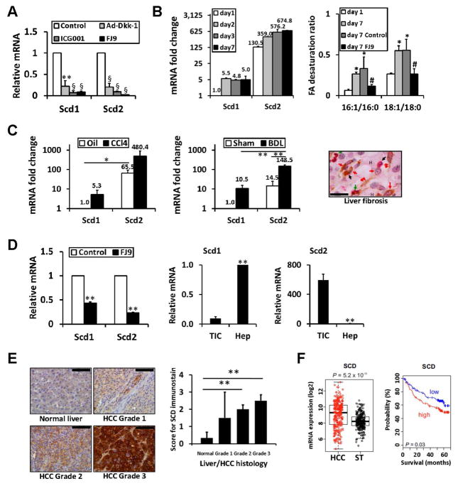
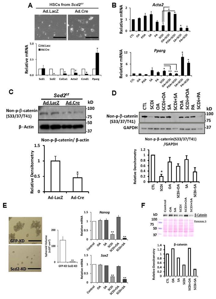
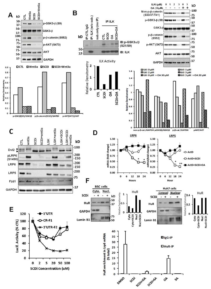
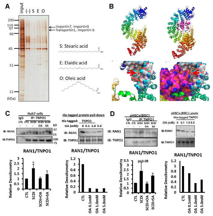
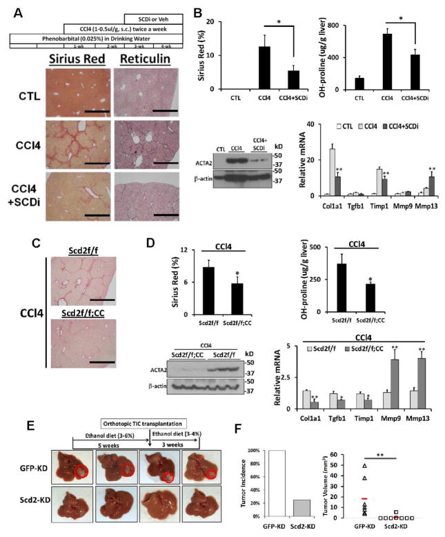
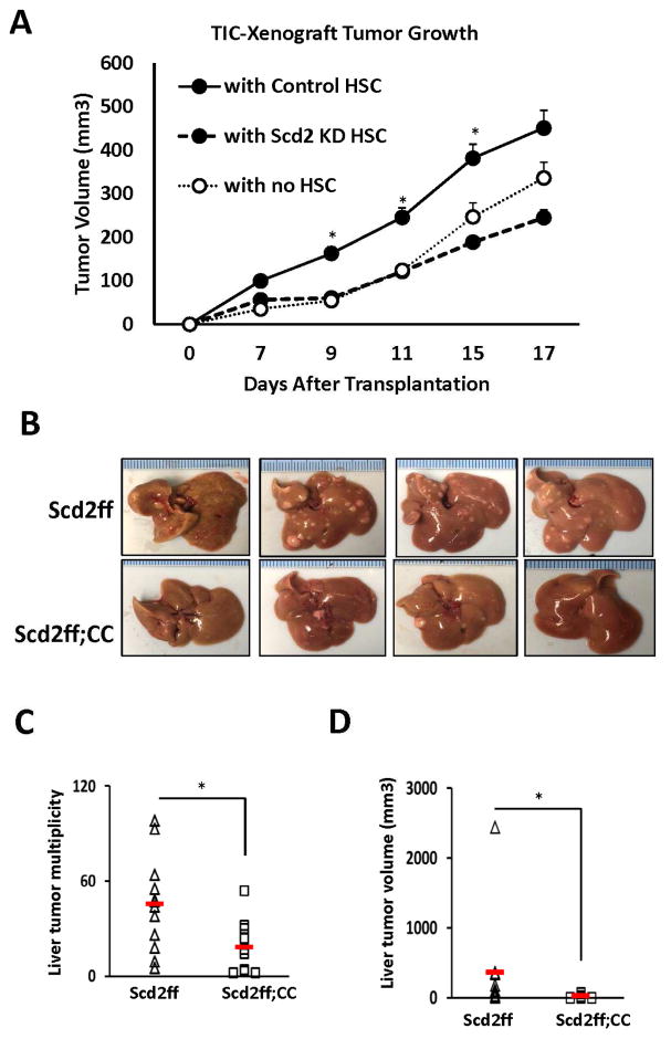
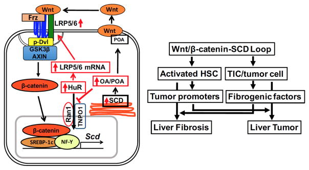
Results: SCD was overexpressed in activated HSC and HCC cells from patients; levels of SCD messenger RNA (mRNA) correlated with HCC stage and patient survival time. In rodent HSCs and TICs, the Wnt effector β-catenin increased sterol regulatory element binding protein 1-dependent transcription of Scd, and β-catenin in return was stabilized by MUFAs generated by SCD. This loop required MUFA inhibition of binding of Ras-related nuclear protein 1 (Ran1) to transportin 1 and reduced nuclear import of elav-like protein 1 (HuR), increasing cytosolic levels of HuR and HuR-mediated stabilization of mRNAs encoding LRP5 and LRP6. Genetic disruption of Scd and pharmacologic inhibitors of SCD reduced HSC activation and TIC self-renewal and attenuated liver fibrosis and tumorigenesis in mice. Conditional disruption of Scd2 in activated HSCs prevented growth of tumors from TICs and reduced the formation of diethyl nitrosamine-induced liver tumors in mice.

Conclusions: In rodent HSCs and TICs, we found SCD expression to be regulated by Wnt-β-catenin signaling, and MUFAs produced by SCD provided a forward loop to amplify Wnt signaling via stabilization of Lrp5 and Lrp6 mRNAs, contributing to liver fibrosis and tumor growth. SCD expressed by HSCs promoted liver tumor development in mice. Components of the identified loop linking HSCs and TICs might be therapeutic targets for liver fibrosis and tumors.

Keywords: Cancer Stem Cells; HCC; Hepatocarcinogenesis; HuR.


Conflict of interest statement

Disclosures: The authors have declared that no conflict of interest exists.

Figures

Fig. 1
Fig. 1. Scd2/SCD upregulated in liver fibrosis and cancers
A. Scd 1 and Scd2, are repressed by three canonical Wnt inhibitors, Dkk1, FJ9, and ICG-001 in cultured rat HSCs, n=4. **p<0.01, §p<0.001. B. Left, Scd1 and Scd2 mRNA in HSCs during culture activation by TaqMan qPCR and expressed as fold change vs. Scd1 expression at day 1. Right, Increases in fatty acid desaturation ratios in day 7 cultured HSCs are abrogated by FJ9. *p<0.05 vs. day 1, #p<0.05 vs. vehicle control at day 7. C. Left and middle, Scd1 and Scd2 mRNA in HSCs from rat CCl4 (n=4) or BDL (n=3) liver fibrosis and respective controls (Oil and Sham) by TaqMan qPCR and expressed as fold change vs. controls. *p<0.05, **p<0.01. Right, HSCs (double red arrow) and myofibroblasts (red arrow) are positive for SCD (brown) and ACTA2 (red) staining in a liver section of alcoholic liver fibrosis patient. Macrophages (black arrow) and PMNs (green arrow) are also present. Hepatocytes are denoted by “H”. Scale bar= 40um. D. FJ9 suppresses Scd1 and Scd2 in mouse liver TICs (left). Scd2 expressed in TICs vs. Scd1 in normal hepatocytes (middle and right) (n=4). **p<0.01. E. SCD induction with the worsening grade of HCC as demonstrated by immunostaining (left) and its semi-quantitation (right) of 17 patients. **p<0.01. Scale bar=100um. F. SCD mRNA expression induced in HCC vs. non-tumor tissue in a patient cohort (n=242) (left) and correlates with mortality when patients are dichotomized by median expression value (right).
Fig. 2
Fig. 2. SCD renders a positive-feedback loop for Wnt/β-catenin
A. Expression of Cre via adenovirus (Ad.Cre, MOI=100) but not LacZ (Ad.LacZ) in HSCs from Scd2ff mice, reverts the cells to quiescent state (top, Scale bar=400um), suppresses Col1a1 and Acta2, and upregulates Pparγ (bottom). Ccnd1 is suppressed by Scd2 ablation (n=4). *p<0.05 vs. Ad.LacZ. B. Acta2 and Pparγ mRNA by qPCR, confirm SCDi effects and rescue with OA and POA but not with SA and PA. *p<0.05. C. Reduced stabilized β-catenin (non-phosphorylated at S33/S37/T41) in Scd2-silenced HSCs (top). A densitometric analysis of six experiments (bottom). *p<0.05. D. SCDi suppression of stabilized β-catenin in HSCs and its rescue with OA and POA but not with SA and PA. A densitometric analysis of three experiments. *p<0.05. E. Reduced spheroid size by Scd2-KD vs. GFP-KD TICs (left and middle) (n=3). **p<0.01. Scale bar=400um. SCDi (400 nM) represses Nanog and Sox2, and this repression is rescued by OA but not SA (right) (n=3). **p<0.01. F. SCDi suppresses β-catenin protein in TICs, the effect rescued by OA.
Fig. 3
Fig. 3. SCD promotes LRP5/6 mRNA stability via ARE binding protein HuR
A. SCDi reduces p(S9)-GSK3β, p(S552)-β-catenin, and p(S473)-AKT in BSCs without or with Wnt3a treatment. B. SCDi inhibits ILK activity in BSCs and this inhibition is rescued by OA (30 μM, left). ILK inhibitor (ILKi) reduces non-p β-catenin, p(S9)-GSK3β, and p(S552)-β-catenin, and p(S473)-AKT in BSC but these effects are not rescued by OA (right). C. Dvl2 phosphorylation induced by exogenous Wnt3a is inhibited by SCDi in BSC. Wnt3a-induced p-LRP6 is abrogated by SCDi due to a loss of LPR6 protein. D. With actinomycin D (ActD, 1 nM), LRP5/6 mRNA is degraded faster in BSC with SCDi vs. control. This effect of SCDi is rescued by OA. *p<0.05, **p<0.01. E. Luc-3′UTR-F2 is dose-dependently suppressed by SCDi to 20% of control in Huh7 cells. F. SCDi reduces HuR protein in cytosol of BSC (left top) and Huh7 cells (right top). SCDi or SA but not OA abrogates HuR binding to Lrp6 3′UTR ARE as determined by RIP analysis (bottom). Error bars represent SEM. *p<0.05
Fig. 4
Fig. 4. OA interferes TNPO1-Ran1 GTPase binding
A. Fatty acid interacting proteins were searched by incubating TIC lysates with nano-beads conjugated with O (oleic acid), S (stearic acid), or E (elaidic acid), differential gel display, and mass spectrometry analysis, revealing TNPO1, importin 5, 7, or β as putative OA-interacting proteins. B. Top, Three-dimensional structure of TNPO1 shown in ribbon representation. The molecule is colored in rainbow color with N-terminus in blue and the C-terminus in red. TNPO1 is shown in complex with Ran1 (in purple, right). Bottom, the Ran1 binding region at the N-terminus is shown in surface model. OA (red arrows) is shown in yellow stick model, and colors on binding surface are according to atoms (N-blue; O-red, C-white, H-cyan). C. Left, Co-IP shows SCDi increases TNPO1-Ran1 binding which is prevented by OA but not by SA in Huh7 cells. Right, OA suppresses the interaction of Ran1 with His-tagged TNPO1 in Huh7 cell lysate. D. Left, SCDi increases TNPO1-Ran1 binding which is prevented by OA but not by SA in BSC. Right, OA suppresses Ran1 interaction with His-tagged TNPO1 in BSC lysate.
Fig. 5
Fig. 5. Importance of Scd2 in liver fibrosis
A. SCDi treatment reduces CCl4 liver fibrosis assessed by Sirius red and reticulin staining vs. vehicle-treated mice (n=6 each). Scale bar=1mm. B. SCDi reduces Sirius red morphometry and liver hydroxyproline content (top), hepatic ACTA2 (bottom left), and Col1a1, Tgfb1, Timp1 mRNA while inducing Mmp9 and Mmp13 (bottom right). C. CCl4-induced liver fibrosis is attenuated in mice with conditional Scd2 knockout (Scd2ff;CC) vs. Scd2ff control mice. D. Conditional Scd2 deficiency inhibits Sirius red staining morphometry and hydroxyproline content (top), hepatic ACTA2 (bottom left), and Col1a1, Tgfb1, Timp1 mRNA while inducing Mmp9 and Mmp13 (bottom right). E and F. Scd2 KD inhibits TIC-induced tumorigenesis in vivo. TICs with Scd2 KD or GFP-KD (control) were orthotopically transplanted in ethanol-fed B6 mice. Red horizontal bars denote group means. **p<0.01. Error bars represent SEM.
Fig. 6
Fig. 6. SCD2 confers HSC-mediated promotion of TIC-dependent tumorigenesis and DEN-induced liver tumor development in mice
A. TIC-initiated tumor growth in nude mice is promoted when co-xenografted (1:1) with control HSCs (n=6) but not with Scd2 KD HSCs (n=6) vs. TICs transplanted with no HSCs (n=6). *p<0.05 vs. TICs with Scd2 KD HSCs. B. Representative photos of DEN-induced and Western alcohol diet-promoted liver tumor in Scd2ff mice which are reduced in conditional Scd2 knockout mice (Scd2ff;CC). C and D. SCD2 deficiency in aHSCs (Scd2ff;CC, n=10 vs. Scd2ff, n=12) significantly reduces multiplicity and tumor volume. Red horizontal bars and error bars denote group means and SEM. *p<0.05.
Fig. 7
Fig. 7. Schematic diagrams of a novel Wnt/β-catenin-SCD-LRP positive loop and cellular crosstak
Left, Wnt/β-catenin-dependent SCD expression leads to HuR-mediated LRP5/6 mRNA stabilization via MUFA (OA, POA) inhibition of HuR nuclear transport by TNPO1 and Ran1. Right, the positive loop individually activates HSCs and TICs and also promotes crosstalk between the two cell types, underlying the link between liver fibrosis and tumor.

References

    1. Fattovich G, Stroffolini T, Zagni I, et al. Hepatocellular carcinoma in cirrhosis: incidence and risk factors. Gastroenterology. 2004;127:S35–50. - PubMed
    1. Coulouarn C, Clément B. Stellate cells and the development of liver cancer: therapeutic potential of targeting the stroma. J Hepatol. 2014;60:1306–9. - PubMed
    1. Monga SP. β-Catenin Signaling and Roles in Liver Homeostasis, Injury, and Tumorigenesis. Gastroenterology. 2015;148:1294–310. - PMC - PubMed
    1. Clevers H, Nusse R. Wnt/β-catenin signaling and disease. Cell. 2012;149:1192–205. - PubMed
    1. Cheng JH, She H, Han YP, et al. Wnt antagonism inhibits hepatic stellate cell activation and liver fibrosis. Am J Physiol Gastrointest Liver Physiol. 2008;294:G39–49. - PubMed

Publication types

MeSH terms