

Case Reports
doi: 10.1136/bcr-2016-218222.
Non-Hodgkin's lymphoma involving a femur bone and bilateral adrenal glands alone with adrenal insufficiency
Affiliations
- PMID: 28143809
- PMCID: PMC5293968
- DOI: 10.1136/bcr-2016-218222
Item in Clipboard
Case Reports
Non-Hodgkin's lymphoma involving a femur bone and bilateral adrenal glands alone with adrenal insufficiency
BMJ Case Rep.
.
Abstract
Primary bone lymphoma and primary adrenal lymphoma are rare clinicopathological entities of non-Hodgkin's lymphoma (NHL). We present the first case of diffuse large B-cell lymphoma with the involvement of a single bone and both adrenal glands alone with adrenal insufficiency. As primary extranodal NHL may have other unusual extranodal lesions, which may present unexplained clinical findings, patients with primary extranodal NHL require careful systemic examination, even when lymphadenopathy is absent.
2017 BMJ Publishing Group Ltd.
Conflict of interest statement
Competing interests: None declared.
Figures
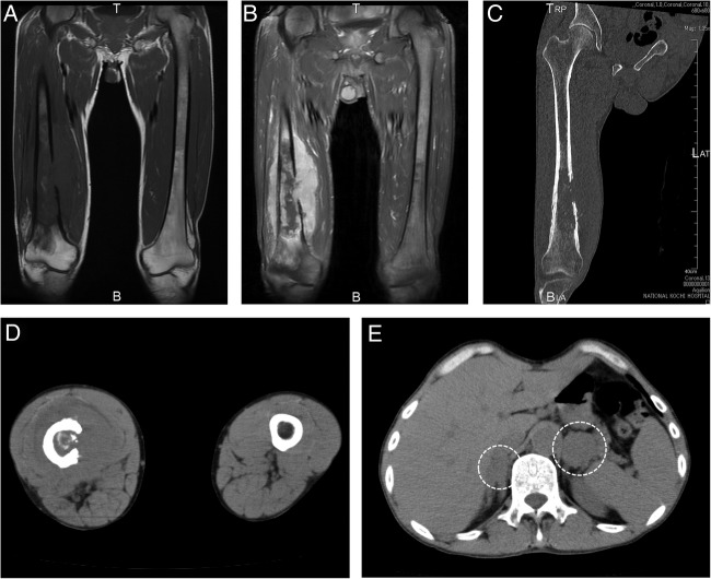
MRI (A, B) and CT (C–E) images obtained at presentation. Coronal T1-weighted (A) and enhanced T1-weighted iterative decomposition of water and fat with echo asymmetry and least-squares estimation (B) MRI and coronal (C) and transverse (D) CT images of the femoral regions show a large osteolytic lesion in the distal region of the right femur infiltrating the adjacent soft tissue. Abdominal CT (E) revealed bilateral adrenal swelling (circled).
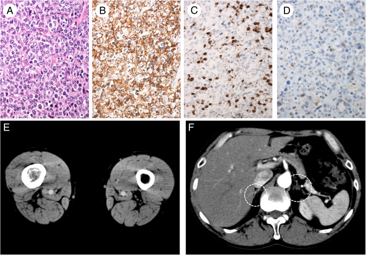
Pathological findings of bone biopsy (A–D) and enhanced CT (E,F) images obtained after treatment. Tissue stained with H&E (A) shows large-sized atypical lymphocytes with diffuse proliferation. The individual cells had nuclei with a coarse chromatin pattern and large nucleoli. Immunochemistry showed that the atypical cells were CD 79a (+) (B), CD3 (−) (C) and CD5 (−) (D). CT images of femoral regions (E) showed disappearance of the right femoral region swelling with reconstruction of the right femur bone cortex. Residual lesion seemed to be present in the medullary cavity of the right femur. Abdominal CT (F) showed disappearance of bilateral adrenal swelling (circled).
References
Publication types
MeSH terms
Substances
LinkOut - more resources
Full Text Sources
Other Literature Sources
Medical
Research Materials