. 2017 Apr 6;129(14):1991-2001.
doi: 10.1182/blood-2016-10-744441. Epub 2017 Jan 31.

Neutrophils acquire the capacity for antigen presentation to memory CD4+ T cells in vitro and ex vivo

Affiliations

Neutrophils acquire the capacity for antigen presentation to memory CD4+ T cells in vitro and ex vivo

Maria Vono et al. Blood. .

Abstract

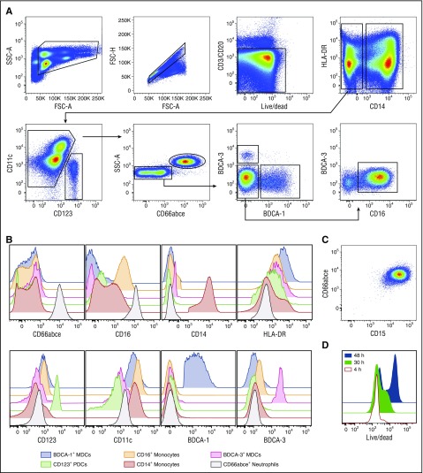
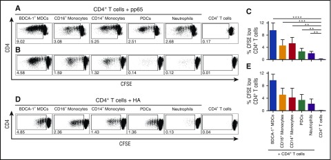
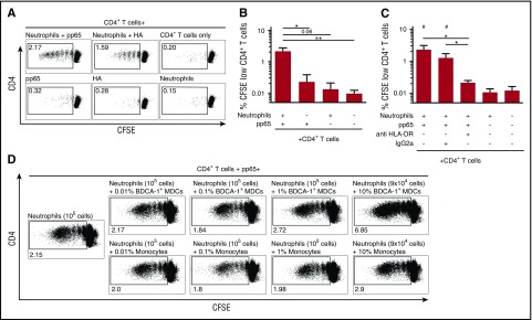
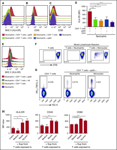
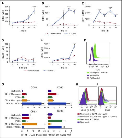
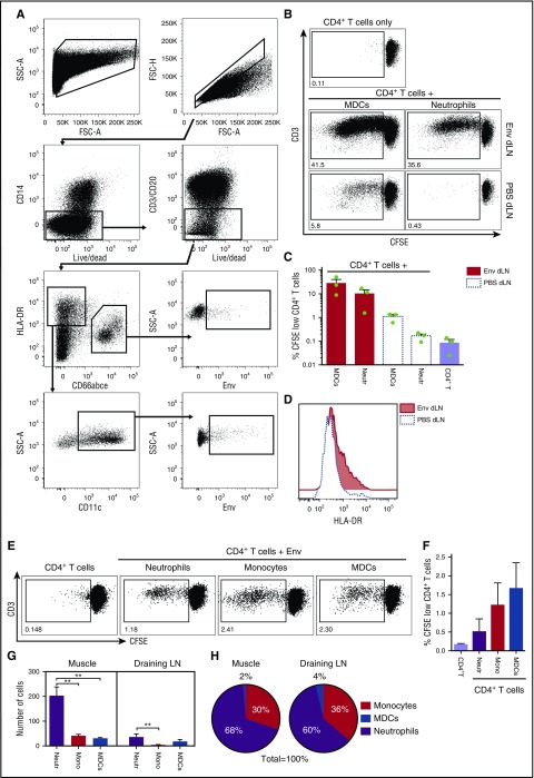
Neutrophils are critical cells of the innate immune system and rapidly respond to tissue injury and infection. Increasing evidence also indicates that neutrophils have versatile functions in contributing to adaptive immunity by internalizing and transporting antigen and influencing antigen-specific responses. Here, we demonstrate that freshly isolated human neutrophils can function as antigen-presenting cells (APCs) to memory CD4+ T cells. Neutrophils pulsed with the cognate antigens cytomegalovirus pp65 or influenza hemagglutinin were able to present the antigens to autologous antigen-specific CD4+ T cells in a major histocompatibility complex class II (MHC-II; HLA-DR)-dependent manner. Although myeloid dendritic cells and monocytes showed superior presenting ability, neutrophils consistently displayed antigen presentation capability. Upregulation of HLA-DR on neutrophils required the presence of the antigen-specific or activated T cells whereas exposure to innate stimuli such as Toll-like receptor ligands was not sufficient. Neutrophils sorted from vaccine-draining lymph nodes from rhesus macaques also showed expression of HLA-DR and were capable of presenting vaccine antigen to autologous antigen-specific memory CD4+ T cells ex vivo. Altogether, the data demonstrate that neutrophils can adapt a function as APCs and, in combination with their abundance in the immune system, may have a significant role in regulating antigen-specific T-cell responses.


Figures

Figure 1.
Figure 1.
Simultaneous isolation of pure populations of human neutrophils, monocytes, and DC subsets by flow cytometry cell sorting. (A) Pure populations of human CD66abce+ neutrophils, CD14+ and CD14CD16+ monocytes, CD123+ PDCs, and CD11c+ MDC subsets (BDCA-1+ and BDCA-3+ subsets) were isolated from the elutriated monocyte-fraction after apheresis by flow cytometry sorting according to the depicted gating strategy. CD14CD16+ monocytes are now indicated as CD16+ monocytes only for simplicity. (B) Representative flow histograms show the expression levels of the indicated markers on the sorted APC populations. (C) Sorted CD66abce+ neutrophils coexpress CD15. (D) Representative flow histograms show the live/dead staining (LIVE/DEAD fixable dead cell stain kit) on sorted neutrophils over time. Neutrophils showed high viability for at least 30 hours and can be maintained in culture longer. A representative donor of 6 is shown. FSC-A, forward scatter area; FSC-H, forward scatter height; SSC-A, side scatter area.
Figure 2.
Figure 2.
Neutrophils can present antigens to antigen-specific memory CD4+T cells but MDCs and monocytes have the highest capacity. Sorted APCs from CMV- or influenza-seropositive donors were pulsed respectively with CMV pp65 or influenza HA and cocultured with autologous CFSE-labeled CD4+ T cells. Their capacity to present antigen to CD4+ T cells was evaluated by measuring proliferating T cells (CFSE dilution). (A,B,D) Proliferating (CFSE low) CD4+ T cells in presence of the indicated APCs from 2 CMV-seropositive donors (A-B) or 1 representative HA-seropositive donor (D) are shown. Bars show percentages of (C) pp65-specific and (E) HA-specific proliferating CD4+ T cells (mean ± SEM, n = 6 and n ≥ 2 healthy human donors, respectively). **P < .01, *** P < .001, **** P < .0001.
Figure 3.
Figure 3.
Human neutrophils present the antigen to antigen-specific memory CD4+T cells in an HLA-DR–dependent manner. Neutrophils from CMV- or influenza-seropositive donors were pulsed with pp65 or HA and cocultured with autologous CFSE-labeled CD4+ T cells for 5 days. (A) Representative plots of HA (where indicated) or pp65-specific proliferating CD4+ T cells for the indicated conditions. (B-C) Bars show percentages of pp65-specific proliferating CD4+ T cells. (C) HLA-DR neutralizing antibody strongly reduced CD4+ T-cell proliferation mediated by neutrophils whereas no reduction was observed in presence of the same concentration of isotype control (mean ± SEM, n = 3-8 donors); *P < .05, **P < .01 and #P < .01 vs CD4+ T cells only. (D) CD4+ T-cell proliferation induced by neutrophils alone or in presence of different amounts of BDCA-1+ MDCs (up to 10%) or CD14+ monocytes (up to 10%).
Figure 4.
Figure 4.
Upregulation of MHC-II (HLA-DR), CD40, and CD80 on neutrophils requires antigen and antigen-specific memory CD4+T cells. Representative histograms show the surface expression levels of HLA-DR (MHC II) (A,E) and the maturation markers CD40 (B) and CD80 (C) on neutrophils cultured for 30 hours alone or with autologous CD4+ T cells for all the indicated conditions. (D) Bar graphs show geometric mean fluorescence intensity (MFI) values of HLA-DR (mean ± SEM, n = 4 donors). (F) Proliferation of allogeneic naive T cells in a mixed lymphocyte reaction induced by neutrophils or monocytes. One representative donor of 3 is shown. (G) IFN-γ/IL-2/TNF production in responding pp65-specific CD4+ T cells stimulated by neutrophils or CD14+ monocytes. One representative donor of 3 is shown. (H) Bar graphs show the surface expression levels of HLA-DR, CD40, and CD80 on neutrophils after stimulation for 30 hours with supernatants (Sup) collected from culture of T cells only (Medium), T cells activated with anti-CD2/CD3/CD28 beads (Beads), or from the neutrophil–T-cell cocultures prepared as for the Ag presentation assay (Ag pres) (mean ± SEM, n = 4). *P< .05, **P < .01, ***P < .001, ****P < .0001. Unstim, unstimulated.
Figure 5.
Figure 5.
TLR7/8-L induces neutrophil activation but is not sufficient for the induction of MHC-II molecules. (A-F) Neutrophils exposed to TLR7/8-L were phenotypically characterized over time by flow cytometry. TLR7/8-L activated neutrophils as detected by upregulation of CD80 (A) and CD83 (B) as well as upregulation of the integrin CD11b (C) over time but was not sufficient for the upregulation of HLA-DR (D). TLR7/8-L enhanced CCR7 levels on neutrophils (E-F). Quantified MFIs of the indicated markers ± SEM on neutrophils over time (A-E) and representative histograms of the surface expression levels of CCR7 from a representative donor of 3 at 30 hours (F) are shown. (G) Surface expression levels of CD80, CD86, CD40, and CD83 on distinct sorted APCs after overnight exposure to TLR7/8-L. Data are presented as ratio of the MFI values between TLR7/8-L–treated and unstimulated cells ± SEM. (H-I) Representative histograms show the surface expression levels of HLA-DR (H) and CD80 (I) on human neutrophils cultured for 30 hours alone or in presence of autologous CD4+ T cells and pp65 with or without TLR7/8-L (2.5 μg/mL); n = 3-5 healthy human donors. *P < .05, **P < .01, ***P < .001, ****P < .0001. FMO, fluorescence minus one.
Figure 6.
Figure 6.
Neutrophils sorted from the dLNs after vaccination present Env to memory CD4+T cells but MDCs have the highest capacity. (A) Neutrophils and MDCs were isolated by flow cytometry sorting from the dLNs of vaccinated rhesus macaques 24 hours postinjection with an Alexa 680–labeled Env or PBS (contralateral arm) according to the depicted gating strategy. (B) Neutrophils isolated from the Env-draining LNs induced well-detectable T-cell proliferation but MDCs showed the highest capacity. Representative plots of proliferating CD4+ T cells. (C) Bars graph show percentages of proliferating CD4+ T cells (mean ± SEM, n = 3 NHPs). (D) Representative histograms show HLA-DR levels on neutrophils sorted from the Env and PBS dLNs. (E) Representative plots show proliferating CD4+ T cells after 5-day cocultures with either neutrophils, monocytes, or MDCs isolated from spleen from the animals above in presence of Env. (F) Bars show percentages of proliferating CD4+ T cells (mean ± SEM, n = 3 NHPs). (G-H) Rhesus macaques were injected with Alexa 680–labeled Env and the cell subsets recruited to the muscle injection site and dLNs were phenotyped and enumerated by flow cytometry. (G) Number of indicated cell subsets mobilized to the Env-injected muscle and dLNs (mean ± SEM, n = 3 NHPs); **P < .01. (H) Proportions of the number of Env+ cell subsets at the injection site and draining LNs. Mono, monocytes; Neutr, neutrophils.

Comment in

References

    1. Amulic B, Cazalet C, Hayes GL, Metzler KD, Zychlinsky A. Neutrophil function: from mechanisms to disease. Annu Rev Immunol. 2012;30:459-489. - PubMed
    1. Galli SJ, Borregaard N, Wynn TA. Phenotypic and functional plasticity of cells of innate immunity: macrophages, mast cells and neutrophils. Nat Immunol. 2011;12(11):1035-1044. - PMC - PubMed
    1. Nathan C. Neutrophils and immunity: challenges and opportunities. Nat Rev Immunol. 2006;6(3):173-182. - PubMed
    1. Thomas CJ, Schroder K. Pattern recognition receptor function in neutrophils. Trends Immunol. 2013;34(7):317-328. - PubMed
    1. Cassatella MA. Neutrophil-derived proteins: selling cytokines by the pound. Adv Immunol. 1999;73:369-509. - PubMed

Publication types

Substances