. 2017 Mar 7;114(10):2604-2609.
doi: 10.1073/pnas.1619809114. Epub 2017 Jan 31.

Oxidative DNA damage is epigenetic by regulating gene transcription via base excision repair

Affiliations

Oxidative DNA damage is epigenetic by regulating gene transcription via base excision repair

Aaron M Fleming et al. Proc Natl Acad Sci U S A. .

Abstract

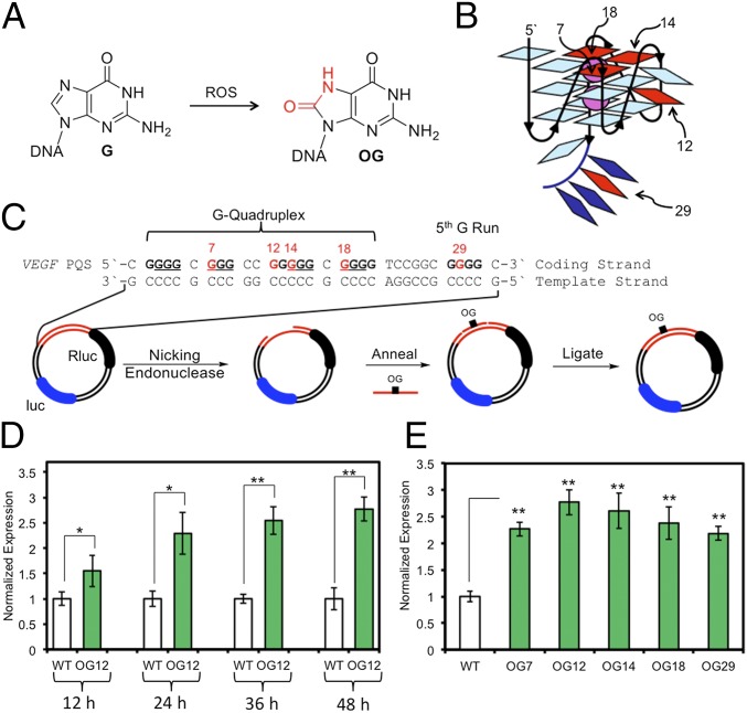
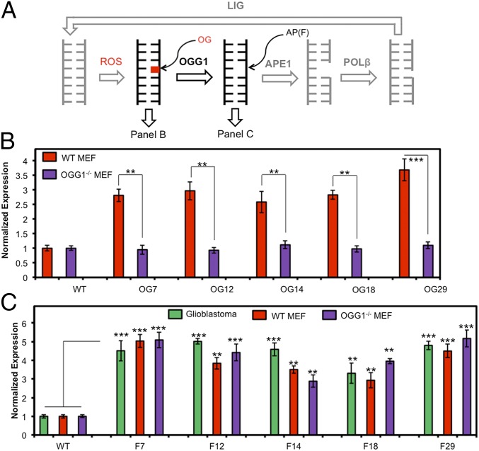
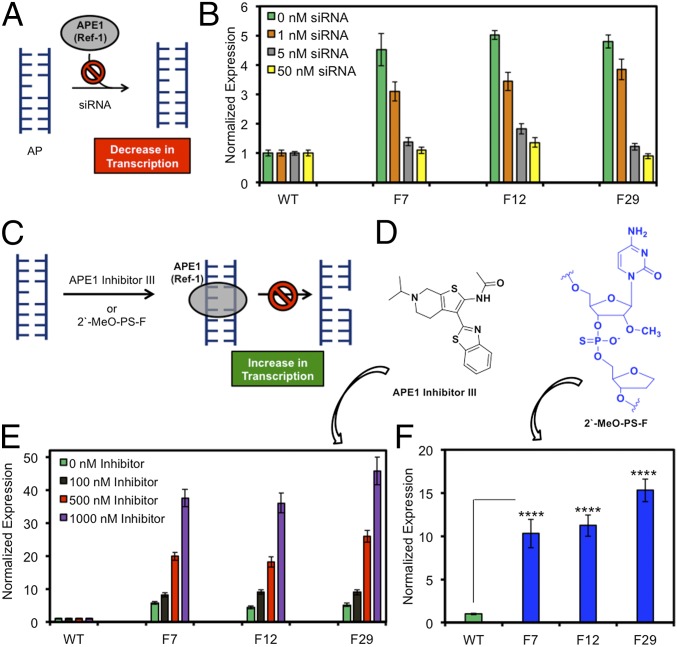
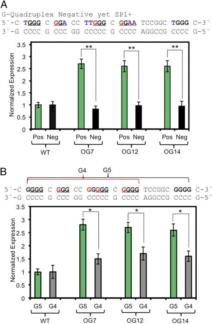
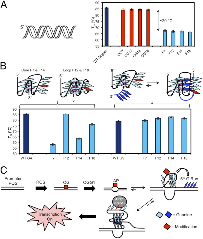
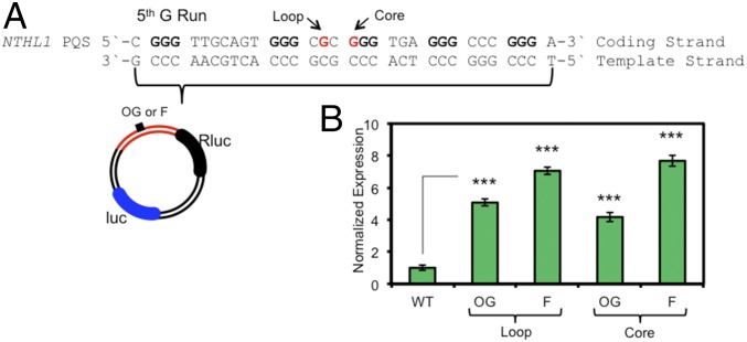
Reactive oxygen species (ROS) have emerged as important cellular-signaling agents for cellular survival. Herein, we demonstrate that ROS-mediated oxidation of DNA to yield 8-oxo-7,8-dihydroguanine (OG) in gene promoters is a signaling agent for gene activation. Enhanced gene expression occurs when OG is formed in guanine-rich, potential G-quadruplex-forming sequences (PQS) in promoter-coding strands, initiating base excision repair (BER) by 8-oxoguanine DNA glycosylase (OGG1), yielding an abasic site (AP). The AP enables melting of the duplex to unmask the PQS, adopting a G-quadruplex fold in which apurinic/apyrimidinic endonuclease 1 (APE1) binds, but inefficiently cleaves, the AP for activation of vascular endothelial growth factor (VEGF) or endonuclease III-like protein 1 (NTHL1) genes. These details were mapped via synthesis of OG and AP analogs at single-nucleotide precision within the promoter of a luciferase reporter system. The reporters were analyzed in human and mouse cells while selectively knocking out or down critical BER proteins to identify the impact on luciferase expression. Identification of the oxidatively modified DNA base OG to guide BER activity in a gene promoter and impact cellular phenotype ascribes an epigenetic role to OG.

Keywords: G-quadruplex; base excision repair; epigenetics; gene regulation; oxidative damage.


Conflict of interest statement

The authors declare no conflict of interest.

Figures

Fig. 1.
Fig. 1.
Oxidation of G in the VEGF PQS induces transcription. (A) G oxidation to OG. (B) VEGF G-quadruplex, labeled with positions studied. (C) VEGF sequence with Gs in the core underlined, reporter system design, and method for site-specific incorporation of DNA modifications. (D) Time-dependent and (E) position-dependent expression at 48 h posttransfection of OG-containing reporters in glioblastoma cells. WT refers to the plasmid containing the VEGF PQS with undamaged Gs. Error bars represent 95% CI on the basis of four or eight replicates. Significance values for each comparison were calculated by a Student’s t test. Significance at *P < 0.05 or **P < 0.01 is indicated.
Fig. 2.
Fig. 2.
BER initiates gene activation when OG is located in a promoter PQS. (A) The BER pathway. (B) Positional dependency in expression of OG-containing reporters in WT and OGG1−/− MEFs. (C) Positional and cell line dependency in expression of F-containing reporters, where F = the stable AP analog THF. Error bars represent 95% CI on the basis of four or eight replicates. Significance values for each comparison were calculated by a Student’s t test. Significance at **P < 0.01 or ***P < 0.001 is indicated.
Fig. 3.
Fig. 3.
Binding of APE1 (Ref-1) to AP in the VEGF PQS promoter element enhances gene transcription in glioblastoma cells. (A) Knockdown of APE1 and prevention of AP binding leads to decreased Rluc expression. (B) Expression levels measured when F-containing reporter plasmids were transfected into APE1 knockdown cells with 0–50 nM siRNA. (C) Mechanism for prevention of APE1 cleavage without impacting binding of an AP leading to increased gene expression. (D) Structures of APE1 inhibitor III and the 2′-MeO-PS-F–modified AP site. (E) Expression level measured when cells were treated with 100–1,000 nM APE1 inhibitor III. (F) Expression level measured when cells were transfected with the poorly cleavable 2′-MeO-PS-F analog. Error bars represent 95% CI on the basis of four or eight replicates. Significance values for each comparison were calculated by a Student’s t test. Significance at ****P < 0.0001 is indicated.
Fig. 4.
Fig. 4.
Gene induction with OG in the VEGF PQS requires the G-quadruplex fold. (A) Expression observed when OG is in a G-quadruplex positive or negative folding sequence context. (B) Requirement of the fifth G run for maximal expression when OG is present. Error bars represent 95% CI on the basis of four or eight replicates. Significance values for each comparison were calculated by a Student’s t test. Significance at *P < 0.05 or **P < 0.01 is indicated.
Fig. 5.
Fig. 5.
Formation of an AP in the VEGF PQS shifts the duplex–quadruplex equilibrium. (A) Tm studies for the VEGF duplex derived from the G4 sequence provide comparisons between WT, OG-containing, and F-containing strands. (B) Tm studies for the VEGF G-quadruplex comparing positional dependency of an F residue in G4 vs. G5 sequences. (C) Proposed mechanism by which initiation of OG removal in the VEGF promoter PQS allows a structural switch to occur for binding of APE1 and activation of transcription.
Fig. 6.
Fig. 6.
Gene activation is observed when OG or F is present in the NTHL1 PQS. (A) The NTHL1 PQS sequence and locations in which OG or F were synthesized. (B) Expression enhancement observed when OG or F is found in the NTHL1 PQS. Error bars represent 95% CI on the basis of four or eight replicates. Significance values for each comparison were calculated by a Student’s t test. Significance at ***P < 0.001 is indicated.

Comment in

References

    1. Hailer-Morrison MK, Kotler JM, Martin BD, Sugden KD. Oxidized guanine lesions as modulators of gene transcription. Altered p50 binding affinity and repair shielding by 7,8-dihydro-8-oxo-2′-deoxyguanosine lesions in the NF-kappaB promoter element. Biochemistry. 2003;42(32):9761–9770. - PubMed
    1. Ramon O, et al. Effects of 8-oxo-7,8-dihydro-2′-deoxyguanosine on the binding of the transcription factor Sp1 to its cognate target DNA sequence (GC box) Free Radic Res. 1999;31(3):217–229. - PubMed
    1. Moore SP, Toomire KJ, Strauss PR. DNA modifications repaired by base excision repair are epigenetic. DNA Repair (Amst) 2013;12(12):1152–1158. - PubMed
    1. Tornaletti S, Maeda LS, Kolodner RD, Hanawalt PC. Effect of 8-oxoguanine on transcription elongation by T7 RNA polymerase and mammalian RNA polymerase II. DNA Repair (Amst) 2004;3(5):483–494. - PubMed
    1. Allgayer J, Kitsera N, Bartelt S, Epe B, Khobta A. Widespread transcriptional gene inactivation initiated by a repair intermediate of 8-oxoguanine. Nucleic Acids Res. 2016;44(15):7267–7280. - PMC - PubMed

Publication types

MeSH terms