. 2017 Jan 31;8(1):e02183-16.
doi: 10.1128/mBio.02183-16.

Trojan Horse Transit Contributes to Blood-Brain Barrier Crossing of a Eukaryotic Pathogen

Affiliations

Trojan Horse Transit Contributes to Blood-Brain Barrier Crossing of a Eukaryotic Pathogen

Felipe H Santiago-Tirado et al. mBio. .

Abstract

The blood-brain barrier (BBB) protects the central nervous system (CNS) by restricting the passage of molecules and microorganisms. Despite this barrier, however, the fungal pathogen Cryptococcus neoformans invades the brain, causing a meningoencephalitis that is estimated to kill over 600,000 people annually. Cryptococcal infection begins in the lung, and experimental evidence suggests that host phagocytes play a role in subsequent dissemination, although this role remains ill defined. Additionally, the disparate experimental approaches that have been used to probe various potential routes of BBB transit make it impossible to assess their relative contributions, confounding any integrated understanding of cryptococcal brain entry. Here we used an in vitro model BBB to show that a "Trojan horse" mechanism contributes significantly to fungal barrier crossing and that host factors regulate this process independently of free fungal transit. We also, for the first time, directly imaged C. neoformans-containing phagocytes crossing the BBB, showing that they do so via transendothelial pores. Finally, we found that Trojan horse crossing enables CNS entry of fungal mutants that cannot otherwise traverse the BBB, and we demonstrate additional intercellular interactions that may contribute to brain entry. Our work elucidates the mechanism of cryptococcal brain invasion and offers approaches to study other neuropathogens.

Importance: The fungal pathogen Cryptococcus neoformans invades the brain, causing a meningoencephalitis that kills hundreds of thousands of people each year. One route that has been proposed for this brain entry is a Trojan horse mechanism, whereby the fungus crosses the blood-brain barrier (BBB) as a passenger inside host phagocytes. Although indirect experimental evidence supports this intriguing mechanism, it has never been directly visualized. Here we directly image Trojan horse transit and show that it is regulated independently of free fungal entry, contributes to cryptococcal BBB crossing, and allows mutant fungi that cannot enter alone to invade the brain.


Figures

FIG 1
FIG 1
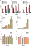
Isolation of loaded macrophages and crossing of the BBB by C. neoformans. (A) Final phase of the sorting strategy used to isolate phagocytes that were healthy (negative for SYTOX dye) and devoid of external fungi (negative for CFW); the complete strategy is shown in Fig. S1. The boxes corresponding to the gates are labeled as follows: gate 1, healthy phagocytes with only internal fungi; gate 2, healthy phagocytes that have external fungi (with or without internal fungi); gate 3, dead or damaged phagocytes. Results are representative of results of 69 independent analyses. (B) Cells collected from the gates shown in panel A (all shown at the same scale; bar = 10 µm). Images are representative of results of 4 independent sorting studies where cells were examined microscopically. (C) Diagram of the BBB model used in this study. (D) Transit of free S. cerevisiae (S. cer) or C. neoformans (C. neo) in the absence or presence of serum; means and standard deviations (SD) of results are shown for one of two similar studies. (E) Mean and SD values over time for transit (bars, left axis) and TEER (points, right axis), for various starting inocula of opsonized C. neoformans. Stars represent TEER values at 0 h. Results from one of two similar studies are shown. (F) Time-dependent transmigration of free fungi, a 1:1 mix of free fungi and empty THPs, and fungus-loaded THPs (1 to 1.49 fungi/host cell). Means and standard errors of the means (SEM) are shown for results of one of seven similar independent experiments. Values plotted for all time course transit assays are cumulative values.
FIG 2
FIG 2
Visualization of Trojan horse crossing. (A) Experimental design for visualization of both sides of the permeable membrane. (B) A field of view showing loaded macrophages associated with the endothelial monolayer (top row) and the bottom of the porous membrane with loaded macrophages alone (bottom row). Both mammalian cell types were stained with CellMask plasma membrane dye (blue); THPs were further stained with DFFDA from the flow sorting (green) and contained mCherry-stained fungi (red). Scale bar, 10 μm. (C) Merged images from a larger field of view, again showing all cell types on the top of the membrane (left), whereas only loaded phagocytes are visible on the bottom (right). Scale bar, 20 μm. Images are representative of multiple fields from two independent experiments, each with three independent time points. The fields shown are from a 1-h time point. We observed loaded macrophages associated with the bottom of the membrane most frequently early in the incubation period (more at 1 h than at 2 to 3 h; not shown); this may represent a time-dependent loss of phagocyte viability or ability to initiate transit. See Movie S1 for another example.
FIG 3
FIG 3
Differential regulation of free and internalized fungal crossing by immune mediators. (A to C) Transmigration of free and internalized fungi in the presence of various chemoattractants, with matched controls. (A) fMLP, N-formyl-methionine-leucyl-phenylalanine peptide (100 nM). DMSO, dimethyl sulfoxide. *, P < 0.0001 (by Sidak’s multiple-comparison test). (B) MCP-1 (also known as CCL2), monocyte chemoattractant protein-1 (100 ng/ml). *, P < 0.03 (by Sidak’s multiple-comparison test). (C) INO, inositol (1 mM). *, P < 0.05 (by Sidak’s multiple-comparison test). (D and E) Transmigration assays using BBB pretreated with TNF-α (10 ng/ml) or IFN-β (1 ng/ml) for 24 h before addition of loaded THPs (*, P < 0.0003 [for comparisons between TNF-α and other treatments by Tukey’s multiple-comparison test]) (D) or of free fungi (*, P < 0.0001 [for comparisons between each compound and PBS by Tukey’s multiple-comparison test]) (E). (F and G) TEER values of model BBBs treated with 10 ng/ml TNF-α (F) or 1 ng/ml IFN-β (G) for 24 h prior to initiation of the study (t = 0 h), at which point the medium was replaced with fresh medium without cytokines (*, P < 0.01; #, P < 0.0002 [all compared to PBS at the same time point by Dunnett’s multiple-comparison test]).
FIG 4
FIG 4
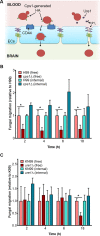
Loaded macrophages provide an alternative route for mutant cryptococci to gain access into the brain. (A) The roles of Cps1 and Ure1 in BBB crossing. HA (green ovals on the surface of the fungi [red]) made by Cps1 is recognized by the endothelial cell (EC) surface receptor CD44 (blue shapes), which triggers endocytosis of the fungal cells. The accumulation of ammonia generated by Ure1 may damage cellular junction proteins, facilitating fungal brain entry. (B and C) Transmigration of free or internalized fungi, comparing wild-type and either cps1Δ (B), or ure1Δ (C) mutants. Means plus SEM are plotted. * denotes P < 0.002 and P < 0.0001 in panels B and C, respectively (both determined by Sidak’s multiple-comparison test).
FIG 5
FIG 5
Visualization of Trojan horse crossing by real-time and electron microscopy. (A) hCMEC monolayers were grown on glass or 0.4% PA pads were grown to confluence and fixed, and adherens junctions were stained with anti-VE-cadherin antibody. Nuclei were stained with propidium iodide. (B and C) Two examples of loaded primary human monocytes crossing endothelia, from Movie S2 (B) and Movie S3 (C); see text for details. (D) TEM of a loaded monocyte in the process of transendothelial migration (left), with a corresponding drawing to identify structures (right). N, nucleus; V, vacuole; F, fungal cell; B, brain endothelial cell.
FIG 6
FIG 6
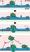
Mechanisms of brain infection by C. neoformans. (A) Depicted are wild-type fungi (red ovals) crossing the BBB either free (1) or within Trojan horse phagocytes (2). Mutant fungi (orange ovals) cannot cross alone (3) but can use Trojan horse transit as an alternative route. See text for details on transendothelial pore formation for Trojan horse transit. (B) Loaded phagocytes potentially contribute to brain invasion by pathways that do not involve true Trojan horse transit (2). Phagocytes can bring the fungus to the CNS internally and then exit the phagocyte by nonlytic exocytosis and cross the BBB as free yeast by transcytosis (1). Finally, we observed two instances of direct cell-to-cell transfer of fungal cells from phagocytes to endothelial cells (3), supporting the hypothesis of a “taxi” mechanism, where loaded phagocytes deliver fungal cells directly into brain endothelial cells. Pink, blood vessel lumen; pale green, brain parenchyma; blue cells, brain endothelial cells; green cells, infected phagocytes; shaded gray rectangle, the extracellular matrix that forms the BBB basal membrane.

References

    1. Brown GD, Denning DW, Gow NA, Levitz SM, Netea MG, White TC. 2012. Hidden killers: human fungal infections. Sci Transl Med 4:165rv13. doi:10.1126/scitranslmed.3004404. - DOI - PubMed
    1. Park BJ, Wannemuehler KA, Marston BJ, Govender N, Pappas PG, Chiller TM. 2009. Estimation of the current global burden of cryptococcal meningitis among persons living with HIV/AIDS. AIDS 23:525–530. doi:10.1097/QAD.0b013e328322ffac. - DOI - PubMed
    1. Fox DS, Djordjevic JT, Sorrell TC. 2011. Signaling cascades and enzymes as cryptococcus virulence factors, p 217–234. In Heitman J, Kozel TR, Kwon-Chung KJ, Perfect J, Casadevall A (ed), Cryptococcus: from human pathogen to model yeast. ASM Press, Washington, DC.
    1. Srikanta D, Santiago-Tirado FH, Doering TL. 2014. Cryptococcus neoformans: historical curiosity to modern pathogen. Yeast 31:47–60. doi:10.1002/yea.2997. - DOI - PMC - PubMed
    1. Serlin Y, Shelef I, Knyazer B, Friedman A. 2015. Anatomy and physiology of the blood-brain barrier. Semin Cell Dev Biol 38:2–6. doi:10.1016/j.semcdb.2015.01.002. - DOI - PMC - PubMed

LinkOut - more resources