. 2017 Jan 31;8(1):e02320-16.
doi: 10.1128/mBio.02320-16.

In Situ Tagged nsp15 Reveals Interactions with Coronavirus Replication/Transcription Complex-Associated Proteins

Affiliations

In Situ Tagged nsp15 Reveals Interactions with Coronavirus Replication/Transcription Complex-Associated Proteins

Jeremiah Athmer et al. mBio. .

Abstract

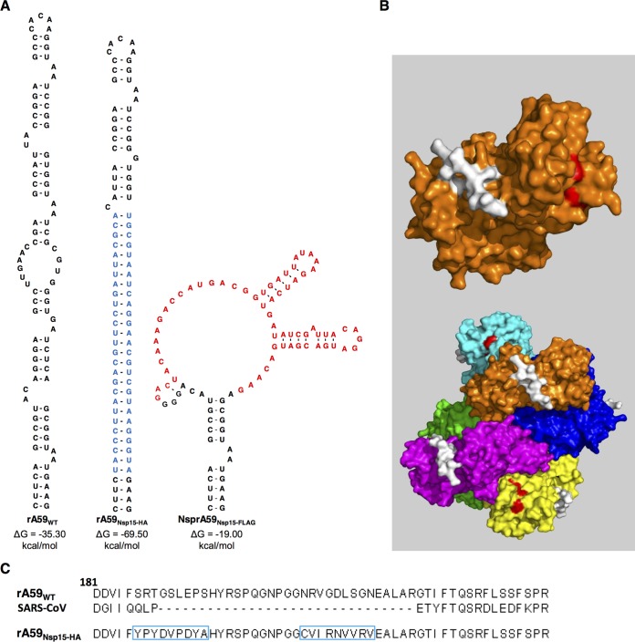
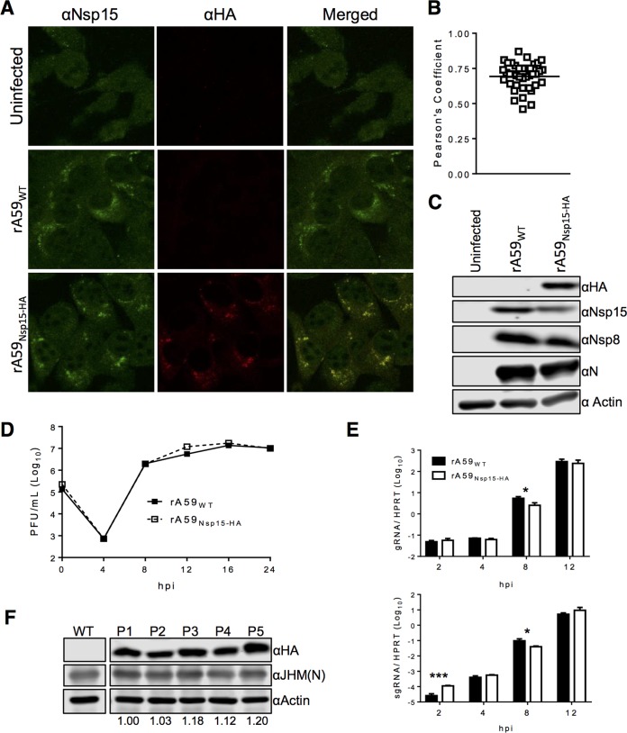
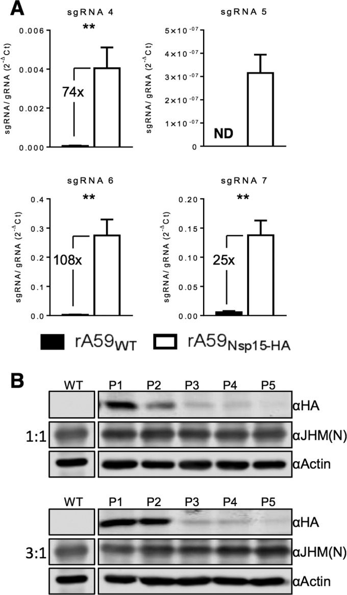
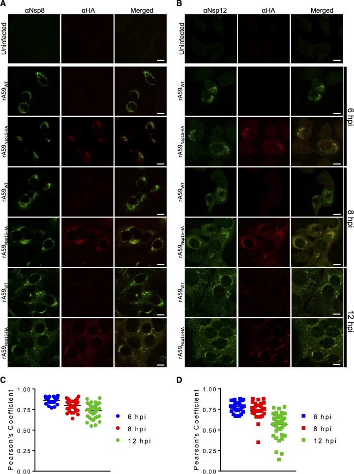
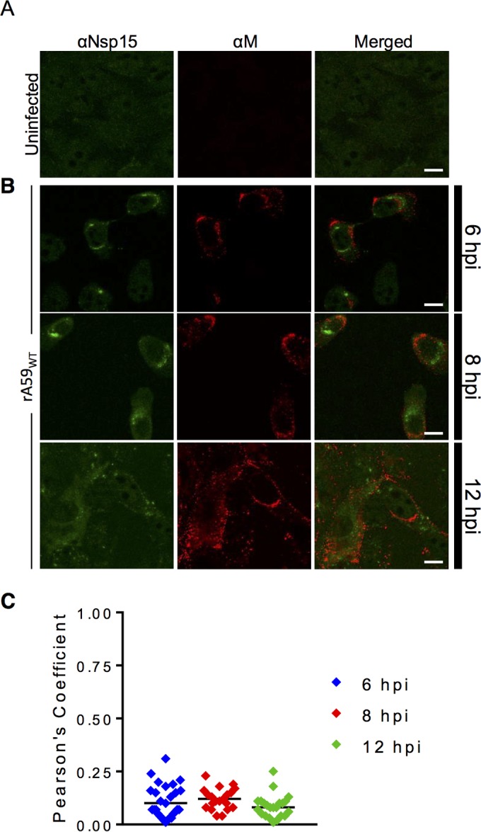
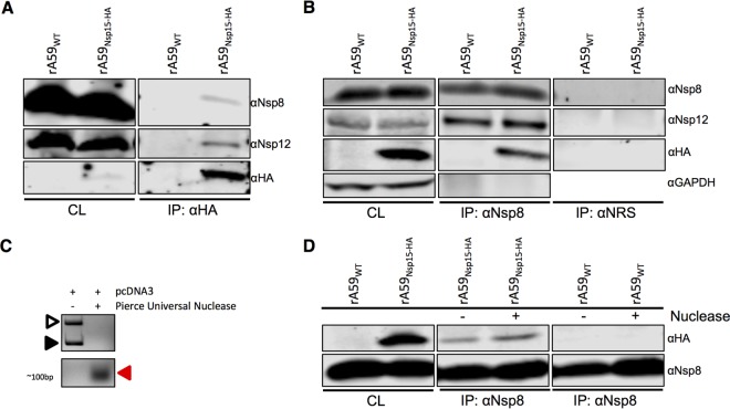
Coronavirus (CoV) replication and transcription are carried out in close proximity to restructured endoplasmic reticulum (ER) membranes in replication/transcription complexes (RTC). Many of the CoV nonstructural proteins (nsps) are required for RTC function; however, not all of their functions are known. nsp15 contains an endoribonuclease domain that is conserved in the CoV family. While the enzymatic activity and crystal structure of nsp15 are well defined, its role in replication remains elusive. nsp15 localizes to sites of RNA replication, but whether it acts independently or requires additional interactions for its function remains unknown. To begin to address these questions, we created an in situ tagged form of nsp15 using the prototypic CoV, mouse hepatitis virus (MHV). In MHV, nsp15 contains the genomic RNA packaging signal (P/S), a 95-bp RNA stem-loop structure that is not required for viral replication or nsp15 function. Utilizing this knowledge, we constructed an internal hemagglutinin (HA) tag that replaced the P/S. We found that nsp15-HA was localized to discrete perinuclear puncta and strongly colocalized with nsp8 and nsp12, both well-defined members of the RTC, but not the membrane (M) protein, involved in virus assembly. Finally, we found that nsp15 interacted with RTC-associated proteins nsp8 and nsp12 during infection, and this interaction was RNA independent. From this, we conclude that nsp15 localizes and interacts with CoV proteins in the RTC, suggesting it plays a direct or indirect role in virus replication. Furthermore, the use of in situ epitope tags could be used to determine novel nsp-nsp interactions in coronaviruses.

Importance: Despite structural and biochemical data demonstrating that the coronavirus nsp15 protein contains an endoribonuclease domain, its precise function during coronavirus infection remains unknown. In this work, we created a novel in situ tagged form of nsp15 to study interactions and localization during infection. This in situ tag was tolerated by MHV and did not affect viral replication. Utilizing this tag, we established that nsp15 localized to sites of replication but not sites of assembly throughout infection. Furthermore, we found that nsp15 interacted with the putative viral primase nsp8 and polymerase nsp12 during CoV infection. The strong association of nsp15 with replication complexes and interactions with replicative CoV enzymes suggest nsp15 is involved in CoV replication. These data and tools developed in this study help elucidate the function of nsp15 during infection and may be used to uncover other novel viral protein interactions.


Figures

FIG 1
FIG 1
Construction of the rA59Nsp15-HA in situ tag. (A, left) RNA secondary structure of the MHV packaging signal (38). (Middle) Mfold predicted RNA secondary structure of MHV P/S with the in situ HA tag and its complement (highlighted blue). (Right) Mfold predicted RNA secondary structure of Nsp15-3×FLAG with an in situ 3×FLAG tag (highlighted in red). (B, top) Surface rendering of an nsp15 monomer with the packaging signal highlighted in white. (Bottom) The MHV nsp15 hexamer with each monomer is depicted with a different color. Amino acids corresponding to the P/S are highlighted in white, and the catalytic triad is highlighted in red on the indicated monomer. The nsp15 crystal structure was retrieved from the PDB database (2GTH) and modified in Pymol (24). (C) MHV and SARS-CoV nsp15 protein sequence surrounding the P/S. The rA59Nsp15 P/S sequence with the HA sequence and its complementary sequence are boxed in blue.
FIG 2
FIG 2
nsp15-HA protein is expressed, and the HA sequence is stable after multiple rounds of passaging in vitro. (A to D) 17Cl-1 cells were infected with rA59Nsp15-HA or rA59WT and fixed at 8 hpi. Fixed cells were costained with anti-nsp15 (green) and anti-HA (red). (B) Pearson’s correlation coefficients for at least 25 individual cells were analyzed and plotted. (C) 17Cl-1 cells were infected with rA59Nsp15-HA or rA59WT, at an MOI of 5, and total cell lysates were harvested at 12 hpi. Cell lysates were then immunoblotted with the indicated antibodies as described in Materials and Methods. (D and E) rA59Nsp15-HA has nearly identical replication kinetics to rA59WT virus on 17Cl-1 cells. (D) 17Cl-1 cells were infected at an MOI of 0.1, and viral progeny were collected at the indicated time points. Input virus was collected following the adsorption step. Virus titers were determined by plaque assay on HeLa-MVR cells (E). 17Cl-1 cells were infected with rA59WT or rA59Nsp15-HA at an MOI of 5. Cells were collected at the indicated time points. Levels of gRNA and sgRNA were determined by RT-qPCR and normalized to hypoxanthine-guanine phosphoribosyltransferase (HPRT). All data are from a single experiment and are representative of two independent experiments. (F) rA59Nsp15-HA was passaged on 17Cl-1 cells at an estimated MOI of 0.1. Virus from each passage was collected and used to infect 17Cl-1 cells for serial continued passaging and immunoblotting. Cell lysates were analyzed by immunoblotting with the indicated antibodies. The ratio of nsp15-HA to N protein was normalized to P1 and is listed below each passage. Scale bars (10 μm) are shown. Error bars indicate range (C) and standard error of the mean (SEM) (D). *, P < 0.05, and ***, P < 0.001, by Students t test.
FIG 3
FIG 3
rA59Nsp15-Ha virus is defective in its ability to selectively package gRNA, resulting in a loss of fitness. (A) Supernatants from rA59Nsp15-HA- and rA59WT-infected 17Cl-1 cells were collected, and cell debris was removed. Virions were pelleted by ultracentrifugation through a 30% sucrose cushion, and viral RNA was isolated. The ratio of sgRNAs to gRNA in viral RNA was measured by RT-qPCR. (B) 17Cl-1 cells were infected with the indicated ratios of rA59Nsp15-HA to rA59WT, with a total MOI of 0.1. Progeny viruses were then passaged at an estimated MOI of 0.1 and collected. Progeny virus from each passage was used to infect 17Cl-1 cells at an MOI of 0.1. Cell lysates were collected at 16 hpi and immunoblotted with the indicated antibodies. Figures 2D and 3B were analyzed in parallel and are imaged from the same immunoblot. **, P < 0.01 by Mann-Whitney U test.
FIG 4
FIG 4
nsp15 strongly colocalizes with nsp8 and nsp12 during infection. (A and B) Uninfected 17Cl-1 cells were costained with anti-nsp8 (green) and anti-HA (red) (A) or anti-nsp12 (green) and anti-HA (red) (B). (C and D) 17Cl-1 cells were infected with rA59Nsp15-HA or rA59WT at an MOI of 1. Cells were fixed and stained at 6 (top), 8 (middle), or 12 (bottom) hpi and costained with either anti-HA and anti-nsp8 (C) or anti-HA and anti-nsp12 (D). Each image is representative of at least 50 cells from two independent experiments. Pearson’s correlation coefficients for at least 25 individual cells were analyzed for each time point for nsp8 (E) and nsp12 (F) and plotted. Scale bars (10 μm) are shown.
FIG 5
FIG 5
nsp15 does not localize to sites of assembly during infection. (A) Uninfected 17Cl-1 cells were costained with anti-nsp15 (green) and anti-M (red). (B) 17Cl-1 cells were infected with rA59WT at an MOI of 1. Cells were fixed and stained at 6 (top), 8 (middle), or 12 (bottom) hpi and costained with anti-nsp15 (green) and anti-M protein (red). (C) Pearson’s correlation coefficients for at least 25 individual cells were analyzed for each time point and plotted.
FIG 6
FIG 6
nsp15 interacts with the RTC proteins nsp8 and nsp12 in an RNA-independent manner. (A, B, and D). 17Cl-1 cells were infected with rA59Nsp15-HA or rA59WT at an MOI of 0.1 and collected at 20 hpi. Cell lysates were subjected to immunoprecipitation with anti-HA (A), anti-nsp8 (B and D), or normal rabbit serum (NRS) (B). Cell lysates (CL) and eluted proteins were immunoblotted with the indicated antibodies. (C) Lysis buffer was spiked with 3 μg of pcDNA3 plasmid with or without nuclease (250 U) and incubated for 8 h. The DNA was analyzed by agarose gel electrophoresis and visualized with ethidium bromide. The solid arrowhead represents linearized plasmid DNA, and the open arrowhead represents the supercoiled DNA. The red arrowhead indicates degraded products of ~100 bp. (D) 17Cl-1 cells were infected as described above, and cell lysates were subjected to immunoprecipitation as described in panel B in the presence or absence of Pierce Universal Nuclease.

References

    1. Fehr AR, Channappanavar R, Perlman S. 2016. Middle East respiratory syndrome: emergence of a pathogenic human coronavirus. Annu Rev Med. doi:10.1146/annurev-med-051215-031152. - DOI - PMC - PubMed
    1. Hilgenfeld R, Peiris M. 2013. From SARS to MERS: 10 years of research on highly pathogenic human coronaviruses. Antiviral Res 100:286–295. doi:10.1016/j.antiviral.2013.08.015. - DOI - PMC - PubMed
    1. Perlman S, Netland J. 2009. Coronaviruses post-SARS: update on replication and pathogenesis. Nat Rev Microbiol 7:439–450. doi:10.1038/nrmicro2147. - DOI - PMC - PubMed
    1. Neuman BW, Chamberlain P, Bowden F, Joseph J. 2014. Atlas of coronavirus replicase structure. Virus Res 194:49–66. doi:10.1016/j.virusres.2013.12.004. - DOI - PMC - PubMed
    1. Angelini MM, Akhlaghpour M, Neuman BW, Buchmeier MJ. 2013. Severe acute respiratory syndrome coronavirus nonstructural proteins 3, 4, and 6 induce double-membrane vesicles. mBio 4:e00524-13. doi:10.1128/mBio.00524-13. - DOI - PMC - PubMed

Publication types

Substances