

Case Report of S1Q3T3 Electrocardiographic Abnormality in a Pregnant Asthmatic Patient During Acute Bronchospasm
- PMID: 28144025
- PMCID: PMC5297401
- DOI: 10.12659/ajcr.901661
Case Report of S1Q3T3 Electrocardiographic Abnormality in a Pregnant Asthmatic Patient During Acute Bronchospasm
Abstract
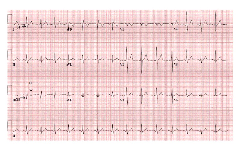
BACKGROUND Asthma is the most common chronic pulmonary disease during pregnancy. Several previous reports have documented reversible electrocardiographic changes during severe acute asthma attacks, including tachycardia, P pulmonale, right bundle branch block, right axis deviation, and ST segment and T wave abnormalities. CASE REPORT We present the case of a pregnant patient with asthma exacerbation in which acute bronchospasm caused S1Q3T3 abnormality on an electrocardiogram (ECG). The complete workup of ECG findings of S1Q3T3 was negative and correlated with bronchospasm. The S1Q3T3 electrocardiographic abnormality can be seen in acute bronchospasm in pregnant women. The other causes like pulmonary embolism, pneumothorax, acute lung disease, cor pulmonale, and left posterior fascicular block were excluded. CONCLUSIONS Asthma exacerbations are of considerable concern during pregnancy due to their adverse effect on the fetus, and optimization of asthma treatment during pregnancy is vital for achieving good outcomes. Prompt recognition of electrocardiographic abnormality and early treatment can prevent adverse perinatal outcomes.
Conflict of interest statement
Conflicts of Interest: None declared
Figures

Similar articles
-
The S1Q3T3 Electrocardiographic Abnormality as a Result of Massive Empyema due to Pyogenic Liver Abscess: A Case Report.Clin Case Rep. 2025 Feb 7;13(2):e70191. doi: 10.1002/ccr3.70191. eCollection 2025 Feb. Clin Case Rep. 2025. PMID: 39926638 Free PMC article.
-
Reversible electrocardiographic changes in severe acute asthma.Thorax. 1977 Jun;32(3):328-32. doi: 10.1136/thx.32.3.328. Thorax. 1977. PMID: 882948 Free PMC article.
-
The electrocardiogram in acute pulmonary embolism.Prog Cardiovasc Dis. 1975 Jan-Feb;17(4):247-57. doi: 10.1016/s0033-0620(75)80016-8. Prog Cardiovasc Dis. 1975. PMID: 123074
-
Acute asthma in pregnancy.Crit Care Clin. 2004 Oct;20(4):731-45, x. doi: 10.1016/j.ccc.2004.05.013. Crit Care Clin. 2004. PMID: 15388199 Review.
-
Asthma and pregnancy.Obstet Gynecol. 2006 Sep;108(3 Pt 1):667-81. doi: 10.1097/01.AOG.0000235059.84188.9c. Obstet Gynecol. 2006. PMID: 16946229 Review.
Cited by
-
S1Q3T3 Electrocardiographic Pattern in a Case of Colonic Ileus: A Case Report.Cureus. 2025 Mar 3;17(3):e79985. doi: 10.7759/cureus.79985. eCollection 2025 Mar. Cureus. 2025. PMID: 40177441 Free PMC article.
-
The S1Q3T3 Electrocardiographic Abnormality as a Result of Massive Empyema due to Pyogenic Liver Abscess: A Case Report.Clin Case Rep. 2025 Feb 7;13(2):e70191. doi: 10.1002/ccr3.70191. eCollection 2025 Feb. Clin Case Rep. 2025. PMID: 39926638 Free PMC article.
-
Massive pleural effusion masquerading as acute pulmonary thromboembolism - An unreported entity.Lung India. 2022 Nov-Dec;39(6):589-592. doi: 10.4103/lungindia.lungindia_663_21. Lung India. 2022. PMID: 36629244 Free PMC article. No abstract available.
-
Left Atrial Myxoma Masquerading as Pulmonary Embolism on Electrocardiogram: A Case Report.J Med Cases. 2021 Dec;12(12):511-515. doi: 10.14740/jmc3775. Epub 2021 Dec 2. J Med Cases. 2021. PMID: 34970376 Free PMC article.
