Review
. 2016 Dec 12:12:2694-2718.
doi: 10.3762/bjoc.12.267. eCollection 2016.

Computational methods in drug discovery

Affiliations
Review

Computational methods in drug discovery

Sumudu P Leelananda et al. Beilstein J Org Chem. .

Abstract

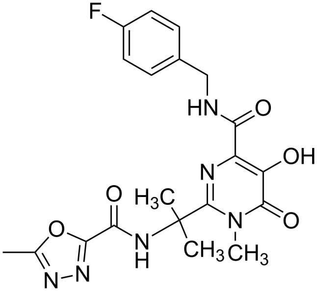
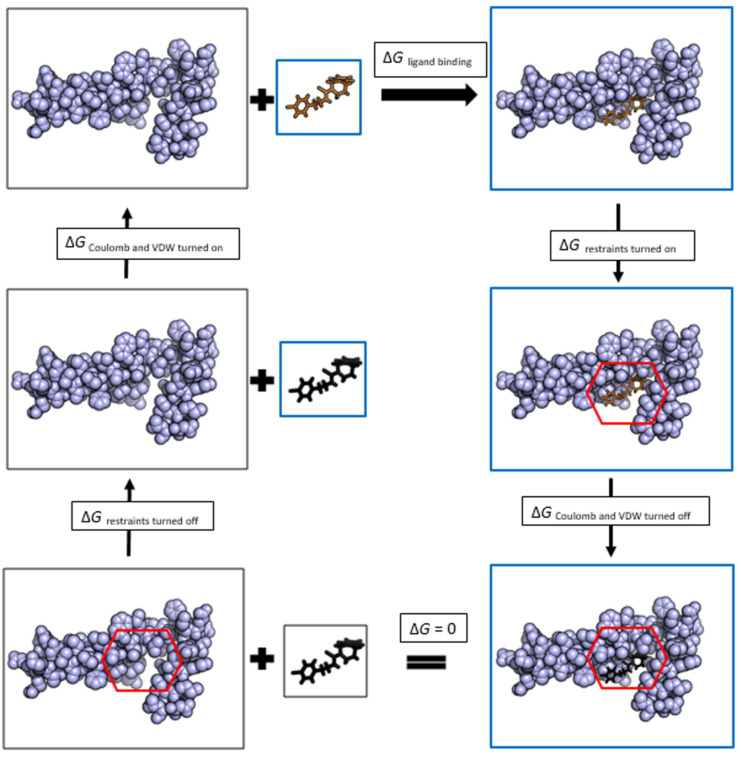
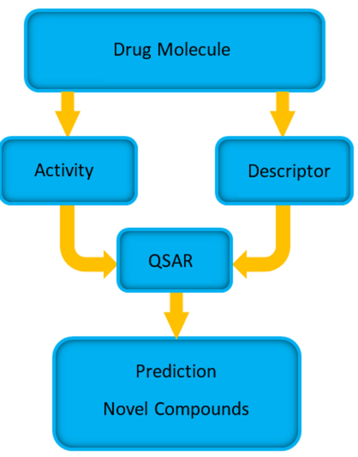
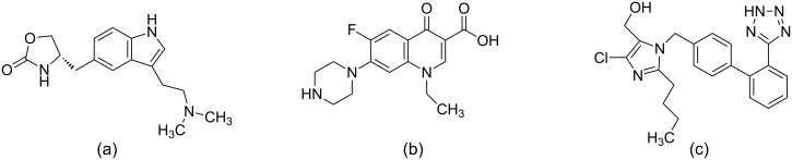
The process for drug discovery and development is challenging, time consuming and expensive. Computer-aided drug discovery (CADD) tools can act as a virtual shortcut, assisting in the expedition of this long process and potentially reducing the cost of research and development. Today CADD has become an effective and indispensable tool in therapeutic development. The human genome project has made available a substantial amount of sequence data that can be used in various drug discovery projects. Additionally, increasing knowledge of biological structures, as well as increasing computer power have made it possible to use computational methods effectively in various phases of the drug discovery and development pipeline. The importance of in silico tools is greater than ever before and has advanced pharmaceutical research. Here we present an overview of computational methods used in different facets of drug discovery and highlight some of the recent successes. In this review, both structure-based and ligand-based drug discovery methods are discussed. Advances in virtual high-throughput screening, protein structure prediction methods, protein-ligand docking, pharmacophore modeling and QSAR techniques are reviewed.

Keywords: ADME; LBDD; QSAR; SBDD; computer-aided drug design; docking; free energy; high-throughput screening; lead optimization; machine learning; pharmacophore; scoring; target flexibility.


Figures

Figure 1
Figure 1
Schematic representation of a computer-aided drug discovery (CADD) pipeline. CADD methods are broadly classified into structure-based and ligand-based methods. Structure-based methods require the 3D information of the target to be known. Ligand-based methods are used when the 3D structure of the target is not known. They use information about the molecules that bind to the target of interest. Hits are identified, filtered and optimized to obtain potential drug candidates that will be experimentally tested in vitro.
Figure 2
Figure 2
FDA approved drugs Saquinavir and Amprenavir for the treatment of HIV infections. (a) The structure of Saquinavir in complex with HIV-1 protease (3OXC) (b) the structure of Amprenavir in complex with HIV-1 protease (3NU3) (c) the molecular structure of Saquinavir and (d) the molecular structure of Amprenavir. Amprenavir and Saquinavir target HIV-1 protease and, in part, have been discovered through structure-based computer aided drug discovery methods.
Figure 3
Figure 3
(a) The crystal structure showing the binding of Dorzolamide (orange) to carbonic anhydrase II (purple) (4M2U) (b) the structure of Dorzolamide. Dorzolamide is an FDA approved drug that targets carbonic anhydrase II to treat patience with glaucoma.
Figure 4
Figure 4
The best ligand binding site identified by SiteHound in HIV-1 protease. The ligand binding pocket is shown in blue spheres and is the known inhibitor binding site of HIV-1 protease.
Figure 5
Figure 5
Binding mode prediction. The known inhibitor Dorzolamide is docked into Carbonic anhydrase II crystal structure (4M2U) (blue) using AutoDock Vina. Four binding poses predicted are shown in green, cyan, red and yellow. The molecular structure of Dorzolamide is shown in Figure 3b.
Figure 6
Figure 6
The molecular structure of Raltegravir. Raltegravir is an FDA approved drug used in the treatment of HIV infection.
Figure 7
Figure 7
An example alchemical thermodynamic cycle for a protein–ligand binding free energy calculation. The protein is shown in blue spheres. The ligand, depicted in solid black, indicates there are no coulombic or van der Waals (VDW) interactions with the environment. The ligand, depicted in solid orange, indicates there are coulombic and VDW interactions with its environment. The systems that are subjected to simulations in each cycle are highlighted in blue boxes. All simulations are run in a water environment. The first step is to add restraints between ligand and the protein in order to keep the ligand confined to the binding pocket and to avoid the ligand leaving the pocket when its interactions are removed. The systems with restraints turned on are indicated by red hexagons. In the next step the coulombic and VDW interactions of the ligand are removed. This step is followed by the removal of the restraints applied to the ligand. Next the coulombic and VDW interactions of the ligand are turned on such that the ligand is in contact with solvent. Summing up the free energy changes along the thermodynamic cycle would give the protein–ligand binding free energy.
Figure 8
Figure 8
Schematic diagram showing the steps involved in QSAR. Known drug molecule activity and descriptor data is obtained and the mathematical model of QSAR is built such that descriptors can predict the activity of each molecule. The predictive power of models are validated and used in predicting activities of novel compounds.
Figure 9
Figure 9
A few drugs discovered with the help of ligand-based drug discovery tools. (a) Zolmitriptan: used as a treatment to migraine (b) Norfloxacin: used in urinary tract infections and (c) Losartan: used to treat hypertension.

References

    1. Tollman P. A Revolution in R&D: How genomics and genetics are transforming the biopharmaceutical industry. 2001.
    1. Yang L, Wang W, Sun Q, Xu F, Niu Y, Wang C, Liang L, Xu P. Bioorg Med Chem Lett. 2016;26:2801–2805. doi: 10.1016/j.bmcl.2016.04.067. - DOI - PubMed
    1. Karthick V, Nagasundaram N, Doss C G P, Chakraborty C, Siva R, Lu A, Zhang G, Zhu H. Infect Dis Poverty. 2016;5:No. 12. doi: 10.1186/s40249-016-0105-1. - DOI - PMC - PubMed
    1. Clark A J, Tiwary P, Borrelli K, Feng S, Miller E B, Abel R, Friesner R A, Berne B J. J Chem Theory Comput. 2016;12:2990–2998. doi: 10.1021/acs.jctc.6b00201. - DOI - PubMed
    1. Chao W R, Yean D, Amin K, Green C, Jong L. J Med Chem. 2007;50:3412–3415. doi: 10.1021/jm070040e. - DOI - PubMed