Application of time-spatial labeling inversion pulse magnetic resonance imaging in the diagnosis of spontaneous intracranial hypotension due to high-flow cerebrospinal fluid leakage at C1-2
- PMID: 28144490
- PMCID: PMC5234307
- DOI: 10.4103/2152-7806.196765
Application of time-spatial labeling inversion pulse magnetic resonance imaging in the diagnosis of spontaneous intracranial hypotension due to high-flow cerebrospinal fluid leakage at C1-2
Abstract
Background: Spontaneous intracranial hypotension (SIH) due to cerebrospinal fluid (CSF) leakage at C1-2 poses diagnostic and therapeutic challenges to spine surgeons. Although computed tomography (CT) myelography has been the diagnostic imaging modality of choice for identifying the CSF leakage point, extradural CSF collection at C1-2 on conventional CT myelography or magnetic resonance imaging (MRI) may often be a false localizing sign.
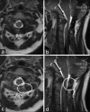
Case description: The present study reports the successful application of time-spatial labeling inversion pulse (T-SLIP) MRI, which enabled the precise identification of the CSF leakage point at C1-2 in a 28-year-old woman with intractable SIH. After identifying the leakage point using both CT myelography and T-SLIP MRI, surgery was performed to seal the CSF leak. Intraoperatively, a pouch suggestive of an extradural arachnoid cyst around the left C2 nerve root was found, which was repaired by packing the pouch with muscle and fibrin glue. Clinical improvement was observed shortly after surgery, and postoperative imaging revealed the disappearance of the CSF leakage.
Conclusions: T-SLIP MRI may provide useful information on the flow dynamics of CSF in SIH patients due to high-flow leakage. However, further experience is required to assess its sensitivity and specificity as an imaging modality for identifying CSF leakage points.
Keywords: C1-2; CT myelography; cerebrospinal fluid leakage; spontaneous intracranial hypotension; time-spatial labeling inversion pulse (T-SLIP) MRI.
Conflict of interest statement
On behalf of all authors, the corresponding author declares no conflict of interest.
Figures


Similar articles
-
Patterns of cerebrospinal fluid (CSF) distribution in patients with spontaneous intracranial hypotension: Assessed with magnetic resonance myelography.J Chin Med Assoc. 2017 Feb;80(2):109-116. doi: 10.1016/j.jcma.2016.02.013. Epub 2016 Oct 12. J Chin Med Assoc. 2017. PMID: 27743810
-
Cerebrospinal fluid leak presented with the C1-C2 sign caused by spinal canal stenosis: a case report.BMC Neurol. 2020 Apr 23;20(1):151. doi: 10.1186/s12883-020-01697-1. BMC Neurol. 2020. PMID: 32326909 Free PMC article.
-
Spontaneous Intracranial Hypotension: A Systematic Imaging Approach for CSF Leak Localization and Management Based on MRI and Digital Subtraction Myelography.AJNR Am J Neuroradiol. 2019 Apr;40(4):745-753. doi: 10.3174/ajnr.A6016. Epub 2019 Mar 28. AJNR Am J Neuroradiol. 2019. PMID: 30923083 Free PMC article.
-
Spontaneous Intracranial Hypotension: Imaging in Diagnosis and Treatment.Radiol Clin North Am. 2019 Mar;57(2):439-451. doi: 10.1016/j.rcl.2018.10.004. Epub 2018 Dec 7. Radiol Clin North Am. 2019. PMID: 30709479 Review.
-
Imaging of the Spontaneous Low Cerebrospinal Fluid Pressure Headache: A Review.Can Assoc Radiol J. 2020 May;71(2):174-185. doi: 10.1177/0846537119888395. Epub 2020 Jan 24. Can Assoc Radiol J. 2020. PMID: 32063004 Review.
References
-
- Cohen-Gadol AA, Mokri B, Piepgras DG, Meyer FB, Atkinson JL. Surgical anatomy of dural defects in spontaneous spinal cerebrospinal fluid leaks. Neurosurgery. 2006;58(4 Suppl 2):238–45. - PubMed
-
- Inamasu J, Guiot BH. Intracranial hypotension with spinal pathology. Spine J. 2006;6:591–9. - PubMed
-
- Inamasu J, Nakamura Y, Orii M, Saito R, Kuroshima Y, Mayanagi K, Ichikizaki K, Doi H. Treatment of spontaneous intracranial hypotension secondary to C-2 meningeal cyst by surgical packing: Case report. Neurol Med Chir. 2004;44:326–30. - PubMed
-
- Inamasu J, Nakatsukasa M. Blood patch for spontaneous intracranial hypotension caused by cerebrospinal fluid leak at C1-2. Clin Neurol Neurosurg. 2007;109:716–9. - PubMed
-
- Ishibe T, Senzoku F, Kamba Y, Ikeda N, Mikawa Y. Time-spatial labeling inversion pulse magnetic rresonance imaging of cystic lesions of the spinal cord. World Neurosurg. 2016;88:693.e13–21. - PubMed
Publication types
LinkOut - more resources
Full Text Sources
Other Literature Sources
Miscellaneous
