

Ultra-delayed lumbar surgical wound hematoma
- PMID: 28144491
- PMCID: PMC5234306
- DOI: 10.4103/2152-7806.196766
Ultra-delayed lumbar surgical wound hematoma
Abstract
Background: There exists an inherent risk of increased venous thromboembolism (VTE) in surgical spine patients, which is independent of their existing risk factors. Prophylaxis and treatment of VTE is an imprecise practice and may have serious complications even well after the initial surgery. Furthermore, there are no clear guidelines on how to manage postoperative spine patients with regards to the timing of anticoagulation.
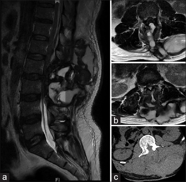
Case description: Here, we present the case of a middle-aged male, status post L2/3 laminectomy and discectomy who developed bilateral below the knee deep venous thrombosis. He was started on Enoxaparin and transitioned to Warfarin and returned with axial back pain, and was found to have a postoperative hematoma almost 3 weeks later in a delayed fashion.
Conclusion: Delayed surgical wound hematoma with neural compression is an important complication to identify and should remain high on the differential diagnosis in patients on warfarin who present with axial spinal pain.
Keywords: Anticoagulation; complication; spinal decompression; venous thromboembolism; wound hematoma.
Conflict of interest statement
There are no conflicts of interest.
Figures
Similar articles
-
Low-molecular-weight heparin prophylaxis 24 to 36 hours after degenerative spine surgery: risk of hemorrhage and venous thromboembolism.Spine (Phila Pa 1976). 2013 Nov 1;38(23):E1498-502. doi: 10.1097/BRS.0b013e3182a4408d. Spine (Phila Pa 1976). 2013. PMID: 23873245
-
High Risk of Symptomatic Venous Thromboembolism After Surgery for Spine Metastatic Bone Lesions: A Retrospective Study.Clin Orthop Relat Res. 2019 Jul;477(7):1674-1686. doi: 10.1097/CORR.0000000000000733. Clin Orthop Relat Res. 2019. PMID: 31135550 Free PMC article.
-
Decreased incidence of venous thromboembolism after spine surgery with early multimodal prophylaxis: Clinical article.J Neurosurg Spine. 2014 Oct;21(4):677-84. doi: 10.3171/2014.6.SPINE13447. Epub 2014 Aug 8. J Neurosurg Spine. 2014. PMID: 25105337
-
Venous thromboembolism in cancer patients.Hosp Pract (1995). 2014 Dec;42(5):24-33. doi: 10.3810/hp.2014.12.1156. Hosp Pract (1995). 2014. PMID: 25485915 Review.
-
Extensive postoperative epidural hematoma after full anticoagulation: case report and review of the literature.J Spinal Cord Med. 2007;30(3):282-7. doi: 10.1080/10790268.2007.11753938. J Spinal Cord Med. 2007. PMID: 17684896 Free PMC article. Review.
References
-
- Stein P, Goldhaber S, Gottschalk A, Hull R, Hyers T, Leeper K, et al. Opinions regarding the diagnosis and management of venous thromboembolic disease. ACCP Consensus Committee on Pulmonary Embolism. American College of Chest Physicians. Chest. 1998;113:499–504. - PubMed
-
- Browd SR, Ragel BT, Davis GE, Scott AM, Skalabrin EJ, Couldwell WT. Prophylaxis for deep venous thrombosis in neurosurgery: A review of the literature. Neurosurg Focus. 2004;17:E1. - PubMed
-
- Carroll SG, Malhotra R, Eustace D, Sharr M, Morcos S. Spontaneous spinal extradural hematoma during pregnancy. J Matern Fetal Med. 1997;6:218–9. - PubMed
-
- Crisi G, Sorgato P, Colombo A, Scarpa M, Falasca A, Angiari P. Gadolinium-DTPA-enhanced MR imaging in the diagnosis of spinal epidural haematoma. Report of a case. Neuroradiology. 1990;32:64–6. - PubMed
Publication types
LinkOut - more resources
Full Text Sources
Other Literature Sources