Review
. 2017 Mar;367(3):427-444.
doi: 10.1007/s00441-016-2545-0. Epub 2017 Jan 31.

Development of the lung

Affiliations
Review

Development of the lung

Johannes C Schittny. Cell Tissue Res. 2017 Mar.

Abstract

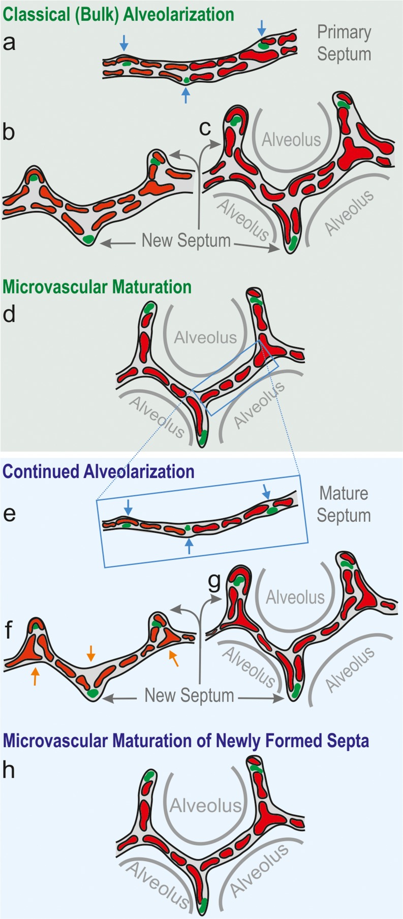
To fulfill the task of gas exchange, the lung possesses a huge inner surface and a tree-like system of conducting airways ventilating the gas exchange area. During lung development, the conducting airways are formed first, followed by the formation and enlargement of the gas exchange area. The latter (alveolarization) continues until young adulthood. During organogenesis, the left and right lungs have their own anlage, an outpouching of the foregut. Each lung bud starts a repetitive process of outgrowth and branching (branching morphogenesis) that forms all of the future airways mainly during the pseudoglandular stage. During the canalicular stage, the differentiation of the epithelia becomes visible and the bronchioalveolar duct junction is formed. The location of this junction stays constant throughout life. Towards the end of the canalicular stage, the first gas exchange may take place and survival of prematurely born babies becomes possible. Ninety percent of the gas exchange surface area will be formed by alveolarization, a process where existing airspaces are subdivided by the formation of new walls (septa). This process requires a double-layered capillary network at the basis of the newly forming septum. However, in parallel to alveolarization, the double-layered capillary network of the immature septa fuses to a single-layered network resulting in an optimized setup for gas exchange. Alveolarization still continues, because, at sites where new septa are lifting off preexisting mature septa, the required second capillary layer will be formed instantly by angiogenesis. The latter confirms a lifelong ability of alveolarization, which is important for any kind of lung regeneration.

Keywords: Alveolarization; Branching morphogenesis; Lung development; Microvascular maturation; Pulmonary acinus.


Conflict of interest statement

The author declares that he has no conflict of interest.

Figures

Fig. 1
Fig. 1
Development of the airways and arteries. The stages of lung development (blue) are correlated to the development of the airways (black) and the arteries (red). On average, an airway of a human lung ends in an alveolar saccule after 23 generations; however, due to the shape of the lung, a range of 18–30 generations has been observed. Pre-acinar arteries are formed out of a capillary plexus surrounding the growing lung buds (vasculogenesis). Intra-acinar arteries grow by angiogenesis (based on Hislop , adapted from Schittny and by courtesy of Springer, Heidelberg)
Fig. 2
Fig. 2
Early human lung development. At E26, post-conceptional, the anlage of the two lungs forms by outpouchings of the foregut on both sides lateral of the anlage of the trachea (a) (Cardoso and Lu 2006). The prospective trachea forms by a distal-to-proximal segregation from the foregut. At E32, the two lung anlages give rise to the two future main bronchi (b). Due to continued branching, the lobar bronchi are formed at E37 (c). Later, at E41, the segmental bronchi follow (d). Organogenesis is completed after the formation of the pleura (d). U upper lobe; m middle lobe; l lower lobe (from Schittny , by courtesy of Springer, Heidelberg)
Fig. 3
Fig. 3
Morphological development of the lung parenchyma during the pseudoglandular, canalicular and saccular stage. The epithelial tubules branch repeatedly during the pseudoglandular stage and penetrate into the surrounding mesenchyme (a, open arrow, branching point). The mesenchyme contains a loose capillary network (a). The epithelium itself is tall and columnar (d). The canalicular stage (b) is characterized (1) by a differentiation of the epithelial cells into type I and type II epithelial cells (e , f), (2) by a widening of the future airways (b), (3) by a multiplication of the capillaries and their first close contacts to the epithelium (b) and (4) by the formation of first future air–blood barriers (ef). During the saccular stage (c), thick immature inter-airspace septa are formed due to a further condensation of the mesenchyme. The immature septa contain a double-layered capillary network, one layer on either side of the septum. The terminal ends of the bronchial tree represent wide spaces and are called saccules (asterisks). (ac modified from Caduff et al. ; d, e modified from Burri and Weibel , f from Woods and Schittny , by courtesy of Cambridge University Press, New York)
Fig. 4
Fig. 4
Formation of the air–blood barrier. During the early canalicular stage (a, rat lung) the epithelium of the terminal airways is still cuboidal and glycogen-rich (closed arrowhead). Already a bit more proximal, the epithelium begins to flatten out (open arrowhead) and starts to form the first optimized future air–blood barriers. During the latter process, the capillaries of the mesenchyme (closed arrow) “move” towards the epithelium (open arrow). In humans (b), remnants of the cuboidal epithelium (closed arrowhead) are still present at the uttermost periphery of the gas exchange region at postnatal day 26, even if alveolarization had already started approximately 6 weeks earlier. This finding illustrates the large overlap between different phases of lung development, especially if peripheral and central parts are compared. Light microscopical images, bar 50 μm. (From Woods and Schittny , by courtesy of Cambridge University Press, New York)
Fig. 5
Fig. 5
3D visualization of distal conducting airways and acini of a rat lung. Distal conducting airways (bronchioles) are shown in green. The bronchioalveolar duct junction is labeled with a red disk. The disks were used as segmentation stoppers in order to separate the conducting from the acinar airways (yellow). The lung tissue is shown in shades of gray. Left panel conducting airways and segmentation stopper; right panel four acini are shown in addition to the structures shown in the left panel; bar 0.5 mm. (From Haberthur et al. , by courtesy of The American Physiological Society, Bethesda, MD, USA)
Fig. 6
Fig. 6
Development of the bronchial tree of the conducting airways of the right middle rat lung lobe. The walls of the conducting airways are shown in gray. The colored spheres represent the entrances of the acini and were used as segmentation stopper. These three-dimensional visualizations show the large similarity of the conducting airways structure at Days 4, 10, 21, 36 and 60 and between different individuals (a). b The bronchial tree at Day 60 embedded in the surrounding lung parenchyma. Due to the monopodial branching pattern of the rat airways, the acinar entrances are not evenly distributed inside any lung lobe. Most of the acinar entrances are located on a virtual cylinder around the main airways and the most peripheral ones facing the pleura with their distal part. As a consequence an outer cortex exists that is free of acinar entrances (altered from Barre et al. , by courtesy of The American Physiological Society, Bethesda, MD, USA)
Fig. 7
Fig. 7
Visualization of alveolarization. 3D visualizations of terminal air spaces were obtained during postnatal rat lung development. At postnatal day 4 (a), large saccules are observed. They were formed by branching morphogenesis. Between days 6 and 36 (ae), many low-rising septa are present, which are indicative for newly-forming septa (arrows). On the same days, higher-rising, mature septa are also visible (arrowheads), while between days 4 and 21 (ad), the size of the terminal airspaces decreases, with an increase of the airspaces observed between days 21 and 60 (e , f). Synchrotron radiation-based X-ray tomographic microscopy was applied for 3D imaging after a classical embedding for electron microscopy. Bar 50 μm. Due to the perspective view, the bar is only correct at the surface of the sample. (From Mund et al. , by courtesy of Springer, Heidelberg)
Fig. 8
Fig. 8
Schematic of classical and continued alveolarization. The interairspace walls present in the saccular stage represent the primary septa. They contain a double-layered immature capillary network (a). Each layer appears as a perforated sheet of capillaries (see also Fig. 9). Smooth muscle cell precursors, elastic fibers and collagen fibrils (green spots) accumulate at sites where new septa (or secondary septa) will be formed (blue arrows, a). The secondary septa form by an upfolding (blue arrows) of one of the two capillary layers (red, b). The resulting newly formed secondary septa (gray arrows) subdivide preexisting airspaces and the alveoli are born (c). At this stage, most of the septa are immature because they possess two capillary layers. During microvascular maturation, the double-layered capillary network fuses to a single-layered one. As a first approximation, microvascular maturation takes place in parallel to alveolarization. Therefore, a significant fraction of new septa are formed starting from a preexisting mature septum containing only a single-layered capillary network (e). Now following the mechanism of continued alveolarization, new alveolar septa are still formed by an upfolding of the capillary layer (red, df), even if the alveolar surface opposing the upfolding is now missing its capillaries (d). This gap is immediately closed by angiogenesis (orange arrows in e , f). In both modes of alveolarization, a sheetlike capillary layer folds up (b , e) in order to form a double-layered capillary network inside the newly formed septum (c, f). Regardless of how and when a new septum is formed, it will mature shortly after by a fusion of the double-layered capillary network. (Altered and extended from Burri ; Burri ; Schittny and Mund ; Woods and Schittny , by courtesy of Cambridge University Press, New York)
Fig. 9
Fig. 9
Late alveolarization. Synchrotron radiation-based X-ray tomographic microscopy was used for a 3D visualization of vascular casts (Vascular Mercox®) of 21-day-old rat lungs. The lumen of the capillaries are shown, which is identical to their inner surface. Inside the cavity of an alveolus, an upfolding of a single-layered capillary network is shown (blue dashed lines in a). These kinds of upfoldings are viewed as the formation of new septa. The tomographic dataset permitted to view the backside of the same septum (b). A local duplication of the existing capillary network was detected at the basis of the up-folding (covering of the blue dashed line in b). While parts of the duplication are already formed (arrowhead), the remaining duplication is just forming, most likely by sprouting angiogenesis (arrow in b). Furthermore, (forming) holes in the vascular cast (green asterisk) are indicative of intussusceptive angiogenesis (Caduff et al. 1986), which is necessary for the enlargement of the capillary layer as the new septum folds up. The entrances of the alveoli are labeled with a yellow dotted line. Bar 10 μm (the magnification varies inside the image due to the foreshortened view). (From Schittny et al. and by courtesy of Springer, Heidelberg)
Fig. 10
Fig. 10
Maturation of the alveolar septa/microvascular maturation. At the beginning of alveolarization, each alveolar surface of an immature septum is served by its own sheetlike capillary layer (a, human lung, postnatal day 26, transmission electron micrograph, capillaries in red). The two capillary layers are separated by a central sheet of interstitial tissue (green). Upon maturation, the two layers of the capillary network start to fuse and the central connective tissue (green) is reduced to a septum interwoven with the single-layered capillary network (b, adult human lung). This process is even better visible in scanning electron micrographs of vascular cast (Mercox®) of rat lungs. While at the start of alveolarization (c, day 4) a double-layered capillary network is present (open arrowhead), towards the end of alveolarization (d, day 44) an optimized single layered capillary network remains. a ,  b schematic: red capillaries; green interstitial tissue; black alveolar epithelial cell type I; blue alveolar epithelial cell type II. Bars (a , b,) 10 μm; (c, d ) 25 μm. (From Schittny ; Woods and Schittny , by courtesy of Cambridge University Press, New York)
Fig. 11
Fig. 11
Comparison of alveolarization and microvascular maturation in rats. The anlage of new septa (dotted line) increases steadily but with a decreasing slope, meaning that at the beginning of alveolarization a higher speed of the formation of new septa was observed as compared to later days (dotted line, filled circles). Based mainly on morphological observations, microvascular maturation was originally defined as a phase following alveolarization. In rats, the stage of microvascular maturation was defined as postnatal days 14–21 (period labeled in gray) but with a large overlap with the stage of alveolarization (Burri 1975). A stereological estimation of microvascular maturation showed that it starts in parallel with alveolarization and levels off until alveolarization ceases at 95% of maturation (solid line, open square). The anlage of new septa was calculated based on the estimation of the length of free septal edge. Day 60 was defined as 100% (Schittny et al. 2008). Microvascular maturation was calculated based on the estimation of the alveolar surface area overlaying a single- (mature) or double-layered (immature) capillary network (Roth-Kleiner et al. 2005). Data from Roth-Kleiner et al. (2005) and Schittny et al. (2008) and own unpublished data
Fig. 12
Fig. 12
Time scale of human lung development. All stages of lung development are overlapping because most processes of lung development are starting centrally and progress into the periphery. The start and ending of microvascular maturation as well as the end of alveolarization are uncertain. Therefore, the bars fade in and out. The embryonic period is not specific for lung development. (Adapted from Schittny and by courtesy of Springer, Heidelberg)

References

    1. Alcorn DG, Adamson TM, Maloney JE, Robinson PM. A morphologic and morphometric analysis of fetal lung development in the sheep. Anat Rec. 1981;201:655–657. doi: 10.1002/ar.1092010410. - DOI - PubMed
    1. Amy RW, Bowes D, Burri PH, Haines J, Thurlbeck WM. Postnatal growth of the mouse lung. J Anat. 1977;124:131–151. - PMC - PubMed
    1. Bachofen M, Weibel ER. Alterations of the gas exchange apparatus in adult respiratory insufficiency associated with septicemia. Am Rev Respir Dis. 1977;116:589–615. doi: 10.1164/arrd.1977.116.4.589. - DOI - PubMed
    1. Barre SF, Haberthur D, Stampanoni M, Schittny JC. Efficient estimation of the total number of acini in adult rat lung. Physiol Rep. 2014;2 doi: 10.14814/phy2.12063. - DOI - PMC - PubMed
    1. Barre SF, Haberthur D, Cremona TP, Stampanoni M, Schittny JC. The total number of acini remains constant throughout postnatal rat lung development. Am J Physiol Lung Cell Mol Physiol. 2016;00325:02016. - PubMed

Publication types