

Detection of liver metastases in cancer patients with geographic fatty infiltration of the liver: the added value of contrast-enhanced sonography
- PMID: 28145108
- PMCID: PMC5381848
- DOI: 10.14366/usg.16041
Detection of liver metastases in cancer patients with geographic fatty infiltration of the liver: the added value of contrast-enhanced sonography
Abstract
Purpose: The aim of this study is to assess the role of contrast-enhanced ultrasonography (CEUS) in the detection of liver metastases in cancer patients with geographic liver fatty deposition on greyscale ultrasonography (US).
Methods: Thirty-seven consecutive cancer patients (24 women and 13 men; age, 33 to 80 years; mean, 58.1 years) with geographic liver fatty deposition, but without any detectable focal liver lesion on greyscale US, underwent sulphur hexafluoride-enhanced US. Two readers reported by consensus the presence, size, and location of any detected lesion. All patients underwent magnetic resonance imaging (MRI) as a confirmatory study. Sensitivity, specificity, positive and negative predictive values (PPV and NPV), and accuracy were calculated.
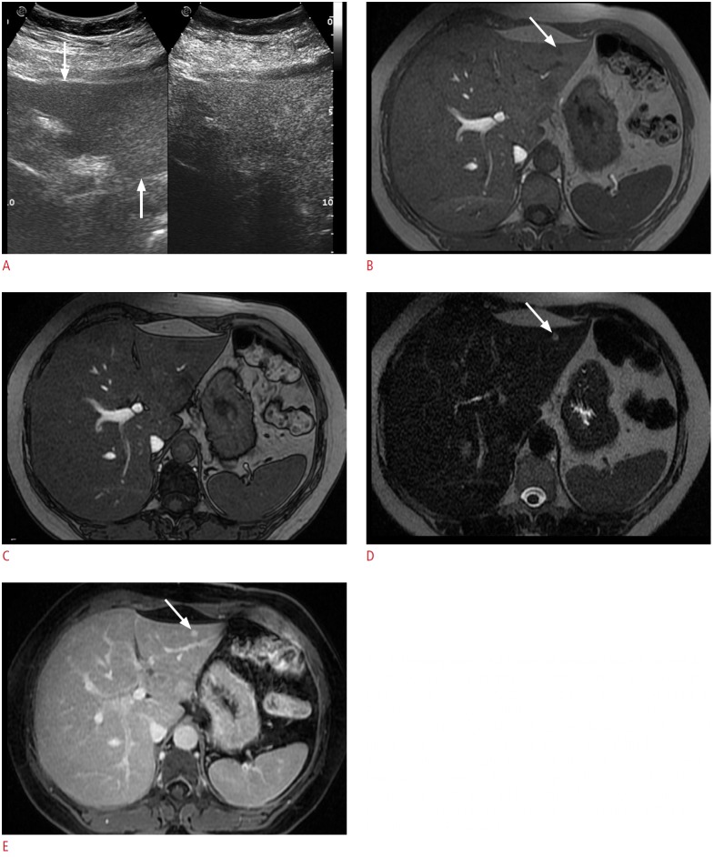
Results: Seven focal liver lesions (size, 4 to 10 mm; mean, 6.1 mm) were detected in 4/37 patients (10.8%): four metastases (size, 5 to 10 mm; mean, 6.7 mm) were detected both by CEUS and MRI, with one hemangioma and two cysts (size range, 4 to 6 mm; mean, 5.3 mm) detected by MRI only. In 1/37 patients (2.7%), CEUS misdiagnosed geographic fatty change as three metastases. In 32/37 patients (86.5%), no lesions were detected. Sensitivity, specificity, PPV, NPV, and accuracy of CEUS were 100% (95% confidence Interval [CI], 1.000 to 1.000), 97.1% (95% CI, 0.914 to 1.027), 75%, 100%, and 97.3%, respectively. No statistically significant differences were found between CEUS and MRI in the detection of focal liver lesions (P=0.480), whereas both of them performed better than baseline US (P<0.001).
Conclusion: CEUS improves the detection of liver metastases in cancer patients with geographic liver fatty deposition on greyscale US.
Keywords: Contrast media; Fatty liver; Liver diseases; Liver neoplasms; Neoplasm metastasis; Ultrasonography.
Conflict of interest statement
No potential conflict of interest relevant to this article was reported.
Figures


Similar articles
-
[Focal liver lesions: clinical usefulness of contrast-enhanced ultrasound in the selection of surgical patients].Chir Ital. 2009 May-Jun;61(3):295-307. Chir Ital. 2009. PMID: 19694231 Italian.
-
Influence of limited examination conditions on contrast-enhanced sonography for characterising liver lesions.Clin Hemorheol Microcirc. 2019;71(2):267-276. doi: 10.3233/CH-189417. Clin Hemorheol Microcirc. 2019. PMID: 30584135
-
Undetermined focal liver lesions on gray-scale ultrasound in patients with fatty liver: characterization with contrast-enhanced ultrasound.J Gastroenterol Hepatol. 2008 Oct;23(10):1511-9. doi: 10.1111/j.1440-1746.2008.05435.x. Epub 2008 Aug 17. J Gastroenterol Hepatol. 2008. PMID: 18713302
-
Malignant focal liver lesions at contrast-enhanced ultrasonography and magnetic resonance with hepatospecific contrast agent.Ultrasound. 2014 May;22(2):91-8. doi: 10.1177/1742271X13513888. Epub 2013 Dec 13. Ultrasound. 2014. PMID: 27433201 Free PMC article.
-
The role of contrast-enhanced ultrasound for focal liver lesion detection: an overview.Ultrasound Med Biol. 2007 Oct;33(10):1515-26. doi: 10.1016/j.ultrasmedbio.2007.04.009. Epub 2007 Jul 6. Ultrasound Med Biol. 2007. PMID: 17618038 Review.
Cited by
-
Hydralazine-augmented contrast ultrasound imaging improves the detection of hepatocellular carcinoma.Med Phys. 2023 Mar;50(3):1728-1735. doi: 10.1002/mp.16232. Epub 2023 Jan 30. Med Phys. 2023. PMID: 36680519 Free PMC article.
-
Contrast-enhanced ultrasound for assessing blood flow modulation of hepatocellular carcinoma by hydralazine.IEEE Int Ultrason Symp. 2022 Oct;2022:10.1109/ius54386.2022.9958467. doi: 10.1109/ius54386.2022.9958467. Epub 2022 Dec 1. IEEE Int Ultrason Symp. 2022. PMID: 37091308 Free PMC article.
-
Focal liver lesions in cirrhosis: Role of contrast-enhanced ultrasonography.World J Radiol. 2022 Apr 28;14(4):70-81. doi: 10.4329/wjr.v14.i4.70. World J Radiol. 2022. PMID: 35646291 Free PMC article. Review.
-
Contrast-enhanced ultrasonography of the liver using SonoVue.Ultrasonography. 2018 Jan;37(1):25-35. doi: 10.14366/usg.17037. Epub 2017 Jul 12. Ultrasonography. 2018. PMID: 28830058 Free PMC article. Review.
-
The Most Appropriate Time Delay after Microbubble Contrast Agent Intravenous Injection to Maximize Liver Metastasis Conspicuity on Contrast-Enhanced Ultrasound.J Med Ultrasound. 2018 Jul-Sep;26(3):128-133. doi: 10.4103/JMU.JMU_12_17. Epub 2018 Sep 14. J Med Ultrasound. 2018. PMID: 30283198 Free PMC article.
References
-
- Quaia E, D'Onofrio M, Palumbo A, Rossi S, Bruni S, Cova M. Comparison of contrast-enhanced ultrasonography versus baseline ultrasound and contrast-enhanced computed tomography in metastatic disease of the liver: diagnostic performance and confidence. Eur Radiol. 2006;16:1599–1609. - PubMed
-
- Rafaelsen SR, Jakobsen A. Contrast-enhanced ultrasound vs multidetector-computed tomography for detecting liver metastases in colorectal cancer: a prospective, blinded, patient-by-patient analysis. Colorectal Dis. 2011;13:420–425. - PubMed
LinkOut - more resources
Full Text Sources
Other Literature Sources
