. 2017 Feb 9;542(7640):255-259.
doi: 10.1038/nature21061. Epub 2017 Feb 1.

Molecular mechanism for the regulation of yeast separase by securin

Affiliations

Molecular mechanism for the regulation of yeast separase by securin

Shukun Luo et al. Nature. .

Erratum in

Abstract

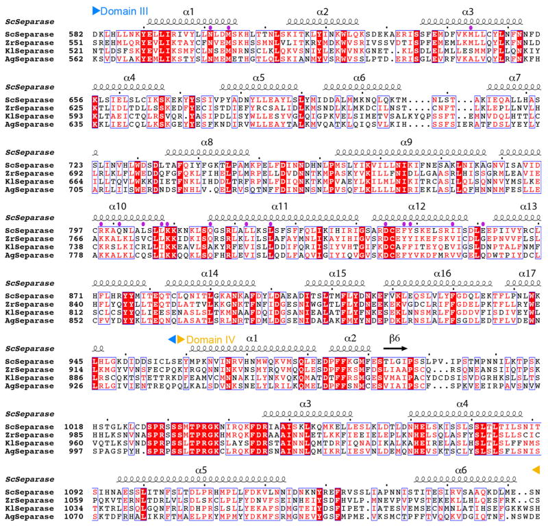
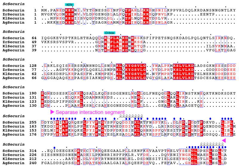
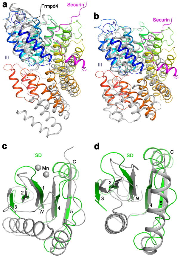
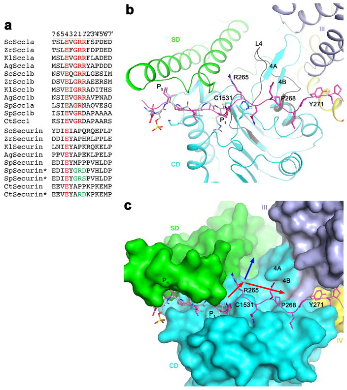
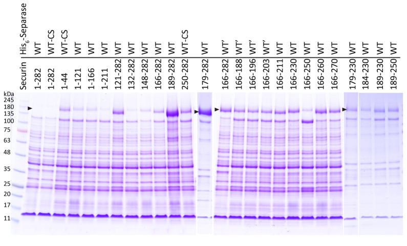
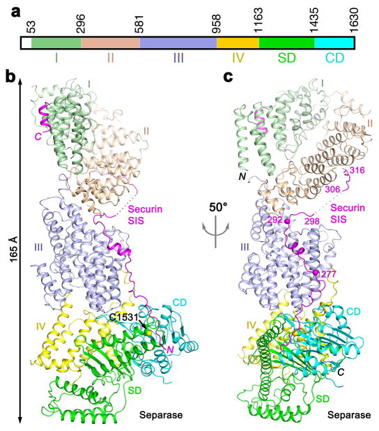
Separase is a cysteine protease with a crucial role in the dissolution of cohesion among sister chromatids during chromosome segregation. In human tumours separase is overexpressed, making it a potential target for drug discovery. The protease activity of separase is strictly regulated by the inhibitor securin, which forms a tight complex with separase and may also stabilize this enzyme. Separases are large, 140-250-kilodalton enzymes, with an amino-terminal α-helical region and a carboxy-terminal caspase-like catalytic domain. Although crystal structures of the C-terminal two domains of separase and low-resolution electron microscopy reconstructions of the separase-securin complex have been reported, the atomic structures of full-length separase and especially the complex with securin are unknown. Here we report crystal structures at up to 2.6 Å resolution of the yeast Saccharomyces cerevisiae separase-securin complex. The α-helical region of separase (also known as Esp1) contains four domains (I-IV), and a substrate-binding domain immediately precedes the catalytic domain and has tight associations with it. The separase-securin complex assumes a highly elongated structure. Residues 258-373 of securin (Pds1), named the separase interaction segment, are primarily in an extended conformation and traverse the entire length of separase, interacting with all of its domains. Most importantly, residues 258-269 of securin are located in the separase active site, illuminating the mechanism of inhibition. Biochemical studies confirm the structural observations and indicate that contacts outside the separase active site are crucial for stabilizing the complex, thereby defining an important function for the helical region of separase.


Conflict of interest statement

The authors declare no competing financial interests.

Figures

Extended Data Fig. 1
Sequence alignment of domains I and II of separase. The secondary structure elements in the S. cerevisiae separase structure are shown and labeled. The boundaries of the domains are indicated. Residues in contact with securin are indicated with the purple dots. Sc: Saccharomyces cerevisiae, Zr: Zygosaccharomyces rouxii, Kl: Kluyveromyces lactis; Ag: Ashbya gossypii. Modified from an output from ESPript .
Extended Data Fig. 2
Sequence alignment of domains III and IV of separase. The secondary structure elements in the S. cerevisiae separase structure are shown.
Extended Data Fig. 3
Sequence alignment of the SD and CD of separase. The catalytic Cys1531 and His1505 residues are indicated with the red dots. Hs: Homo sapiens; Sp: Schizosaccharomyces pombe; Ct: Chaetomium thermophilum.
Extended Data Fig. 4
Sequence alignment of securin. The separase interaction segment (SIS) is indicated. Residues in contact with separase are indicated with the blue dots. Residue 263 is equivalent to the P1 residue of separase substrates, and is indicated with the red asterisk. Residues 317–360 of S. cerevisiae securin are disordered in the current structures and are poorly conserved in sequence.
Extended Data Fig. 5
Overlay of the structures of the separase-securin complexes. (a). The complex formed by residues 71–1630 of separase and 258–373 of securin is shown in color, and that by residues 51–1630 of separase and 258–373 of securin in gray. Residues 73–80 from another molecule of separase in the crystal is shown in orange, forming a β-sheet with the N-terminal segment of separase. (b). panel a viewed after a 50° rotation around the vertical axis.
Extended Data Fig. 6
Additional structural information on the separase-securin complex. (a). 2Fo–Fc electron density for helices 4 and 5 of domain I at 2.6 Å resolution, contoured at 1σ. Helices 6 and 7 are also shown for reference. The directions of the helices are indicated with the red arrows. (b). The β4A-β4B segment of CD (cyan) is immediately after the catalytic Cys1531 residue, and has interactions with domain III (light blue). The equivalent segment in the C. thermophilum SD-CD free enzyme structure is a loop (L4), shown in dark gray. (c). Interactions between residues 290–296 of securin SIS (magenta) with domains III (light blue) and II (light brown) of separase. (d). Interactions between the C-terminal segment of securin SIS (magenta) and domain I of separase (green). Deletion of the first 155 residues of separase would remove helix α3 in this binding site.
Extended Data Fig. 7
Structural homologs of domains III and SD of separase. (a). Overlay of the structures of domain III of separase (color ramp from N- (blue) to C-terminus (red)) and the TPR domain of LGN (gray, PDB entry 4WNG; 11% sequence identity, Z score of 14.4) . The Frmpd4 ligand (black) of LGN is bound to a different region of the structure compared to securin. (b). Overlay of the structures of domain III of separase and the subunit 7 of the APC/C (PDB entry 5G04; 5% identity, 14.3 Z score) . (c). Overlay of the structures of the SD of separase (green) and a part of the PIWI domain of Argonaute (gray, PDB entry 4N76; 10% sequence identity, 5.5 Z score. As a comparison, matching this β-sheet to that in C. thermophilum separase produced a Z score of 5.7.) ,. Residues in the helical insert between β3 and β4 of separase are removed for clarity. (d). Overlay of the structures of the SD of separase and the YqgF domain of Tex (gray, PDB entry 3BZK; 4% sequence identity, 5.5 Z score) .
Extended Data Fig. 8
Possible binding groove for the P′ residues of the substrate. (a). Alignment of the separase cleavage sites in Scc1 substrates. The two cleavage sites in each protein are named a and b. The equivalent residues in securin are also shown. The asterisks indicate securin mutants (mutations in green) that become substrates of separase. The P and P′ residues are labeled at the top, and the cleavage site is indicated with the vertical line. (b). The overall binding mode of residues 258–271 of securin in separase. The β4A-β4B segment of CD (cyan) is a loop (L4, dark gray) in the C. thermophilum SD-CD structure. (c). A groove in the active site of separase (red arrows) can accommodate the P′ residues. The blue arrow indicates another groove in this region, but the binding mode of securin suggests that the groove indicated by the red arrows is more likely.
Extended Data Fig. 9
Biochemical characterizations of the interactions between separase and securin. Z. rouxii separase was co-expressed with various segments of Z. rouxii securin, with truncations at the N- and/or C-terminus. The insoluble fraction was run on SDS PAGE. The position of separase (with an N-terminal His tag) is indicated with the black arrowhead. WT: full-length Z. rouxii separase; WT-CS: full-length Z. rouxii separase with C1497S mutation; WT′: Z. rouxii separase with an internal deletion of residues 952–1010, corresponding to a poorly conserved, disordered loop in domain IV of the S. cerevisiae separase structure. For gel source data, see Supplementary Figure 1.
Figure 1
Figure 1
Crystal structure of the yeast separase-securin complex. (a). Domain organization of S. cerevisiae separase. The domains are labeled and given different colors. (b). Overall structure of the yeast separase-securin complex. The domains in separase are colored according to panel a, and the securin SIS is in magenta. The side chain of the catalytic Cys1531 residue of separase is shown as a sphere model. (c). Overall structure of the complex viewed after a 50° rotation around the vertical axis. Two of the phosphorylation sites in the securin SIS (Ser277 and Ser292) , are indicated with the spheres and labeled. The structure figures were produced with PyMOL (www.pymol.org).
Figure 2
Figure 2
Structures of the domains in separase. (a). Structure of domains I and II of separase. The directions of helices 4 and 5 in domain I are indicated with the red arrows. (b). Structure of domain III of separase. The helices are colored ramping from blue at the N-terminus to red at C-terminus. (c). Structure of domains IV, SD and CD of separase, and overlay of the structure of the SD-CD of C. thermophilum separase (gray) . The active site is indicated with the red asterisk. The red arrowhead indicates the region where the β4A-β4B hairpin has a different conformation and is partly disordered in C. thermophilum separase, and the purple box highlights the region where domain IV provides an extra strand (labeled 6) to the β-sheet of SD.
Figure 3
Figure 3
Interactions between separase and securin. (a). Molecular surface of separase, colored by the domains. The active site of separase is indicated with the red asterisk. The view is same as Fig. 1c. (b). Omit Fo–Fc electron density at 3.0 Å resolution for residues 258–269 of securin, contoured at 2σ. (c). Interactions between residues 258–265 of securin SIS (magenta) with the active site of separase. The side chains of residues in the interface are shown as stick models and labeled. The bound position of a substrate-mimic inhibitor to C. thermophilum separase is shown in gray . (d). Close-up of the active site region showing the differences between the bound position of securin (magenta) and that of the inhibitor to C. thermophilum separase (gray) . (e). Interactions between residues 271–288 of securin SIS (magenta) with domain III (light blue) of separase.
Figure 4
Figure 4
Biochemical characterizations of the interactions between separase and securin. (a). Domain organizations of S. cerevisiae and Z. rouxii securin. The KEN motif and the destruction box (D-box) are also indicated. (b). Z. rouxii separase (with an N-terminal His tag) was co-expressed with various segments of Z. rouxii securin. The eluates from nickel columns were separated by SDS gel electrophoresis. The positions of separase and securin are indicated with the black and red arrowheads, respectively. WT: full-length Z. rouxii separase; WT-CS: full-length Z. rouxii separase C1497S mutant; WT′: Z. rouxii separase with an internal deletion of residues 952–1010, corresponding to a poorly conserved, disordered loop in domain IV of the S. cerevisiae separase structure. For gel source data, see Supplementary Figure 1. (c). Summary of the expression results in panel b. The solubility levels of separase for various securin segments are indicated.

References

    1. Uhlmann F, Wernic D, Poupart MA, Koonin EV, Nasmyth K. Cleavage of cohesin by the CD clan protease separin triggers anaphase in yeast. Cell. 2000;103:375–386. - PubMed
    1. Hauf S, Waizenegger IC, Peters JM. Cohesin cleavage by separase required for anaphase and cytokinesis in human cells. Science. 2001;293:1320–1323. - PubMed
    1. Nasmyth K. Segregating sister genomes: the molecular biology of chromosome separation. Science. 2002;297:559–565. - PubMed
    1. Peters JM. The anaphase-promoting complex: proteolysis in mitosis and beyond. Mol Cell. 2002;9:931–943. - PubMed
    1. Uhlmann F. Separase regulation during mitosis. Biochem Soc Trans. 2003;70:243–251. - PubMed

Publication types

MeSH terms