. 2018 Jan;175(2):232-245.
doi: 10.1111/bph.13735. Epub 2017 Mar 5.

The clinically used PARP inhibitor olaparib improves organ function, suppresses inflammatory responses and accelerates wound healing in a murine model of third-degree burn injury

Affiliations

The clinically used PARP inhibitor olaparib improves organ function, suppresses inflammatory responses and accelerates wound healing in a murine model of third-degree burn injury

Akbar Ahmad et al. Br J Pharmacol. 2018 Jan.

Abstract

Background and purpose: The PARP inhibitor olaparib has recently been approved for human use for the therapy of cancer. Considering the role of PARP in critical illness, we tested the effect of olaparib in a murine model of burn injury, in order to begin exploring the feasibility of repurposing olaparib for the therapy of burn patients.

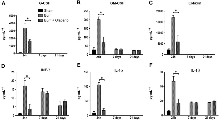
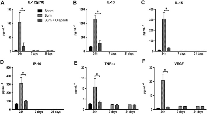
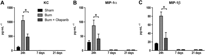
Experimental approach: Mice were subjected to scald burn injury and randomized into vehicle or olaparib (10 mg·kg-1 ·day-1 i.p.) groups. Outcome variables included indices of organ injury, clinical chemistry parameters, plasma levels of inflammatory mediators (at 24 h, 7 and 21 days) and burn wound size (at 21 days).

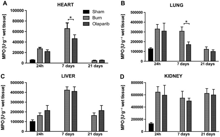
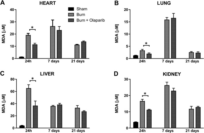
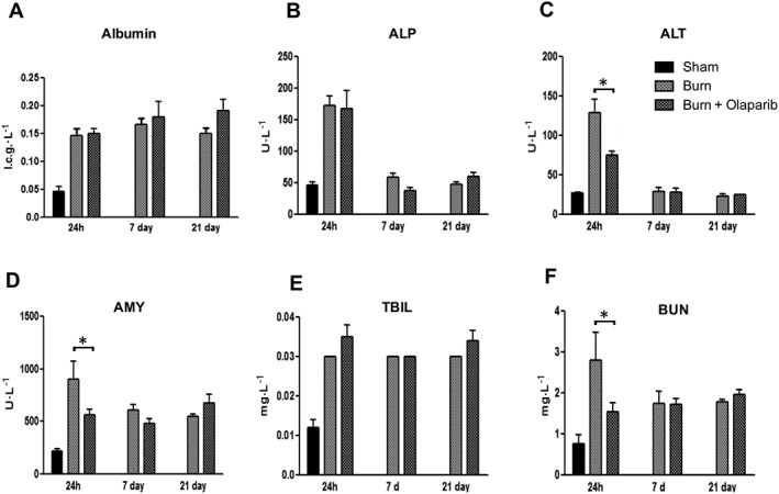
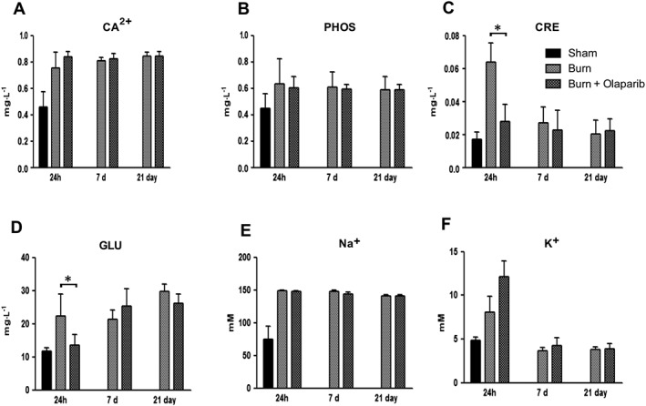
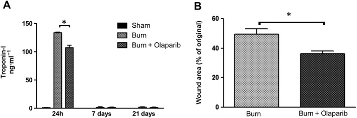
Key results: Olaparib reduced myeloperoxidase levels in heart and lung homogenates and reduced malondialdehyde levels in all tissues 24 h post-burn. Olaparib also reduced circulating alkaline aminotransferase, amylase and blood urea nitrogen and creatinine levels, indicative of protection against hepatic, pancreatic and renal dysfunction. Pro-inflammatory mediator (TNF-α, IL-1β, IFN-γ, GCSF, GM-CSF, eotaxin, KC, MIP-1-α and IL-3, 6 and 12) levels as well as the levels of several mediators that are generally considered anti-inflammatory (IL-4, 10 and 13) were reduced by olaparib. Plasma troponin-I levels (an indicator of skeletal muscle damage) was also attenuated by olaparib. Finally, olaparib stimulated wound healing.

Conclusions and implications: The clinically approved PARP inhibitor olaparib improves organ function, suppresses inflammatory responses and accelerates wound healing in murine burn injury. The data raise the potential utility of olaparib for severe burn injury.

Linked articles: This article is part of a themed section on Inventing New Therapies Without Reinventing the Wheel: The Power of Drug Repurposing. To view the other articles in this section visit http://onlinelibrary.wiley.com/doi/10.1111/bph.v175.2/issuetoc.


Figures

Figure 1
Figure 1
Effect of olaparib treatment on burn‐induced increases in heart, lung, liver and kidney MPO levels. (A) Heart, (B) lung, (C) liver and (D) kidney MPO levels are shown in sham‐control mice and in mice subjected to burn injury and in burn mice treated with olaparib (10 mg·kg−1, i.p.) once a day for 1, 6 and 20 days. Compared to non‐burn sham control, burn produced a significant increase (P < 0.05) in MPO levels in the heart and lung at 24 h and 7 days, in the liver at 7 days and in the kidney at 24 h, 7 and 21 days. Significant effect of olaparib during burn injury in the heart and lung is shown at 7 days (*P < 0.05). Data are shown as mean ± SEM of 10 animals for each group.
Figure 2
Figure 2
Effect of olaparib treatment on burn‐induced increases in heart, lung, liver and kidney MDA levels. (A) Heart, (B) lung, (C) liver and (D) kidney MDA levels are shown in sham‐control mice and in mice subjected to burn injury and in burn mice treated with olaparib (10 mg·kg−1, i.p.) once a day for 1, 6 and 20 days. Compared to non‐burn sham control, burn produced a significant increase (P < 0.05) in MDA levels in all tissues at all three time points studied. Significant effect of olaparib during burn injury in all four tissues studied is shown at 24 h (*P < 0.05). Data are shown as mean ± SEM of 10 animals for each group.
Figure 3
Figure 3
Effect of olaparib treatment in burn‐induced increases in selected parameters of organ injury. Various physiological and organ injury marker levels, albumin, alkaline phosphatase (ALP), alanine aminotransferase (ALT), amylase (AMY), total bilirubin (TBIL), and blood urea nitrogen (BUN) are shown in sham‐control mice and in mice subjected to burn injury and in burn mice treated with olaparib (10 mg·kg−1, i.p.) once a day for 1, 6 and 20 days. Compared to non‐burn sham control, burn produced a significant increase (P < 0.05) in albumin, AMY, bilirubin and BUN levels at all three time points studied and in ALT and ALP levels at 24 h. Significant effect of olaparib on ALT, ALT and BUN levels during burn injury is shown at 24 h (*P < 0.05). Data are shown as mean ± SEM of 10 animals for each group.
Figure 4
Figure 4
Effect of olaparib treatment on burn‐induced increases in selected parameters of organ injury. Various physiological and organ injury marker levels (plasma calcium [Ca2+], plasma phosphate [PHOS], plasma creatinine [CRE], plasma glucose [GLU], plasma sodium [Na+] and plasma potassium [K+]) are shown in sham‐control mice and in mice subjected to burn injury and in burn mice treated with olaparib (10 mg·kg−1, i.p.) once a day for 1 day, 6 days and 20 days. Compared to non‐burn sham control, burn produced a significant increase (P < 0.05) in calcium and sodium levels at all three time points studied and in creatinine, glucose and potassium levels at 24 h. Significant effect of olaparib on creatinine and glucose levels during burn injury is shown at 24 h (*P < 0.05). Data are shown as mean ± SEM of 10 animals for each group.
Figure 5
Figure 5
Effect of olaparib treatment on burn‐induced increases in selected plasma inflammatory mediator levels. Plasma levels of G‐CSF, GM‐CSF, eotaxin, IFN‐γ, IL‐1α and IL‐1β are shown in sham‐control mice and in mice subjected to burn injury and in burn mice treated with olaparib (10 mg·kg−1, i.p.) once a day for 1, 6 and 20 days. Compared to non‐burn sham control, burn produced a significant increase (P < 0.05) in GM‐CSF, eotaxin, IFN‐γ, IL‐1α and IL‐1β levels of vehicle‐treated (but not olaparib‐treated) mice at 24 h, and in IFN‐γ and IL‐1β levels in both vehicle‐ and olaparib‐treated mice at 7 and 21 days. Significant effect of olaparib on all six mediators studied during burn injury is shown at 24 h (*P < 0.05). Data are shown as mean ± SEM of 10 animals for each group.
Figure 6
Figure 6
Effect of olaparib treatment on burn‐induced increases in selected plasma inflammatory mediator levels. Plasma levels of IL‐2, IL‐3, IL‐4, IL‐6, IL‐10 and IL‐12(p40) are shown in sham‐control mice and in mice subjected to burn injury and in burn mice treated with olaparib (10 mg·kg−1, i.p.) once a day for 1, 6 and 20 days. Compared to non‐burn sham control, burn produced a significant increase (P < 0.05) in IL‐3 levels at 24 h and in all other mediator levels at all time points studied. Significant effect of olaparib on all six mediators studied during burn injury is shown at 24 h (*P < 0.05). Data are shown as mean ± SEM of 10 animals for each group.
Figure 7
Figure 7
Effect of olaparib treatment on burn‐induced increases in selected plasma inflammatory mediator levels. Plasma levels of IL‐12 (p70), Il‐13, IL‐15, IP‐10, TNF‐α and VEGF are shown in sham‐control mice and in mice subjected to burn injury and in burn mice treated with olaparib (10 mg·kg−1, i.p.) once a day for 1, 6 and 20 days. Compared to non‐burn sham control, burn produced a significant increase (P < 0.05) in plasma levels of all six mediators studied in the vehicle‐treated (but not olaparib‐treated) mice at 24 h. Significant effect of olaparib on all six mediators studied during burn injury is shown at 24 h (*P < 0.05). Data are shown as mean ± SEM of 10 animals for each group.
Figure 8
Figure 8
Effect of olaparib treatment on burn‐induced increases in selected plasma chemokine levels. Plasma levels of KC, MIP‐1α and MIP‐1β are shown in sham‐control mice and in mice subjected to burn injury and in burn mice treated with olaparib (10 mg·kg−1, i.p.) once a day for 1, 6 and 20 days. Compared to non‐burn sham control, burn produced a significant increase (P < 0.05) in plasma levels of KC at 24 h in both vehicle‐treated and olaparib‐treated and in the plasma levels of MIP‐1α and MIP‐1β at 24 h in vehicle‐treated (but not olaparib‐treated) mice. Significant effect of olaparib on all three mediators studied during burn injury is shown at 24 h (*P < 0.05). Data are shown as mean ± SEM of 10 animals for each group.
Figure 9
Figure 9
Olaparib reduces the burn‐induced increases in plasma troponin‐I levels and reduces burn wound area. (A) Plasma troponin‐I levels are shown in sham‐control mice and in mice subjected to burn injury and in burn mice treated with olaparib (10 mg·kg−1, i.p) once a day for 1, 6 and 20 days. Compared to non‐burn sham control, burn produced a significant increase (P < 0.05) in plasma troponin‐I levels at 24 h; olaparib exerted a significant inhibitory effect (*P < 0.05). Data are shown as mean ± SEM of 10 animals per group. (B) % wound area (as percentage of the original burn wound area) is shown on Day 21 in mice subjected to burn injury and in burn mice treated with olaparib (10 mg·kg−1, i.p) once a day for 20 days. *P < 0.05 shows a significant reduction in the wound area in olaparib‐treated animals. Data are shown as mean ± SEM of 10 animals per group.

References

    1. Ahmad A, Druzhyna N, Szabo C (2016). Delayed treatment with sodium hydrosulfide improves regional blood flow and alleviates cecal ligation and puncture (CLP)‐induced septic shock. Shock 46: 183–193. - PMC - PubMed
    1. Ahmad SF, Zoheir KM, Ansari MA, Korashy HM, Bakheet SA, Ashour AE (2015). The role of poly(ADP‐ribose) polymerase‐1 inhibitor in carrageenan‐induced lung inflammation in mice. Mol Immunol 63: 394–405. - PubMed
    1. Ahmad A, Szabo C (2016). Both the H2S biosynthesis inhibitor aminooxyacetic acid and the mitochondrially targeted H2S donor AP39 exert protective effects in a mouse model of burn injury. Pharmacol Res 113: 348–355. - PubMed
    1. Alano CC, Garnier P, Ying W, Higashi Y, Kauppinen TM, Swanson RA (2010). NAD+ depletion is necessary and sufficient for poly(ADP‐ribose) polymerase‐1‐mediated neuronal death. J Neurosci 30: 2967–2978. - PMC - PubMed
    1. Alexander SPH, Benson HE, Faccenda E, Pawson AJ, Sharman JL, McGrath JC et al (2015). The Concise Guide to PHARMACOLOGY 2015/16: Overview. Brit J Pharmacol 172: 5729–5743. - PMC - PubMed

Publication types