. 2017 Jan 31;18(5):1090-1099.
doi: 10.1016/j.celrep.2017.01.010.

Visualizing Changes in Cdkn1c Expression Links Early-Life Adversity to Imprint Mis-regulation in Adults

Affiliations

Visualizing Changes in Cdkn1c Expression Links Early-Life Adversity to Imprint Mis-regulation in Adults

Mathew Van de Pette et al. Cell Rep. .

Abstract

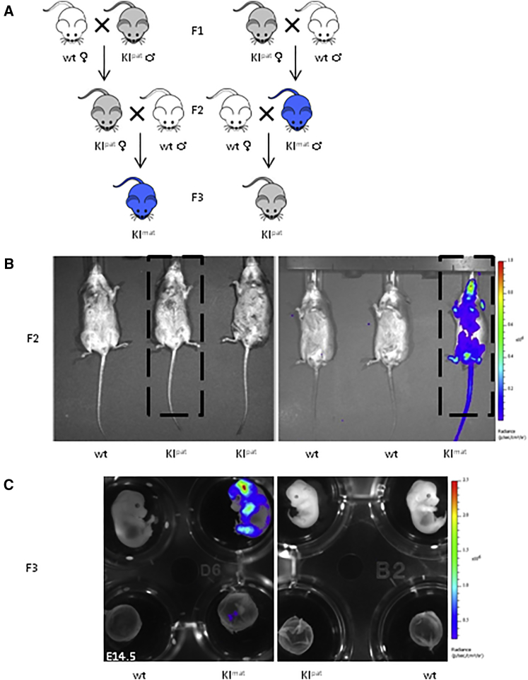
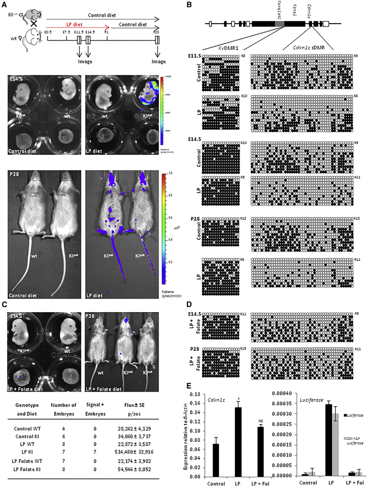
Imprinted genes are regulated according to parental origin and can influence embryonic growth and metabolism and confer disease susceptibility. Here, we designed sensitive allele-specific reporters to non-invasively monitor imprinted Cdkn1c expression in mice and showed that expression was modulated by environmental factors encountered in utero. Acute exposure to chromatin-modifying drugs resulted in de-repression of paternally inherited (silent) Cdkn1c alleles in embryos that was temporary and resolved after birth. In contrast, deprivation of maternal dietary protein in utero provoked permanent de-repression of imprinted Cdkn1c expression that was sustained into adulthood and occurred through a folate-dependent mechanism of DNA methylation loss. Given the function of imprinted genes in regulating behavior and metabolic processes in adults, these results establish imprinting deregulation as a credible mechanism linking early-life adversity to later-life outcomes. Furthermore, Cdkn1c-luciferase mice offer non-invasive tools to identify factors that disrupt epigenetic processes and strategies to limit their long-term impact.

Keywords: Cdkn1c; bioluminescence; environmental stress; imprinting; luciferase reporter mice.


Figures

None
Graphical abstract
Figure 1
Figure 1
Visualizing Cdkn1c Gene Expression In Vivo Using Bioluminescence (A) Scheme of alternative knockin (KI) strategies used to generate Cdkn1c-FLucLacZ and Cdkn1c-FLuc embryonic stem cells (ESCs) and reporter mouse lines, in which sequences coding for the T2A peptide, the open reading frame of FLuc, a second T2A peptide, and the open reading frame of LacZ were inserted between the last amino acid and the translation termination codon in exon 3 (…KRLREGRG…; Cdkn1c-FLucLacZ) or IRES elements and the open reading frame of FLuc was inserted into a unique HindIII in the 3′ UTR (Cdkn1c-FLuc). (B) Low-level bioluminescence (blue-green) in Cdkn1c-FLucLacZ and Cdkn1c-FLuc ESCs was detected in clones with a presumed maternal insertion (KImat), but not in clones with a paternal insertion (KIpat) or in wild-type ESCs (wt) (scale bar represents levels of bioluminescence). (C) Total Cdkn1c expression (left), determined by RT-PCR, was increased in ESC clones with either a KImat (dashed line) or a KIpat (solid line) insertion over 21 days of embryoid body differentiation. Luciferase expression (right), determined by RT-PCR, was detected uniquely in KImat clones. Samples were normalized to β-actin and expressed as the mean ± SE. (D) Bioluminescent imaging of representative P28 female Cdkn1c-FLucLacZ mice. Luciferase activity was observed in Cdkn1c-FLucLacZ KImat, with very low/negligible signals detectable upon paternal inheritance (KIpat) or in wild-type mice (wt). Strongest signal was evident in the skin, with low level signal detected in the internal organs. (E) Bioluminescence detected in pregnancies with maternal inheritance of Cdkn1c-FLucLacZ (KImat, left) in utero, but not paternal inheritance (KIpat, right; less than twice background). Lower panels show bioluminescence imaging of dissected E11.5 embryos, where luciferase activity was seen in head and back of KImat embryos and placental tissue and quantified (flux). All Cdkn1c-FLucLacZ embryos imaged showed predicted parent-of-origin-specific bioluminescent activity. (F) Total Cdkn1c gene expression in embryos (E11.5) was determined by RT-PCR, and levels were similar in samples from wild-type and where Cdkn1c-FLucLacZ was transmitted maternally (KImat) or paternally (KIpat; left). Luciferase (black) and Cdkn1c-Luciferase (gray) transcripts were detected uniquely from KImat. Samples were normalized to β-actin and expressed as the mean ± SE. (G) Scheme of the mouse IC2 imprinting domain, showing the two DMRs that regulate Cdkn1c imprinted expression (KvDMR1 and Cdkn1c sDMR) and the position of bi-allelic (white), maternally expressed (dark gray), and paternally expressed (light gray) genes. Bisulfite analysis showing DNA methylation at KvDMR1 and Cdkn1c sDMR is similar in KImat and wt embryos at E11.5 (closed circles, methylated; open circles, un-methylated; where number indicates fully un-methylated strands).
Figure 2
Figure 2
Correct Imprint Resetting of Cdkn1c-FLucLacZ across Generations (A) Diagram showing predicted expression and inheritance of a maternally expressed imprinted gene (such as Cdkn1c) or transgene (Cdkn1c-FLucLacZ) in reciprocal crosses across three generations. Wild-type mice are shown in white, expression through maternal inheritance is shown in blue, and inheritance of a silent imprint (Cdkn1c-FLucLacZ; KIpat) is indicated in gray. (B) Experimental evidence of imprint resetting; bioluminescent imaging of adult F2 mice, Cdkn1c-FLucLacZ KIpat females (left box), and KImat males (right box), showing predicted parent-of-origin-specific luciferase activity (blue). Highlighted animals were then used to generate F3 (as outlined in A). (C) Bioluminescent image of E14.5 embryos, generated from the indicated transgenic parents; signal was detected upon maternal inheritance of luciferase in embryos (upper panel) and in placental tissue (lower panel; left), which had been silent in the previous generation. Conversely, paternal inheritance of the previously active luciferase was sufficient to silence the previously active allele (right).
Figure 3
Figure 3
Silencing of Paternal Cdkn1c-FLucLacZ Is Transiently Released by In Utero Exposure to Epigenetic Drugs (A) Embryos carrying silent (paternally inherited) Cdkn1c-FLucLacZ were generated by mating wild-type (wt) females with homozygous Cdkn1c-FLucLacZ males. Pregnant females were treated with trichostatin A (TSA) or 5′ azacytidine (5′Aza) alone or together at the times indicated. Offspring were imaged at E14.5, at birth (P1), and at 4 weeks of age (P28). (B) Low-level bioluminescence was occasionally detected in 5′Aza- and TSA-alone treated pregnancies, whereas stronger and consistent signal (blue) was detected in combination-treated (Combi) embryos in utero (left) or individually dissected embryos (right) in the head and back. (C) Cdkn1c-FLucLacZ-derived bioluminescent activity was consistently elevated in E14.5 embryos exposed to combined drug treatment. (D) Bisulfite analysis of DNA methylation at the KvDMR1 and Cdkn1c sDMR in the brain of E14.5 Cdkn1c-FLucLacZ KIpat embryos shows reduced methylation in embryos exposed to combination drug treatment versus controls (closed circles, methylated; open circles, un-methylated). (E) Variable increases in luciferase activity (blue, flux) characterize combination-drug-treated Cdkn1c-FLucLacZ KIpat animals at P1 (right), with no signal detected in vehicle-treated controls (left). (F) Luciferase activity was no longer detected in Cdkn1c-FLucLacZ KIpat mice at P28 that had been exposed to combination drug treatment in utero. (G) Bisulfite analysis of DNA methylation at the KvDMR1 and Cdkn1c sDMR in the brain of P28 Cdkn1c-FLucLacZ KIpat mice shows that previously ablated methylation is restored by adulthood to normal levels (closed circles, methylated; open circles, un-methylated).
Figure 4
Figure 4
Stable Silencing of Paternally Inherited Cdkn1c-FLucLacZ during Life Course Depends upon the Availability of Methyl Donors In Utero (A) Offspring with a silent (paternally inherited) Cdkn1c-FLucLacZ were generated by mating wild-type (wt) females with heterozygous Cdkn1c-FLucLacZ males. Upon detection of a vaginal plug, a group of pregnant females were switched to a calorie-matched but low-protein (LP) diet for the duration of their pregnancy, with mothers and litters returning to a normal diet after birth. Pregnancies were imaged/examined at the times indicated (E11.5, E14.5, and P28). No mis-expression of luciferase was observed at day E11.5 (Figure S2), irrespective of diet; however, by E14.5, luciferase activity was detected selectively in embryos of mothers fed LP diet (upper right) and expression continued as these matured into adults (lower right). No signal was detected in animals fed normal (control) diet at any time. (B) Comparative bisulfite analysis of DNA methylation at the Cdkn1c locus in the brain of Cdkn1c-FLucLacZ KIpat animals (E11.5, E14.5, and P28) born to mothers fed control versus LP diet during pregnancy. Cdkn1c sDMR becomes hypo-methylated in LP conditions in utero, and methylation is not restored subsequently (closed circles, methylated; open circles, un-methylated). Methylation at the KvDMR1 is unaltered. (C) Pregnant females as in (A) were fed LP diet supplemented with increased folate. Bioluminescent imaging of embryos (E14.5) from mothers fed LP + folate showed reduced mis-expression of Cdkn1c-FLucLacZ KIpat as compared with those fed LP alone (A), with luciferase activity remaining low or negligible as they matured into adults (P28; upper right; image scales same as A). (D) Bisulfite analysis showing DNA methylation at KvDMR1 and Cdkn1c sDMR in E14.5 (upper) and P28 (lower) animals born to mothers fed LP + folate diet. Progressive hypo-methylation of the Cdkn1c sDMR was buffered against by the increased dietary folate. (E) Total Cdkn1c gene expression in E14.5 brain was determined by RT-PCR, and levels were elevated in samples from LP-exposed litters (p < 0.033), compared to control and LP + folate litters (p < 0.38). Luciferase (black) and Cdkn1c-Luciferase (gray) transcripts were detected using RT-PCR uniquely in LP brain samples, demonstrating loss of imprinting. Samples were normalized to β-actin and expressed as the mean ± SE.

References

    1. Andrews S.C., Wood M.D., Tunster S.J., Barton S.C., Surani M.A., John R.M. Cdkn1c (p57Kip2) is the major regulator of embryonic growth within its imprinted domain on mouse distal chromosome 7. BMC Dev. Biol. 2007;7:53. - PMC - PubMed
    1. Auclair G., Guibert S., Bender A., Weber M. Ontogeny of CpG island methylation and specificity of DNMT3 methyltransferases during embryonic development in the mouse. Genome Biol. 2014;15:545. - PMC - PubMed
    1. Bartolomei M.S., Ferguson-Smith A.C. Mammalian genomic imprinting. Cold Spring Harb. Perspect. Biol. 2011;3:a002592. - PMC - PubMed
    1. Bhogal B., Arnaudo A., Dymkowski A., Best A., Davis T.L. Methylation at mouse Cdkn1c is acquired during postimplantation development and functions to maintain imprinted expression. Genomics. 2004;84:961–970. - PubMed
    1. Caspary T., Cleary M.A., Baker C.C., Guan X.J., Tilghman S.M. Multiple mechanisms regulate imprinting of the mouse distal chromosome 7 gene cluster. Mol. Cell. Biol. 1998;18:3466–3474. - PMC - PubMed

Publication types