. 2017 Feb 21;8(8):12558-12575.
doi: 10.18632/oncotarget.14894.

Inhibition of CDK8 mediator kinase suppresses estrogen dependent transcription and the growth of estrogen receptor positive breast cancer

Affiliations

Inhibition of CDK8 mediator kinase suppresses estrogen dependent transcription and the growth of estrogen receptor positive breast cancer

Martina S J McDermott et al. Oncotarget. .

Abstract

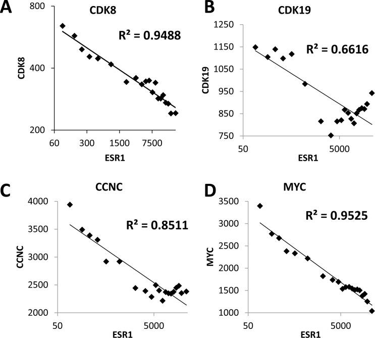
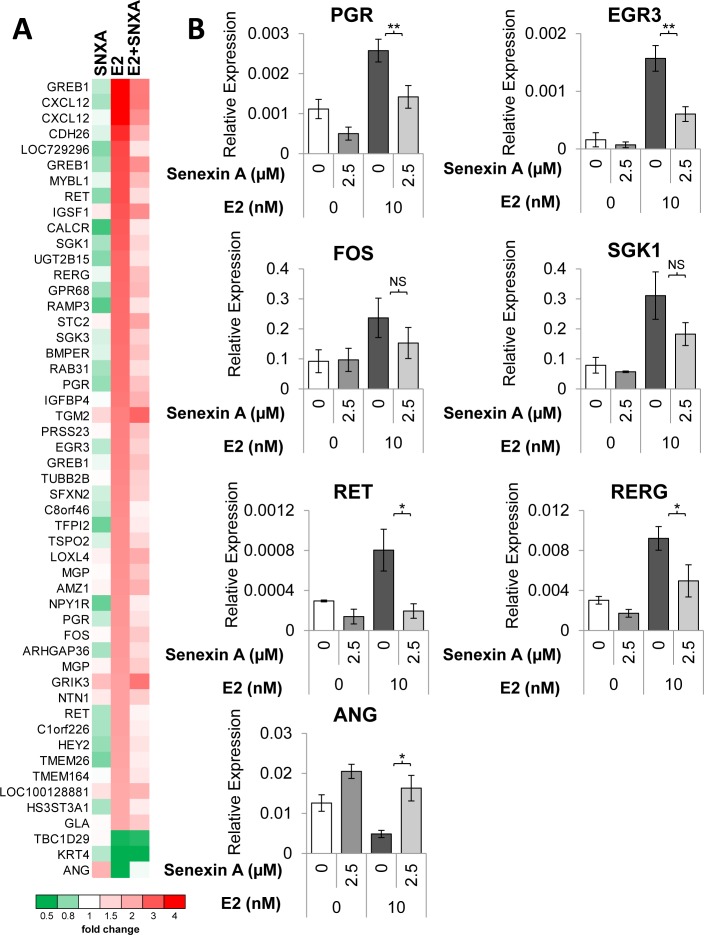
Hormone therapy targeting estrogen receptor (ER) is the principal treatment for ER-positive breast cancers. However, many cancers develop resistance to hormone therapy while retaining ER expression. Identifying new druggable mediators of ER function can help to increase the efficacy of ER-targeting drugs. Cyclin-dependent kinase 8 (CDK8) is a Mediator complex-associated transcriptional regulator with oncogenic activities. Expression of CDK8, its paralog CDK19 and their binding partner Cyclin C are negative prognostic markers in breast cancer. Meta-analysis of transcriptome databases revealed an inverse correlation between CDK8 and ERα expression, suggesting that CDK8 could be functionally associated with ER. We have found that CDK8 inhibition by CDK8/19-selective small-molecule kinase inhibitors, by shRNA knockdown or by CRISPR/CAS9 knockout suppresses estrogen-induced transcription in ER-positive breast cancer cells; this effect was exerted downstream of ER. Estrogen addition stimulated the binding of CDK8 to the ER-responsive GREB1 gene promoter and CDK8/19 inhibition reduced estrogen-stimulated association of an elongation-competent phosphorylated form of RNA Polymerase II with GREB1. CDK8/19 inhibitors abrogated the mitogenic effect of estrogen on ER-positive cells and potentiated the growth-inhibitory effects of ER antagonist fulvestrant. Treatment of estrogen-deprived ER-positive breast cancer cells with CDK8/19 inhibitors strongly impeded the development of estrogen independence. In vivo treatment with a CDK8/19 inhibitor Senexin B suppressed tumor growth and augmented the effects of fulvestrant in ER-positive breast cancer xenografts. These results identify CDK8 as a novel downstream mediator of ER and suggest the utility of CDK8 inhibitors for ER-positive breast cancer therapy.

Keywords: CDK8; breast cancer; estrogen independence; estrogen receptor; transcription.


Conflict of interest statement

CONFLICT OF INTEREST

EVB is a consultant, MC is a contract PI and a consultant and IBR is the President of Senex Biotechnology, Inc.

Figures

Figure 1
Figure 1. CDK8/19 expression inversely correlates with estrogen receptor expression
Correlation between the expression of ESR1 (ERα) and A. CDK8, B. CDK19, C. CCNC and D. MYC in 3,491 breast cancers. Each graph shows the average values for 35 bins of 100 samples, arranged by ESR1 expression.
Figure 2
Figure 2. Effects of CDK8/19 inhibition on ER
A. Luciferase expression from ER-dependent consensus promoter in T47D-ER/Luc reporter cells deprived of estrogen for 72 hours prior to treatment with 10 nM estradiol (E2) and increasing concentrations of Senexin A for 18 hours. RLU refers to relative luciferase units as a percentage of control. B. Western blot analysis of phospho-ER (Ser118) and ER expression in T47D-ER/Luc reporter cells deprived of estrogen for 72 hours prior to treatment with 10 nM estradiol (E2) in the presence or absence of 2.5 µM Senexin A for 15 mins and 6 hours. Loading was normalized using GAPDH and densitometry was performed using ImageLab software.
Figure 3
Figure 3. Effects of Senexin A on ER-regulated gene expression
A. q-PCR analysis of ER-responsive gene expression of GREB1, CXCL12, and TFF1 in estrogen-deprived MCF7, BT474 and T47D-ER/Luc cells treated with E2 (10 nM) and Senexin A (2.5 µM) (SNXA) for 6 and 12 hours. * denotes P < 0.05 and ** denotes P < 0.01. B. q-PCR analysis of GREB1, CXCL12, and TFF1 gene expression in MCF7 cells cultured in regular media (without prior estrogen deprivation) following treatment with 10 nM estradiol (E2) and 2.5 µM Senexin A (SNXA) alone and in combination for 12 hr.
Figure 4
Figure 4. Effects of CDK8 knockdown and knockout on ER-regulated gene expression
A. shRNA knockdown of CDK8 in BT474 cells (BT474 shCDK8) and CRISPR-Cas9 knockout of CDK8 in BT474 cells (BT474 CRISPR-CDK8) analyzed for protein expression by western blotting. B. q-PCR analysis of the effects of E2 on GREB1 gene expression in estrogen-deprived BT474 shCDK8 compared to BT474 PLKO.1 and in BT474 CRISPR-CDK8 compared to BT474 CRISPR-CTRL cells.
Figure 5
Figure 5. Transcriptomic analysis of the effects of CDK8/19 inhibition on ER signaling
A. Heatmap of the expression of genes altered > 2-fold (p < 0.05) by estrogen treatment, based on microarray analysis performed on estrogen-deprived MCF7 cells treated with either 10 nM estradiol (E2), 2.5 µM Senexin A (SNXA) or a combination of E2 and Senexin A (E2+SNXA) for 12hr. B. q-PCR validation of estrogen regulated genes in MCF7 cells treated with E2 (10 nM) and Senexin A (2.5 µM) for 12 hours. * denotes P < 0.05 and ** denotes P < 0.01.
Figure 6
Figure 6. Chromatin immunoprecipitation (ChIP) analysis of ER regulation by CDK8/19
ChIP for ER A., CDK8 B. and RNA-PolII S2P C. was performed on estrogen-deprived MCF7 cells treated with E2 (10 nM) and Senexin A (2.5 µM) for 12 hours. ChIP was followed by q-PCR of different regions of GREB1 and GAPDH. The schematics in A. show transcriptional start site (TSS), estrogen receptor binding elements (EREs) and primer binding locations used in A. and B. (indicated by black bars, with primer positions are listed as the distance from the TSS (Kb)). The schematics in C show the primer binding locations as the distance from the TSS of the respective gene (Kb). * denotes P < 0.05.
Figure 7
Figure 7. Anti-mitogenic effects of CDK8/19 inhibition in ER-positive breast cancer cells
A. Effect of 10 nM estradiol (E2) in the presence and absence of 5 µM Senexin A on the growth of estrogen deprived BT474, T47D-ER/Luc, MCF7 and MCF7-Veh cells over 5 days. B. Effects of CDK8/19 inhibitors on the growth of MCF7, BT474 and T47D-ER/Luc cells in estrogen-containing media. Cells were treated with a range of Senexin A and Senexin B concentrations and the cell growth was measured by cell counting on days 3, 5 and 7 after treatment.
Figure 8
Figure 8. Effects of CDK8/19 inhibition on the emergence of estrogen independent cells
Long term growth of MCF7, BT474 and T47D-ER/Luc cells in estrogen deprived media in the presence of Senexin A (5 µM), GDC0941 (1 µM), Lapatinib (1 µM), RAD001 (200 nM) and Senexin B (5 µM). Media and drug were replenished every 3-4 days. Cells were fixed and stained with crystal violet after 15 days for MCF7, 20 days for BT474 and 39 days for T47D-ER/Luc. Additional treated plates of cells were counted and cell counts expressed relative to untreated cells.
Figure 9
Figure 9. Effects of a CDK8/19 inhibitor and fulvestrant on ER-positive breast cancer cell growth
in vitro and in vivo. A. Growth inhibitory effects of Senexin B, fulvestrant and a 50:1 mixture of Senexin B and fulvestrant in MCF7, BT474 and T47D-ER/Luc. B. Tumor volume changes, C. relative mouse body weight changes, and D. terminal tumor weights of xenografts generated by subcutaneous injection MCF7 cells in NSG mice (n = 11-13 per group), treated with vehicle control, Senexin B (100 mg/kg, twice daily), fulvestrant (5 mg/kg, twice weekly) or a combination of Senexin B and fulvestrant, over 40 days. Data are expressed as Mean ± SEM. E. q-PCR analysis of GREB1 gene expression in RNA extracted from MCF7 xenograft tumors.

Comment in

  • CDK8: a new breast cancer target.
    Crown J. Crown J. Oncotarget. 2017 Feb 28;8(9):14269-14270. doi: 10.18632/oncotarget.15354. Oncotarget. 2017. PMID: 28209918 Free PMC article. No abstract available.

Similar articles

Cited by

References

    1. The American Cancer Society What are the key statistics about breast cancer? 2016
    1. Clark GM, Osborne CK, McGuire WL. Correlations between estrogen receptor, progesterone receptor, and patient characteristics in human breast cancer. Journal of Clinical Oncology. 1984;2:1102–1109. - PubMed
    1. Keen JC, Davidson NE. The biology of breast carcinoma. Cancer. 2003;97:825–833. - PubMed
    1. Lim E, Metzger-Filho O, Winer EP. The natural history of hormone receptor-positive breast cancer. Oncology (Williston Park, N.Y.) 2012;26:688–94. 696. - PubMed
    1. Tsai MJ, O'Malley BW. Molecular mechanisms of action of steroid/thyroid receptor superfamily members. Annual Review of Biochemistry. 1994;63:451–486. - PubMed

MeSH terms